. 2012 May 11;336(6082):728-32.
doi: 10.1126/science.1216338.

Structural basis for DNA damage-dependent poly(ADP-ribosyl)ation by human PARP-1

Affiliations

Structural basis for DNA damage-dependent poly(ADP-ribosyl)ation by human PARP-1

Marie-France Langelier et al. Science. .

Abstract

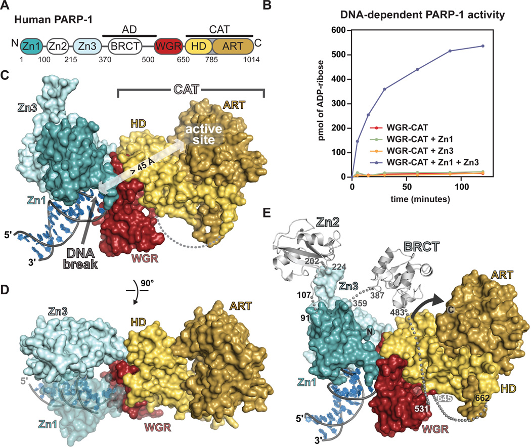
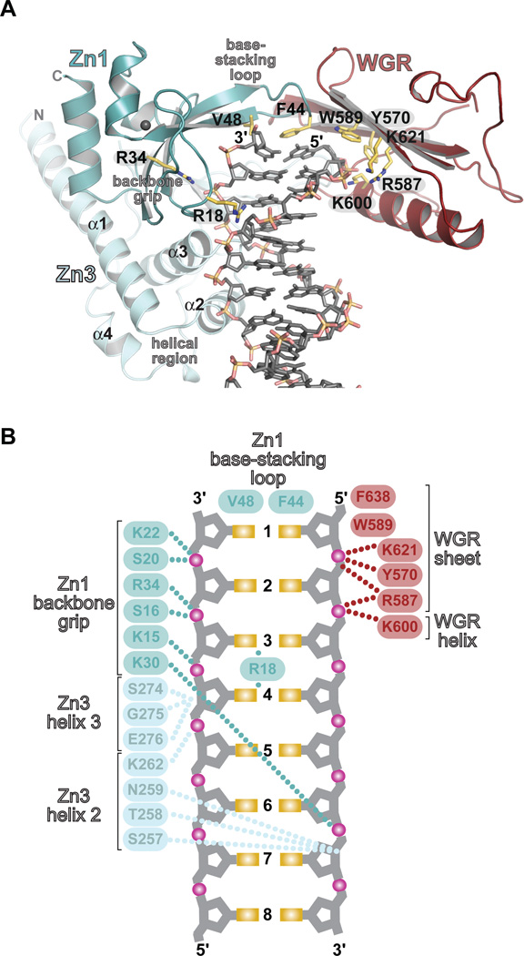
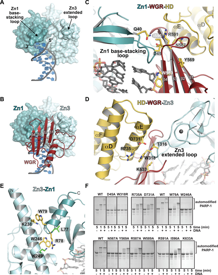
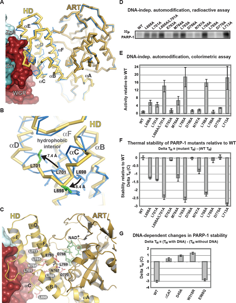
Poly(ADP-ribose) polymerase-1 (PARP-1) (ADP, adenosine diphosphate) has a modular domain architecture that couples DNA damage detection to poly(ADP-ribosyl)ation activity through a poorly understood mechanism. Here, we report the crystal structure of a DNA double-strand break in complex with human PARP-1 domains essential for activation (Zn1, Zn3, WGR-CAT). PARP-1 engages DNA as a monomer, and the interaction with DNA damage organizes PARP-1 domains into a collapsed conformation that can explain the strong preference for automodification. The Zn1, Zn3, and WGR domains collectively bind to DNA, forming a network of interdomain contacts that links the DNA damage interface to the catalytic domain (CAT). The DNA damage-induced conformation of PARP-1 results in structural distortions that destabilize the CAT. Our results suggest that an increase in CAT protein dynamics underlies the DNA-dependent activation mechanism of PARP-1.


Figures

Fig. 1
Fig. 1. Overview of the PARP-1/DNA crystal structure
(A) Modular domain architecture of human PARP-1. (B) Colorimetric assay of PARP-1 DNA-dependent automodification using the indicated domain combinations (see also Fig. S1 A,B). (C) Surface representation of the PARP-1/DNA structure. (D) A 90° rotation was applied to the view in C. (E) The Zn2 and BRCT domains (light grey) were manually positioned on the PARP-1/DNA structure using the structures of Zn2 and BRCT (PDB codes 3odc 2cok, respectively). The arrow accents the close proximity of AD and CAT.
Fig. 2
Fig. 2. PARP-1 forms a multi-domain DNA binding interface
Zn1, Zn3, and WGR collectively form an interface with DNA. (A) Key Zn1–, Zn3–, and WGR–DNA contacts are highlighted. (B) Schematic representation of PARP-1 contacts with DNA.
Fig. 3
Fig. 3. DNA-induced interdomain contacts are critical for DNA-dependent PARP-1 activation
(A) Surface representation of Zn1 and Zn3 bound to DNA. WGR and CAT have been omitted for clarity. (B) The Zn1 base-stacking loop, the Zn3 extended loop, and the 5'-terminated DNA strand form a binding site for WGR. (C) The Zn1–WGR–HD interface. (D) The HD-WGR–Zn3 interface. (E) The Zn3–Zn1 interface. Residues targeted for mutagenesis are labeled (yellow). Residues identified in a random screen for inactive mutants (20) are labeled (green). (F) SDS-PAGE assay of DNA-dependent PARP-1 automodification activity. WT and the indicated mutants were monitored for a shift in migration due to the covalent addition of PAR.
Fig. 4
Fig. 4. Distortion of the HD structure modulates PARP-1 catalytic activity
(A) Cα trace of CAT structure in the absence of DNA and regulatory domains (PDB code 1a26; blue), and CAT in the PARP-1/DNA structure (yellow/brown). (B) Detailed view of Leu698 and Leu701 contributions to the HD hydrophobic interior of the isolated CAT (blue), and their re-positioning in the PARP-1/DNA structure (yellow with green side chains). (C) Ribbon representation of CAT in the PARP-1/DNA complex. Residues mutated are drawn in pink and labeled: HD interior hydrophobic core (white), HD exterior (black). The position of NAD+ was modeled for reference. (D) Radioactive assay of DNA-independent PARP-1 automodification using 32P-NAD+. (E) Colorimetric assay of DNA-independent PARP-1 automodification. Values represent quantification of the 90-minute time point of a time course experiment (Fig. S7) and are an average of three independent experiments with associated standard deviations. (F) Relative thermal stability of PARP-1 mutants obtained by differential scanning fluorimetry. Changes in thermal stability (ΔTM) were calculated as shown. (G) Relative change in thermal stability upon DNA binding. ΔTM were calculated as shown.

Comment in

References

    1. D'Amours D, Desnoyers S, D'Silva I, Poirier GG. Biochem J. 1999 Sep 1;342(Pt 2):249. - PMC - PubMed
    1. Krishnakumar R, Kraus WL. Mol Cell. 2010 Jul 9;39:8. - PMC - PubMed
    1. Fong PC, et al. N Engl J Med. 2009 Jul 9;361:123. - PubMed
    1. Rouleau M, Patel A, Hendzel MJ, Kaufmann SH, Poirier GG. Nat Rev Cancer. 2010 Apr;10:293. - PMC - PubMed
    1. Langelier MF, Planck JL, Roy S, Pascal JM. J Biol Chem. 2011 Mar 25;286:10690. - PMC - PubMed

Publication types

Associated data