. 2012 Jul;122(2):295-307.
doi: 10.1111/j.1471-4159.2012.07787.x. Epub 2012 Jun 6.

Bi-directional regulation of CaMKIIα phosphorylation at Thr286 by NMDA receptors in cultured cortical neurons

Affiliations

Bi-directional regulation of CaMKIIα phosphorylation at Thr286 by NMDA receptors in cultured cortical neurons

Xianju Zhou et al. J Neurochem. 2012 Jul.

Abstract

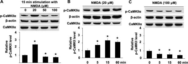
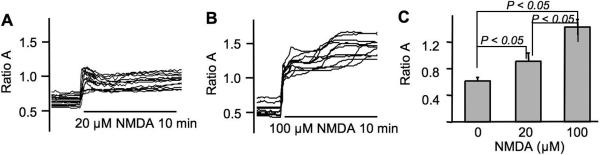
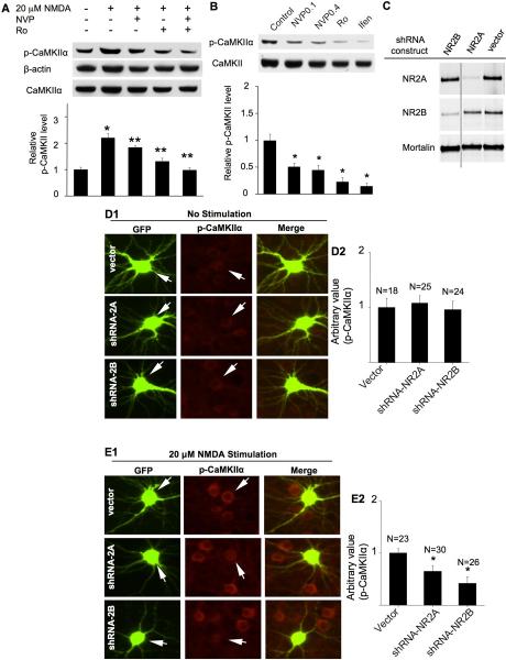
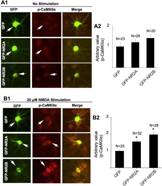
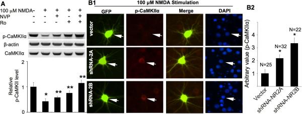
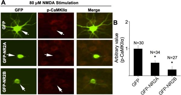
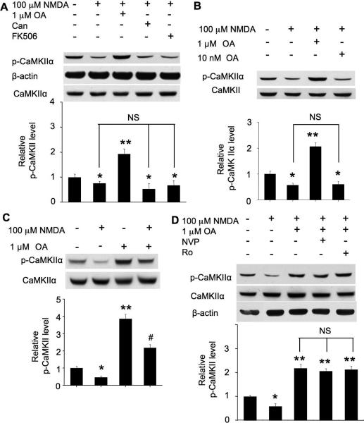
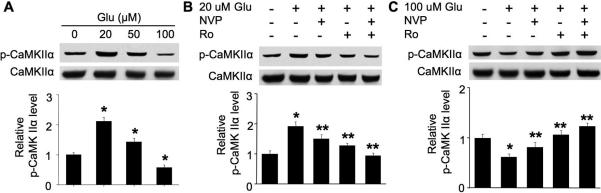
The N-methyl-D-aspartate (NMDA) receptor (NMDAR)-stimulated autophosphorylation of calmodulin-dependent kinase IIα at Thr286 may regulate many aspects of neuroplasticity. Here, we show that low NMDA concentration (20 μM) up-regulated Thr286 phosphorylation, and high concentration (100 μM) caused dephosphorylation. We next modulated the strength of NMDAR activation by manipulating NMDAR 2A subunit (NR2A) and NMDAR 2B subunit (NR2B), which represent the major NMDAR subtypes in forebrain regions. Pharmacological inhibition and molecular knockdown of NR2A or NR2B blocked 20 μM NMDA-induced phosphorylation. Conversely, over-expression of NR2A or NR2B enhanced phosphorylation by 20 μM NMDA. The 100 μM NMDA-induced dephosphorylation was suppressed by inhibition or knockdown of NR2A or NR2B, and enhanced by over-expression of NR2A or NR2B. Compared to NR2A, NR2B showed a higher impact on the NMDA-stimulated bi-directional regulation of Thr286 phosphorylation. We further found that activation of NR2A and NR2B by 100 μM NMDA-induced dephosphorylation through protein phosphatases (PP) that are inhibited by high concentration okadaic acid (1 μM), but not by PP2A and PP2B inhibitors. This novel function of NMDAR in dynamic regulation of calmodulin-dependent kinase IIα activity provides new evidence to support the current understanding that, depending on the degree of activation, NMDAR may lead to different and even opposing effects on intracellular signaling.


Figures

Fig. 1
Fig. 1
NMDA-mediated phosphorylation and dephosphorylation of CaMKIIα at Thr286 are concentration-dependent. A. DIV 14 neurons were stimulated by 20 μM or 50 μM or 100 μM NMDA for 15 min. Cellular extracts were analyzed by Western blot using antibodies against phospho-CaMKIIα (at Thr286), total CaMKIIα andβ-actin. B. DIV 14 neurons were stimulated by 20 μM NMDA for 5 min, 15 min, and 60 min. Phosphorylation of Thr286 was analyzed by Western blot. C. DIV 14 neurons were stimulated by 100 μM NMDA for 5 min, 15 min, and 60 min. Phosphorylation of Thr286 was analyzed by Western blot. For all treatment, NMDA was co-applied, TTX, CNQX, and nifedipine (as described in the method section). The top panels are representative Western blots. The bottom panels are qualifications of phospho-CaMKIIα at Thr286 from three independent experiments for each group (normalized to β-actin signal; the CaMKIIα signal is also shown, indicating that there is no obvious change after NMDA treatment). Data were quantified from multiple samples, and expressed as average +/− SEM. *: p < 0.05 between the treated samples and the control samples.
Fig. 2
Fig. 2
NMDA at low and high concentration causes significant elevation in intracellular calcium. The relative intracellular Ca2+ level was determined by Fura ratio (F340/380) with calcium imaging of 14–16 DIV cortical neurons. The ratio of F340/380 was collected from multiple neurons during the 10 min stimulation with 20 μM (A) or 100 μM NMDA (B). The quantification (average +/− SEM) is shown in C.
Fig. 3
Fig. 3
Role of NR2A- and NR2B-containing NMDAR in the up-regulation of CaMKIIα phosphorylation. DIV 14 cultures were pre-treated with TTX (1 μM), CNQX (40 μM), and nifedipine (5 μM) for 30 min for all experiments before NMDA treatment (15 min for A and 10 min for E). A. Pre-treatment with NVP-AAM077 (0.1 μM) or Ro 25-6981 (0.5 μM) were used to preferentially block the activation of NR2A or NR2B, respectively. B. DIV 14 neurons were treated with NVP-AAM077 (0.1 μM or 0.4 μM as indicated) or Ro25-6981 (0.5 μM) or ifenprodil (3 μM), as indicated, for 30 min. The level of phosphorylated CaMKIIα and total CaMKIIα was determined by Western blot. Top panels: representative images from three independent experiments. Bottom panels: quantification for Thr286 phosphorylation. *: p < 0.05 between the control and the NMDA-treated groups. **: p < 0.05 between the NMDA-treated and the inhibitor-pretreated groups. NVP: NVP-AAM077. Ro: Ro 25-6981. Ifen: ifenprodil. C. Knockdown of NR2A and NR2B in neurons. Neurons were transduced with lentivirus expressing shRNA-2Aa or shRNA-2Bi constructs. The expression level of NR2A, NR2B, and Mortalin (as a non-target control protein) was determined by Western blot. D and E. cortical neurons were co-transfected with GFP and the shRNA vector or shRNA-NR2Aa or shRNA-NR2Bi construct, as indicated, on DIV 12. On DIV 16, neurons were pre-treated with TTX (1 μM), CNQX (20 μM) and nifedipine (5 μM), and then fixed and co-stained for phosphorylated CaMKIIα (at Thr286) and GFP. In E, the neurons were fixed after a 10 min treatment with 20 μM NMDA. The level of Thr286 phosphorylation in representative neurons transfected with vector, or shRNA-NR2Aa, or shRNA-NR2Bi (as indicated by the arrows) is shown in D1 (no stimulation) and E1 (stimulated by 20 μM NDMA). The level of Thr286 phosphorylation in shRNA-transfected neurons was compared to that of the surrounding non-transfected neurons. For quantifications shown in D2 and E2, the level of Thr286 in neurons transfected with GFP and shRNA vector was defined as 1, and the immuno-intensity in neurons transfected with GFP and shRNA-2Aa or shRNA-2Bi was normalized to vector controls. The quantification is presented as average +/− SEM. *: p < 0.05, by ANOVA.
Fig. 4
Fig. 4
Overexpression of NR2A and NR2B enhances NMDA-induced phosphorylation at Thr286. Cortical neurons were transfected with plasmid expressing GFP or GFP-NR2A or GFP-NR2B at DIV 12. At DIV 16, non-stimulated neurons (A) or NMDA (20 μM for 10 min)-stimulated neurons (B) were fixed and co-stained for phosphorylated CaMKIIα (at Thr286) and GFP. The level of Thr286 phosphorylation in representative neurons transfected with GFP, or GFP-NR2A, or GFP-NR2B (as indicated by the arrows) is shown in A1 and B1. For quantifications shown in A2 and B2, the level of Thr286 phosphorylation in neurons transfected with GFP was defined as 1, and the immuno-intensity in neurons transfected with GFP-NR2A or GFP-NR2B was normalized to the GFP controls. The quantification is presented as average +/− SEM. *: p < 0.05, by ANOVA.
Fig. 5
Fig. 5
Pharmacological inhibition or knockdown of NR2A and NR2B attenuates NMDA-induced CaMKIIα dephosphorylation at Thr286. DIV 14 cultures were pre-treated with TTX (1 μM), CNQX (40 μM), and nifedipine (5 μM) for 30 min for all experiments before the 10 min NMDA (100 μM) treatment. A. Pre-treatment with NVP-AAM077 (0.1 μM) or Ro 25-6981 (0.5 μM) was used to preferentially block the activation of NR2A or NR2B, respectively. The level of phosphorylated CaMKIIα and total CaMKIIα was determined by Western blot. Top panels: representative results from three independent experiments. Bottom panels: quantification for Thr286 phosphorylation. *: p < 0.05 between the control and the NMDA-treated groups. **: p <0.05 between the NMDA-treated and the inhibitor-pretreated groups. NVP: NVP-AAM077. Ro: Ro 25-6981. B. cortical neurons were co-transfected with GFP and the shRNA vector or shRNANR2Aa or shRNA-NR2Bi construct, as indicated, at DIV 12. At DIV 16, neurons were pre-treated with TTX (1 μM), CNQX (20 μM) and nifedipine (5 μM). Neurons were fixed after a 10 min treatment with 100 μM NMDA. The level of Thr286 phosphorylation in representative neurons transfected with vector, or shRNA-NR2Aa, or shRNA-NR2Bi (as indicated by the arrows) is shown in B1. The quantification is presented in B2 as average +/− SEM. *: p < 0.05, by ANOVA.
Fig. 6
Fig. 6
Overexpression of NR2A or NR2B exaggerates CaMKIIα dephosphorylation after stimulation with high concentration of NMDA. Cortical neurons were transfected with plasmid expressing GFP or GFP-NR2A or GFP-NR2B at DIV 12. At DIV 16, neurons were first pre-treated with TTX (1 μM), CNQX (20 μM) and nifedipine (5 μM), and stimulated with 80 μM NMDA for 10 min. Neurons were then fixed and co-stained for phosphorylated CaMKIIα and GFP. The level of Thr286 phosphorylation in representative neurons transfected with GFP, or GFP-NR2A, or GFP-NR2B (as indicated by the arrows) is shown in A. The quantification is presented as average +/− SEM in B. *: p < 0.05, by ANOVA.
Fig. 7
Fig. 7
Role of protein phosphatases in NMDA-induced dephosphorylation at Thr286. Cultures were pre-treated with TTX (1 μM), CNQX (40 μM), and nifedipine (5 μM) for 30 min for all experiments before NMDA application. A. PP inhibitors, OA (1 μM) or cantharidin (100 nM) or FK506 (1 μM), was applied for 10 min before stimulation with 100 μM NMDA. Samples were collected 15 min after NMDA treatment, and Thr286 phosphorylation was examined by Western blot. B. The effects of 1 μM and 10 nM OA on NMDA (100 μM)-induced dephosphorylation. C. Neurons were treated as described in A. The effects of OA on Thr286 phosphorylation were examined with NMDA-stimulated or non-stimulated neurons, as indicated. D. Neurons were treated with NMDA similarly as in A. OA (1 μM) was applied with or without NVP-AAM077 (0.1 μM) or Ro 25-6981 (0.5 μM), as indicated, before NMDA. 15 min following NMDA treatment, samples were harvested and analyzed for Thr286 phosphorylation by Western blot. The top panels are representative Western blot results from three independent experiments. The bottom panels are quantification for the relative level of Thr286 phosphorylation. *: p < 0.05 when comparing with the control group. **: p < 0.05 when comparing with either control or NMDA-treated groups. #: p<0.05 when comparing with all other groups. NS: not statistically different between these groups. Can: cantharidin. NVP: NVP-AAM077. Ro: Ro 25-6981.
Fig. 8
Fig. 8
Role of NR2A and NR2B in bi-directional regulation of Thr286 phosphorylation induced by endogenous NMDAR agonist glutamate. DIV 14 neurons were first pre-treated with TTX, CNQX, and nifedipine for 30 min, and followed by stimulation with glutamate for 15 min. Samples were collected and analyzed for phosphorylation at Thr286 by Western blot. A. Glutamate at low concentrations (20 and 50 μM) up-regulates phosphorylation, and 100 μM glutamate causes dephosphorylation. B. Pharmacological inhibition of NR2A (by 0.1 μM NVPAAM077) or NR2B (by Ro 25-6981 at 0.5 μM) attenuates 20 μM glutamate-induced phosphorylation. C. Pharmacological inhibition of NR2A (by 0.1 μM NVP-AAM077) or NR2B (by Ro 25-6981 at 0.5 μM) attenuates 100 μM glutamate-induced dephosphorylation. The top panels are representative Western blot results from three independent experiments. The bottom panels are quantification for the relative level of Thr286 phosphorylation. *: p < 0.05 when comparing with the control group. **: p < 0.05 when comparing with the glutamate-treated groups.

Similar articles

Cited by

References

    1. Al-Hallaq RA, Conrads TP, Veenstra TD, Wenthold RJ. NMDA diheteromeric receptor populations and associated proteins in rat hippocampus. J Neurosci. 2007;27:8334–8343. - PMC - PubMed
    1. Barria A, Malinow R. NMDA receptor subunit composition controls synaptic plasticity by regulating binding to CaMKII. Neuron. 2005;48:289–301. - PubMed
    1. Bayer KU, De Koninck P, Leonard AS, Hell JW, Schulman H. Interaction with the NMDA receptor locks CaMKII in an active conformation. Nature. 2001;411:801–805. - PubMed
    1. Berberich S, Punnakkal P, Jensen V, Pawlak V, Seeburg PH, Hvalby O, Kohr G. Lack of NMDA receptor subtype selectivity for hippocampal long-term potentiation. J Neurosci. 2005;25:6907–6910. - PMC - PubMed
    1. Buard I, Coultrap SJ, Freund RK, Lee YS, Dell'Acqua ML, Silva AJ, Bayer KU. CaMKII “autonomy” is required for initiating but not for maintaining neuronal long-term information storage. J Neurosci. 2010;30:8214–8220. - PMC - PubMed

Publication types

MeSH terms