

A description of the lumbar interfascial triangle and its relation with the lateral raphe: anatomical constituents of load transfer through the lateral margin of the thoracolumbar fascia
- PMID: 22582887
- PMCID: PMC3512280
- DOI: 10.1111/j.1469-7580.2012.01517.x
A description of the lumbar interfascial triangle and its relation with the lateral raphe: anatomical constituents of load transfer through the lateral margin of the thoracolumbar fascia
Abstract
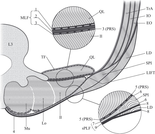
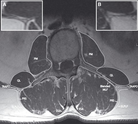
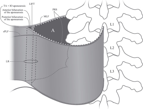
Movement and stability of the lumbosacral region is contingent on the balance of forces distributed through the myofascial planes associated with the thoracolumbar fascia (TLF). This structure is located at the common intersection of several extremity muscles (e.g. latissimus dorsi and gluteus maximus), as well as hypaxial (e.g. ventral trunk muscles) and epaxial (paraspinal) muscles. The mechanical properties of the fascial constituents establish the parameters guiding the dynamic interaction of muscle groups that stabilize the lumbosacral spine. Understanding the construction of this complex myofascial junction is fundamental to biomechanical analysis and implementation of effective rehabilitation in individuals with low back and pelvic girdle pain. Therefore, the main objectives of this study were to describe the anatomy of the lateral margin of the TLF, and specifically the interface between the fascial sheath surrounding the paraspinal muscles and the aponeurosis of the transversus abdominis (TA) and internal oblique (IO) muscles. The lateral margin of the TLF was exposed via serial reduction dissections from anterior and posterior approaches. Axial sections (cadaveric and magnetic resonance imaging) were examined to characterize the region between the TA and IO aponeurosis and the paraspinal muscles. It is confirmed that the paraspinal muscles are enveloped by a continuous paraspinal retinacular sheath (PRS), formed by the deep lamina of the posterior layer of the TLF. The PRS extends from the spinous process to transverse process, and is distinct from both the superficial lamina of the posterior layer and middle layer of the TLF. As the aponeurosis approaches the lateral border of the PRS, it appears to separate into two distinct laminae, which join the anterior and posterior walls of the PRS. This configuration creates a previously undescribed fat-filled lumbar interfascial triangle situated along the lateral border of the paraspinal muscles from the 12th rib to the iliac crest. This triangle results in the unification of different fascial sheaths along the lateral border of the TLF, creating a ridged-union of dense connective tissue that has been termed the lateral raphe (Spine, 9,1984, 163). This triangle may function in the distribution of laterally mediated tension to balance different viscoelastic moduli, along either the middle or posterior layers of the TLF.
© 2012 The Authors. Journal of Anatomy © 2012 Anatomical Society.
Figures

Similar articles
-
Organization of the fascia and aponeurosis in the lumbar paraspinal compartment.Surg Radiol Anat. 2018 Nov;40(11):1231-1242. doi: 10.1007/s00276-018-2087-0. Epub 2018 Aug 31. Surg Radiol Anat. 2018. PMID: 30171298
-
The thoracolumbar fascia: anatomy, function and clinical considerations.J Anat. 2012 Dec;221(6):507-36. doi: 10.1111/j.1469-7580.2012.01511.x. Epub 2012 May 27. J Anat. 2012. PMID: 22630613 Free PMC article. Review.
-
The functional coupling of the deep abdominal and paraspinal muscles: the effects of simulated paraspinal muscle contraction on force transfer to the middle and posterior layer of the thoracolumbar fascia.J Anat. 2014 Oct;225(4):447-62. doi: 10.1111/joa.12227. Epub 2014 Aug 20. J Anat. 2014. PMID: 25139243 Free PMC article.
-
Anatomical and functional relationships between external abdominal oblique muscle and posterior layer of thoracolumbar fascia.Clin Anat. 2018 Oct;31(7):1092-1098. doi: 10.1002/ca.23248. Epub 2018 Oct 26. Clin Anat. 2018. PMID: 30113090
-
The anatomical myofascial continuum between the neck and eyes.Clin Anat. 2022 Apr;35(3):340-346. doi: 10.1002/ca.23835. Epub 2022 Feb 1. Clin Anat. 2022. PMID: 35043988 Review.
Cited by
-
Local anesthetic spread into the paravertebral space with two types of quadratus lumborum blocks: a crossover volunteer study.J Anesth. 2019 Feb;33(1):26-32. doi: 10.1007/s00540-018-2578-5. Epub 2018 Nov 9. J Anesth. 2019. PMID: 30413879 Clinical Trial.
-
Anatomical study of paratenons and fascia lata connections in the posteromedial knee region.Surg Radiol Anat. 2022 Jun;44(6):821-827. doi: 10.1007/s00276-022-02927-6. Epub 2022 Mar 22. Surg Radiol Anat. 2022. PMID: 35316382
-
The feasibility and impact of instrument-assisted manual therapy (IAMT) for the lower back on the structural and functional properties of the lumbar area in female soccer players: a randomised, placebo-controlled pilot study design.Pilot Feasibility Stud. 2020 Apr 16;6:47. doi: 10.1186/s40814-020-00592-3. eCollection 2020. Pilot Feasibility Stud. 2020. PMID: 32322406 Free PMC article.
-
Anatomic connections of the diaphragm: influence of respiration on the body system.J Multidiscip Healthc. 2013 Jul 25;6:281-91. doi: 10.2147/JMDH.S45443. Print 2013. J Multidiscip Healthc. 2013. PMID: 23940419 Free PMC article.
-
Organization of the fascia and aponeurosis in the lumbar paraspinal compartment.Surg Radiol Anat. 2018 Nov;40(11):1231-1242. doi: 10.1007/s00276-018-2087-0. Epub 2018 Aug 31. Surg Radiol Anat. 2018. PMID: 30171298
References
-
- Armstrong O, Hamel A, Grignon B, et al. Lumbar hernia: anatomical basis and clinical aspects. Surg Radiol Anat. 2008;30:533–537. discussion 609–510. - PubMed
-
- Astarcioglu H, Sokmen S, Atila K, et al. Incarcerated inferior lumbar (Petit’s) hernia. Hernia. 2003;7:158–160. - PubMed
-
- Bailey FR, Miller AM. Textbook of Embryology. 3rd edn. New York: William Wood; 1916.
-
- Barker PJ, Briggs CA. Attachments of the posterior layer of lumbar fascia. Spine. 1999;24:1757–1764. - PubMed
-
- Barker PJ, Brigg CA, Bogeski G. Montreal, QC, Canada: 2001. Muscle attachments of the lumbar spine; pp. 238–239. 4th Interdisciplinary World Congress on Low Back and Pelvic Pain.
MeSH terms
LinkOut - more resources
Full Text Sources
Medical
Research Materials
