. 2012 Jun 5;51(22):4453-62.
doi: 10.1021/bi3003204. Epub 2012 May 24.

Characterization of the [2Fe-2S] cluster of Escherichia coli transcription factor IscR

Affiliations

Characterization of the [2Fe-2S] cluster of Escherichia coli transcription factor IscR

Angela S Fleischhacker et al. Biochemistry. .

Abstract

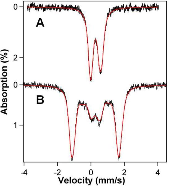
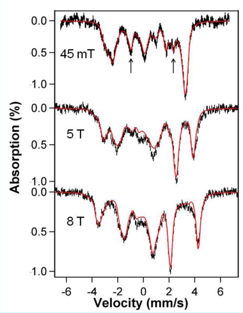
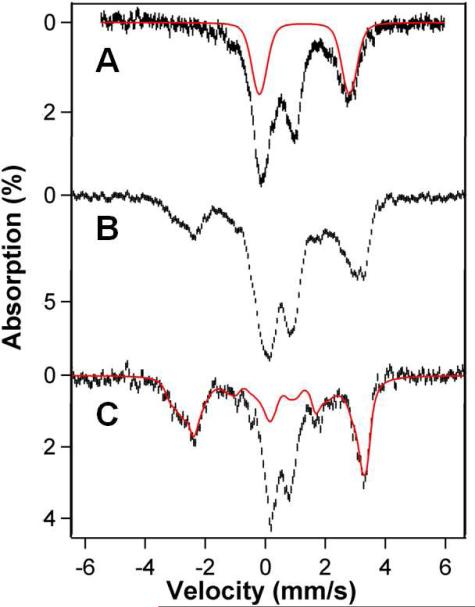
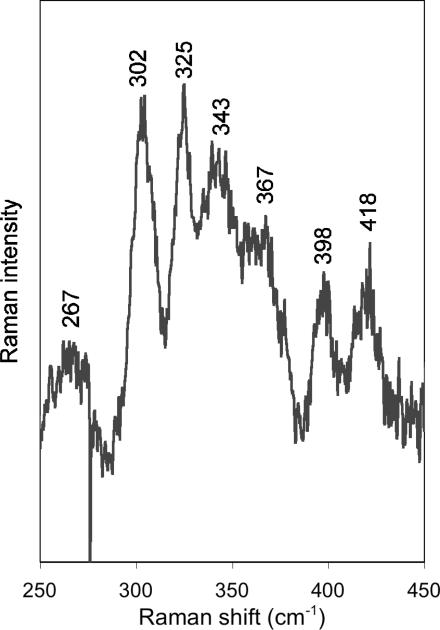
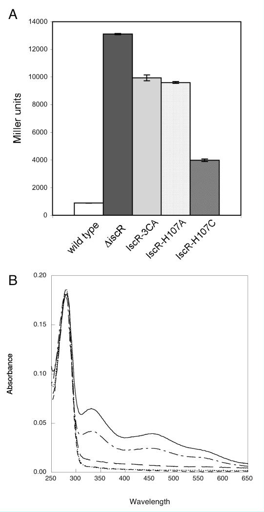
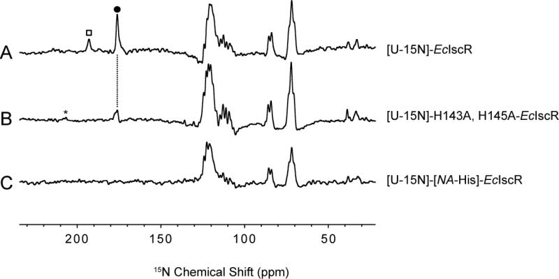
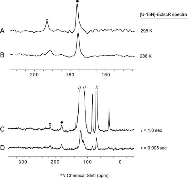
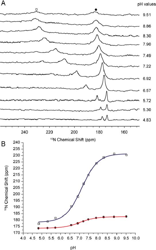
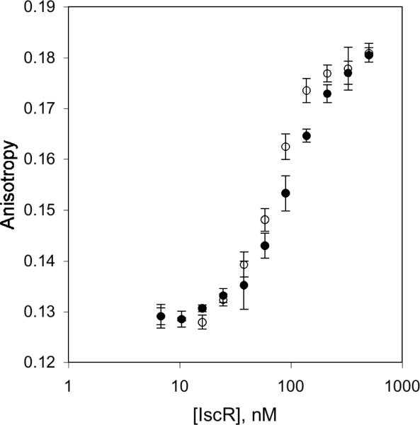
IscR is an Fe-S cluster-containing transcription factor involved in a homeostatic mechanism that controls Fe-S cluster biogenesis in Escherichia coli. Although IscR has been proposed to act as a sensor of the cellular demands for Fe-S cluster biogenesis, the mechanism by which IscR performs this function is not known. In this study, we investigated the biochemical properties of the Fe-S cluster of IscR to gain insight into the proposed sensing activity. Mössbauer studies revealed that IscR contains predominantly a reduced [2Fe-2S](+) cluster in vivo. However, upon anaerobic isolation of IscR, some clusters became oxidized to the [2Fe-2S](2+) form. Cluster oxidation did not, however, alter the affinity of IscR for its binding site within the iscR promoter in vitro, indicating that the cluster oxidation state is not important for regulation of DNA binding. Furthermore, characterization of anaerobically isolated IscR using resonance Raman, Mössbauer, and nuclear magnetic resonance spectroscopies leads to the proposal that the [2Fe-2S] cluster does not have full cysteinyl ligation. Mutagenesis studies indicate that, in addition to the three previously identified cysteine residues (Cys92, Cys98, and Cys104), the highly conserved His107 residue is essential for cluster ligation. Thus, these data suggest that IscR binds the cluster with an atypical ligation scheme of three cysteines and one histidine, a feature that may be relevant to the proposed function of IscR as a sensor of cellular Fe-S cluster status.


Figures

Figure 1
Figure 1
4.2 K Mössbauer spectra of anaerobically isolated IscR that has been exposed to air recorded in zero field (A) and for B = 8.0 T (B). Solid lines are simulations assuming a cluster with S = 0 using the nested set of parameters: ΔEQ(1) = -0.48 mm/s, δ(1) = 0.27 mm/s, η(1) = 0.5; ΔEQ(2) = +0.72 mm/s, δ(2) = 0.30 mm/s, η(2) = 0.5.
Figure 2
Figure 2
4.2 K Mössbauer spectra of dithionite reduced IscR recorded in parallel applied field as indicated. The solid red lines are a spectral simulation based on eq 1 using the parameters listed in Table 1. For the simulation of the 45 mT spectrum we have added a doublet for a minor contaminant (8%, arrows) with ΔEQ = 3.35 mm/s and δ = 0.70 mm/s.
Figure 3
Figure 3
Mössbauer spectra of whole cells recorded at 4.2 K in parallel applied fields on 45 mT. (A) Spectrum of whole E. coli cells without overexpression of IscR. Solid red line outlines contribution of a collection of high-spin Fe2+ species. (B) Spectrum of whole cells overexpressing wild type IscR. (C) Spectrum obtained by subtracting from (B) the spectrum of (A), assuming that it represents 50% of the Fe in sample. The procedure gives a good view of the features of the IscR cluster. The red line is the spectral simulation (representing 32% of Fe) from Figure 2 A (without the 8% contaminant).
Figure 4
Figure 4
77 K resonance Raman spectrum of anaerobically isolated IscR (47% occupancy, ~3 mM), obtained with laser excitation at 488 nm and a power of 100 mW at the sample (resolution is ~6 cm-1). The average of nine scans is presented here, and the contributions from ice lattice vibrations were removed by subtracting a properly scaled spectrum of buffer obtained under identical conditions.
Figure 5
Figure 5
(A) Expression levels of the iscR promoter fused to lacZ were determined in strains containing wild type IscR (white bar), ΔiscR (black bar), IscR-C92A/C98A/C104 (IscR-3CA, gray bar), IscR-H107A (light patterned bar), or IscR-H107C (dark patterned bar). Strains were grown under anaerobic conditions in MOPS minimal media containing 0.2% glucose and were assayed for β-galactosidase activity per OD600 (Miller units). (B) Optical spectra of IscR variants. The spectra of wild type IscR (solid line), IscR-E43A (— - —), IscR-C92A/C98A/C104A (— —), IscR-H107A (- - -), and IscR-H107C (. . . .) were obtained under anaerobic conditions in 10 mM HEPES, pH 7.4, with 200 mM KCl at room temperature (10 μM protein). All variants were isolated under anaerobic conditions as described in the Methods.
Figure 6
Figure 6
15N-NMR spectra of ~2 mM reduced IscR samples at pH 6.4 collected at 298 K under rapid pulsing conditions. (A) [U-15N]-IscR(WT) produced from cells grown on 15NH4Cl as the sole nitrogen source. (B) [U-15N]-IscR(H143A, H145A) produced from cells grown on 15NH4Cl as the sole nitrogen source. (C) [U-15N]-IscR(WT) produced from cells grown on 15NH4Cl in the presence of unlabeled L-histidine. Together, these spectra show that the indicated signals (□ and •) in (A) arise from histidine residues and that the signal labeled with a closed dot (•) is from His107, the one His residue not removed by substitution in the sample whose spectrum is shown in (B). The peak labeled with an open square (□) appeared in other spectra of [U-15N]-IscR(H143A, H145A), not shown. The peak labeled with an asterisk (*) is from 14N15N in nitrogen gas in air.
Figure 7
Figure 7
Evidence from 15N NMR spectra that the labeled signals (□ and •) of reduced [U-15N]-IscR(WT) arise from nuclei affected by electron-nuclear (hyperfine) interactions. (A, B) The chemical shifts of the labeled peaks exhibit anti-Curie shifts as a function of temperature as expected for an Fe-S cluster ligand (delay value 5.0 ms). (C, D) Spectra taken as two different delay values (1.0 s and 5.0 ms) between the data collection pulses suggest that the labeled peaks correspond to rapidly relaxing nuclei as expected for a cluster ligand.
Figure 8
Figure 8
(A) Dependence of 15N NMR spectra of 2 mM reduced [U-15N]-IscR(WT) collected at 298 K on the pH of the sample. (B) Plot of pH dependence of the chemical shifts of peak 1 (□) and peak 2 (•). The data were fitted to theoretical curves, which yielded pKa1 = 7.12 ± 0.03 (blue line) and pKa2 = 7.04 ± 0.02 (red line). The uncertainties are those from curve fitting. We consider these pKa values to be equal within experimental error.
Figure 9
Figure 9
In vitro DNA binding assays. As isolated IscR was exposed to oxygen and then tested for its ability to bind to double stranded DNA containing the iscR binding site in the absence (filled circles) or presence (open circles) of 10 μM dithionite.

References

    1. Giel JL, Rodionov D, Liu MZ, Blattner FR, Kiley PJ. IscR-dependent gene expression links iron-sulphur cluster assembly to the control of O2-regulated genes in Escherichia coli. Mol. Microbiol. 2006;60:1058–1075. - PubMed
    1. Schwartz CJ, Giel JL, Patschkowski T, Luther C, Ruzicka FJ, Beinert H, Kiley PJ. IscR, an Fe-S cluster-containing transcription factor, represses expression of Escherichia coli genes encoding Fe-S cluster assembly proteins. Proc. Natl. Acad. Sci. U. S. A. 2001;98:14895–14900. - PMC - PubMed
    1. Frazzon J, Dean DR. Feedback regulation of iron-sulfur cluster biosynthesis. Proc. Natl. Acad. Sci. U. S. A. 2001;98:14751–14753. - PMC - PubMed
    1. Yeo WS, Lee JH, Lee KC, Roe JH. IscR acts as an activator in response to oxidative stress for the suf operon encoding Fe-S assembly proteins. Mol. Microbiol. 2006;61:206–218. - PubMed
    1. Nesbit AD, Giel JL, Rose JC, Kiley PJ. Sequence-specific binding to a subset of IscR-regulated promoters does not require IscR Fe-S cluster ligation. J. Mol. Biol. 2009;387:28–41. - PMC - PubMed

Publication types

MeSH terms

LinkOut - more resources