. 2012 May 14;14(3):R77.
doi: 10.1186/bcr3187.

Downregulation of the tumor-suppressor miR-16 via progestin-mediated oncogenic signaling contributes to breast cancer development

Affiliations

Downregulation of the tumor-suppressor miR-16 via progestin-mediated oncogenic signaling contributes to breast cancer development

Martin A Rivas et al. Breast Cancer Res. .

Abstract

Introduction: Experimental and clinical evidence points to a critical role of progesterone and the nuclear progesterone receptor (PR) in controlling mammary gland tumorigenesis. However, the molecular mechanisms of progesterone action in breast cancer still remain elusive. On the other hand, micro RNAs (miRNAs) are short ribonucleic acids which have also been found to play a pivotal role in cancer pathogenesis. The role of miRNA in progestin-induced breast cancer is poorly explored. In this study we explored progestin modulation of miRNA expression in mammary tumorigenesis.

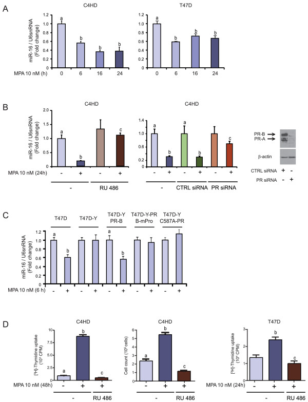
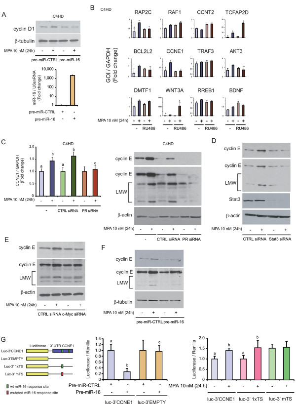
Methods: We performed a genome-wide study to explore progestin-mediated regulation of miRNA expression in breast cancer. miR-16 expression was studied by RT-qPCR in cancer cell lines with silenced PR, signal transducer and activator of transcription 3 (Stat3) or c-Myc, treated or not with progestins. Breast cancer cells were transfected with the precursor of miR-16 and proliferation assays, Western blots or in vivo experiments were performed. Target genes of miR-16 were searched through a bioinformatical approach, and the study was focused on cyclin E. Reporter gene assays were performed to confirm that cyclin E 3'UTR is a direct target of miR-16.

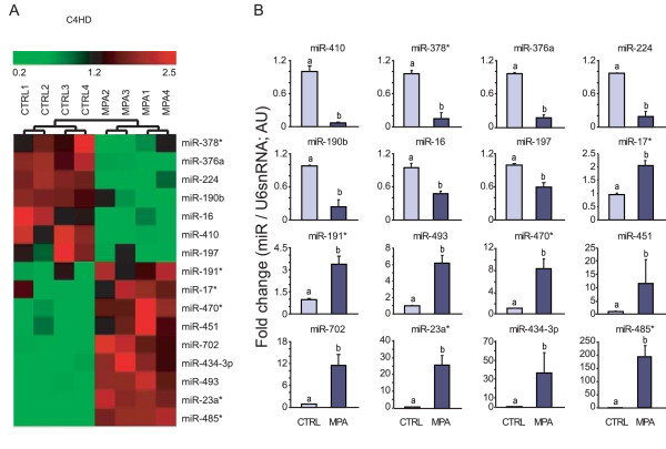
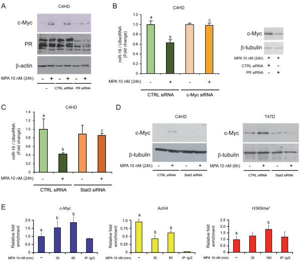
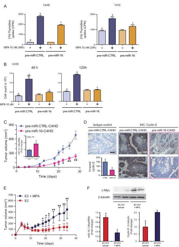
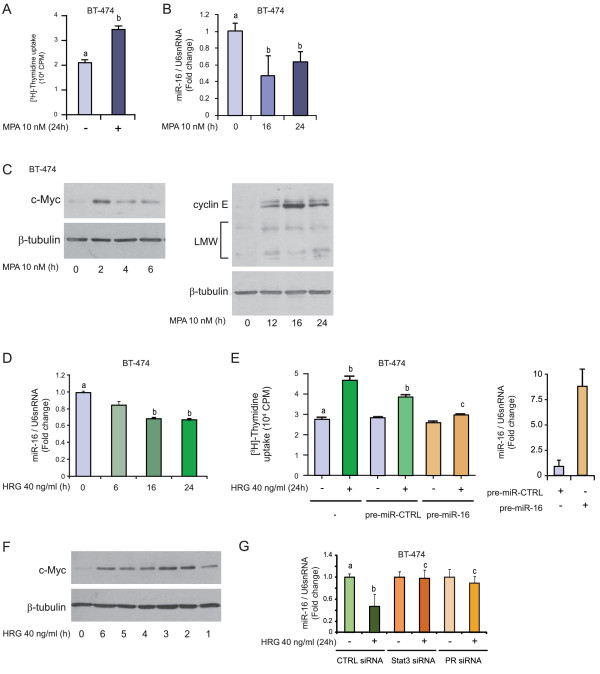
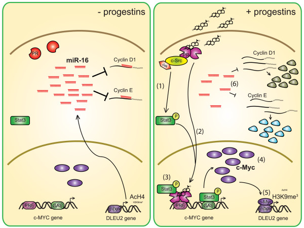
Results: We found that nine miRNAs were upregulated and seven were downregulated by progestin in mammary tumor cells. miR-16, whose function as a tumor suppressor in leukemia has already been shown, was identified as one of the downregulated miRNAs in murine and human breast cancer cells. Progestin induced a decrease in miR-16 levels via the classical PR and through a hierarchical interplay between Stat3 and the oncogenic transcription factor c-Myc. A search for miR-16 targets showed that the CCNE1 gene, encoding the cell cycle regulator cyclin E, contains conserved putative miR-16 target sites in its mRNA 3' UTR region. We found that, similar to the molecular mechanism underlying progestin-modulated miR-16 expression, Stat3 and c-Myc participated in the induction of cyclin E expression by progestin. Moreover, overexpression of miR-16 abrogated the ability of progestin to induce cyclin E upregulation, revealing that cyclin E is a novel target of miR-16 in breast cancer. Overexpression of miR-16 also inhibited progestin-induced breast tumor growth in vitro and in vivo, demonstrating for the first time, a role for miR-16 as a tumor suppressor in mammary tumorigenesis. We also found that the ErbB ligand heregulin (HRG) downregulated the expression of miR-16, which then participates in the proliferative activity of HRG in breast tumor cells.

Conclusions: In this study, we reveal the first progestin-regulated miRNA expression profile and identify a novel role for miR-16 as a tumor suppressor in progestin- and growth factor-induced growth in breast cancer.


Figures

Figure 1
Figure 1
Progestins modulate miRNAs expression in breast cancer cells. A, Heat map depicting the expression profile of miRNA genes with changes ≥2 fold after 24 hours of MPA treatment. Total RNA was extracted from primary cultures of C4HD cells treated with 10 nM MPA or left untreated for 24 hours and used for miRNA profiling with the Applied Biosystems Mouse Low Density qPCR miRNA Array (n = 4). The small nuclear RNA U6 (U6 snRNA) was used as an endogenous control to normalize the results. B, Average fold changes of miRNAs significantly modulated by MPA (P < 0.05, n = 4) in primary cultures of C4HD cells. Each graph depicts the fold change of a specific miRNA in C4HD cells treated with MPA or left untreated; all values were normalized to U6 snRNA. The data shown represent the means of three independent experiments ± SEM (P < 0.01 for b versus a). MPA, medroxyprogesterone acetate; SEM, standard error of the mean.
Figure 2
Figure 2
Progestins induce miR-16 downregulation via the classical PR. A, C4HD and T47D cells were treated with 10 nM MPA for the times shown. B, C4HD cells were either pretreated with 10 nM RU486 (left panel) or transfected with 100 nM PR and control (CTRL) siRNAs, and were then stimulated with MPA for 24 hours or left untreated (middle panel). The western blot (WB) in the right panel shows the effect of siRNAs on PR expression in C4HD cells. The experiment shown was performed with PR siRNA #1, but the same results were obtained with PR siRNA #2. C, T47D and T47D-Y cells were treated with MPA for the indicated time or T47D-Y cells were transiently transfected with the PR-B isoform (T47D-Y-PR-B), PR-BmPro mutant (T47D-Y-PR-BmPro cells) or the C587A-PR mutant (T47D-YC587A-PR cells) before MPA stimulation. In A to C, miR-16 expression levels were determined by RT-qPCR. The fold change of miR-16 expression levels upon MPA treatment was calculated by normalizing the absolute levels of miR-16 to those of U6 snRNA, which was used as internal control, and setting the value of untreated cells to 1. D, C4HD cells were treated with 10 nM MPA for 48 hours (left panel) or T47D cells were treated with 10 nM MPA for 24 hours (right panel) and the incorporation of [3H]-thymidine was used as a measure of DNA synthesis. The middle panel shows cell counts for C4HD cells that were treated with 10 nM MPA for 48 hours and then stained with Trypan blue dye. Experiments shown in A to D were repeated in triplicate with similar results. The data shown represent the means of three independent experiments ± SEM (P < 0.001 for b versus a and c versus b). MPA, medroxyprogesterone acetate; PR, progesterone receptor; SEM, standard error of the mean.
Figure 3
Figure 3
Progestin induces miR-16 downregulation via c-Myc and Stat3. A, C4HD cells were treated with MPA or transfected with PR siRNAs or CTRL siRNAs before MPA stimulation. Western blot (WB) was performed with anti-c-Myc or anti-PR antibodies and filters were reprobed with an anti-β-actin antibody. The experiment shown was performed with PR siRNA #1, but the same results were obtained with PR siRNA #2. B, C4HD cells were transfected with c-Myc and CTRL siRNAs and were then treated with MPA. miR-16 levels were studied by RT-qPCR, and data analysis was performed as described in Figure 2. The WB in the right side of the figure shows the effects of siRNAs on c-Myc expression in C4HD cells. The experiment shown was performed with c-Myc siRNA #5, but the same results were obtained with c-Myc siRNA #6. C, C4HD cells were transfected with Stat3 siRNAs or CTRL siRNAs and then treated with MPA for 24 hours. miR-16 levels were studied by RT-qPCR, and data analysis was performed as described in Figure 2. The experiment shown was performed with Stat3 siRNA #1, but the same results were obtained with PR siRNA #3. D, C4HD and T47D cells were transfected with Stat3 siRNAs or CTRL siRNAs before MPA stimulation and WBs were performed with anti-c-Myc antibodies. Filters were reprobed with an anti-β-tubulin antibody. The experiment shown was performed with Stat3 siRNA #1, but the same results were obtained with PR siRNA #3. E, Recruitment of c-Myc, and H4 acetylation (AcH4) or trimethylation of lysine 9 histone H3 (H3K9me3) levels at the promoter of the DLEU2 gene was studied by ChIP. Amounts of immunoprecipitated DNA were normalized to inputs and are reported relative to the untreated control group, which was set to 1 (P < 0.001 for b versus a). Experiments shown in A to E were repeated in triplicate with similar results. Data shown represent the means of three independent experiments ± SEM (P < 0.001 for b versus a and c versus b). ChIP, chromatin immunoprecipitation; MPA, medroxyprogesterone acetate; SEM, standard error of the mean.
Figure 4
Figure 4
Cyclin E is a target of miR-16 in breast cancer cells. A, Upper panel, C4HD cells were transfected with pre-miR-16 or pre-miR-control (CTRL) before MPA stimulation. WB was performed with an anti-cyclin D1 antibody, and filters were reprobed with an anti-β-tubulin antibody. Bottom panel, as a control of transfection efficiency, miR-16 levels are shown in pre-miR-16-C4HD and pre-miR-CTRL-C4HD cells. B, C4HD cells were treated with MPA or pretreated with 10 nM RU486 before MPA stimulation, and mRNA expression levels of candidate miR-16 target genes were determined by RT-qPCR. The fold change of mRNA expression levels was calculated by normalizing the absolute levels of the gene-of-interest (GOI) mRNA to GAPDH levels, which were used as an internal control, and setting the value of untreated cells to 1. RAP2C, member of RAS oncogene family; RAF1, v-raf-1 murine leukemia viral oncogene homolog 1; CCNT2, cyclin T2; TCFAP2D, transcription factor AP-2 delta; BCL2L2, BCL2-like 2; CCNE1, cyclin E; TRAF3, TNF receptor-associated factor 3; Akt3, v-akt murine thymoma viral oncogene homolog 3; DMTF1, cyclin D binding myb-like transcription factor 1; WNT3A, wingless-type MMTV integration site family, member 3A; RREB1, ras responsive element binding protein 1; BDNF, brain-derived neurotrophic factor. C, C4HD cells were transfected with PR and CTRL siRNAs and then treated with MPA for 24 hours. Left panel, cyclin E mRNA levels were studied by RT-qPCR and data analysis was performed as described in Figure 2B. Right panel, WB was performed with an anti-cyclin E antibody and filters were reprobed with an anti-β-actin antibody. Longer exposures showing the expression of the low molecular weight (LMW) cyclin E isoforms are shown in the middle panel. The experiment shown was performed with PR siRNA #1, but the same results were obtained with PR siRNA #2. D, C4HD cells were transfected with Stat3 or CTRL siRNAs and were then treated with MPA or remained untreated. WB was performed as described in C. As a control for siRNA efficiency, the membranes were probed with an anti-Stat3 antibody. The experiment shown was performed with Stat3 siRNA #1, but the same results were obtained with PR siRNA #3. E, C4HD cells were transfected with c-Myc or CTRL siRNAs and then treated with MPA. WB was performed as in Figure 4C. The experiment shown was performed with c-Myc siRNA #5, but the same results were obtained with c-Myc siRNA #6. F, C4HD cells were transfected with pre-miR-16 or pre-miR-CTRL before MPA stimulation and WB was performed as in C. G, A scheme depicting the different constructions used is shown in the left panel. C4HD cells were transfected with a construct carrying the CCNE1 3' UTR cloned downstream of the firefly luciferase reporter gene (luc-3'CCNE1), middle panel, or with a construct that carried a minimal region of CCNE1 3'UTR which comprised only one of the miR-16 responding sites either wild type (luc-3' 1×TS) or mutated (luc-3' mTS), right panel. As a control, cells were transfected with a construct that lacks the 3' UTR cloned downstream of the luciferase gene (luc-3'EMPTY). Cells were co-transfected with pre-miR-16 or pre-miR-CTRL (middle panel) or treated with 10 nM MPA for 24 hours (right panel). Firefly luciferase activity was measured as described in the Methods. Renilla luciferase was used for normalization. The experiments shown in A to G were repeated in triplicate with similar results. The data shown represent the means of three independent experiments ± SEM (P < 0.001 for b versus a and c versus b). MPA, medroxyprogesterone acetate; PR, progesterone receptor; SEM, standard error of the mean; WB, western blot.
Figure 5
Figure 5
miR-16 is a tumor suppressor in progestin-induced breast cancer growth in vitro and in vivo. A, miR-16 inhibits in vitro progestin-induced breast cancer growth. C4HD or T47D cells were transfected with pre-miR-CTRL or pre-miR-16. After 48 hours of transfection, cells were treated with 10 nM MPA or left untreated, and proliferation was measured by [3H]-thymidine incorporation as described in Figure 2D. B, C4HD cells were transfected with pre-miR-CTRL or pre-miR-16. After 48 hours, cells were treated with 10 nM MPA for 48 or 120 hours or left untreated, and proliferation was measured by cell count as described in Figure 2D. The experiments shown in A and B were repeated four times with similar results. The data shown represent the means of the data from three independent experiments ± SEM (P < 0.001 for b versus a and c versus b). C, miR-16 inhibits in vivo progestin-induced breast cancer growth. C4HD cells were transfected with pre-miR-CTRL or pre-miR-16 for 48 hours and then injected subcutaneously (s.c.) into BALB/c mice at 2 × 10⁶ cells/mouse. Mice were simultaneously injected with a 40 mg MPA depot. Tumor volume was calculated as described in the Methods. Each point represents the mean volume ± SEM of six independent tumors for both experimental groups. The experiment shown in C was repeated twice with similar results. *P < 0.01 or **P < 0.001 versus control. Inset, levels of pre-miR-16 in pre-miR-CTRL-C4HD and pre-miR16-C4HD tumors were studied by RT-qPCR at day 14; data analysis was performed as described in Figure 2. D, Cyclin E is an in vivo target of miR-16. Immunohistochemistry (IHC) for cyclin E in pre-miR-CTRL-C4HD and pre-miR-16-C4HD tumors (400×). Representative images are shown. As control, IHC was performed using an irrelevant rabbit antibody. Scale bar, 50 μM. Inset, average H-score, used to quantify the levels of cyclin E in pre-miR-CTRL-C4HD and pre-miR-16-C4HD tumors. E, BT-474.m1 cells were injected s.c. into nude mice at 20 × 106 cells/mouse. Mice were simultaneously injected with a 0.72 mg E2 depot. Seven days after cell injection, half of the mice were injected with a 40 mg MPA depot (arrow). Tumor volume was calculated as described in Methods. Each point represents the mean volume ± SEM of six independent tumors for both experimental groups. *P < 0.01 or **P < 0.001 versus control. F, c-Myc WB was performed in whole protein extracts from BT-474 tumors growing into mice treated or not with MPA (upper panel). WB from two representative animals from each group is shown. miR-16 levels were measured in RNA from BT-474 tumors from mice treated or not with MPA (lower-left panel). Quantification of cyclin E from WB performed on whole protein extracts from BT-474 tumors from mice treated or not with MPA (lower-right panel). MPA, medroxyprogesterone acetate; SEM, standard error of the mean; WB, western blot.
Figure 6
Figure 6
miR-16 is a tumor suppressor in HRG-induced breast cancer growth. A, BT-474 cells were treated with 10 nM MPA for 24 hours and proliferation was measured by [3H]-thymidine incorporation as described in Figure 2D. B, BT-474 cells were treated with 10 nM MPA for the times shown. miR-16 expression levels were determined by RT-qPCR, and data analysis was performed as described in Figure 2A. C, BT-474 cells were treated with MPA for the times shown and WB was performed with an anti-c-Myc antibody (left panel) or with an anti-cyclin E antibody (right panel) and filters were reprobed with an anti-β-tubulin antibody. In the WB, cyclin E LMW isoforms are shown. D, BT-474 cells were treated with 40 ng/ml HRG for the times shown and miR-16 levels were measured as described in Figure 2A. E, BT-474 cells were transfected with pre-miR-16 or pre-miR-CTRL, and proliferation was evaluated by [3H]-thymidine uptake as described in Figure 2D after 24 hours of HRG treatment. In the right panel, as a control for transfection efficiency, miR-16 levels are shown in pre-miR-16- and pre-miR-CTRL-transfected BT-474 cells. F, BT-474 cells were treated with 40 ng/ml HRG for the times shown, and WB was performed with anti-c-Myc antibody and filters were reprobed with an anti-β-tubulin antibody. G, BT-474 cells were transfected with Stat3, c-Myc and CTRL siRNAs and then treated with HRG for 24 hours. miR-16 levels were studied by RT-qPCR, and data analysis was performed as described in Figure 2. The experiment shown was performed with Stat3 siRNA #3 and c-Myc siRNA #5, but the same results were obtained with Stat3 siRNA #1 and c-Myc siRNA #6. Experiments shown in A to G were repeated in triplicate with similar results. The data shown represent the means of three independent experiments ± SEM (P < 0.001 for b versus a and c versus b). HRG, heregulin; LMW, low molecular weight; MPA, medroxyprogesterone acetate; SEM, standard error of the mean; WB, western blot.
Figure 7
Figure 7
Model of MPA-induced miR-16 downregulation and cell-cycle control. In the absence of progestin stimulation, steady state levels of miR-16 repress the translation of key mRNAs required for cell-cycle progression, such as cyclin D1 and cyclin E mRNAs (left panel). Binding of progestins to PR (right panel) induces Stat3 activation via the activation of c-Src and Jak kinases (Step 1) [18]. At the same time, progestin-activated PR translocates to the nucleus where it binds to PREs, such as the c-Myc PRE (Step 2) [69]. In addition, Stat3 migrates to the nucleus and binds to its response elements (GAS sites) and is also known to act as a PR co-activator (Step 3) [71]. The latter event results in the upregulation of the oncogenic transcription factor c-Myc (Step 4), which represses miR-16 expression by binding to E-box response elements and inducing chromatin remodeling (decrease of AcH4 and increase of H3K9me3, Step 5) [36,73,74]. Decreased levels of miR-16 would result in an increased expression of its targets, including cyclin D1 and cyclin E, and would lead to cell growth (Step 6). PR, progesterone receptor; PREs, progesterone response elements.

Similar articles

Cited by

References

    1. Lange CA, Richer JK, Horwitz KB. Hypothesis: progesterone primes breast cancer cells for cross-talk with proliferative or antiproliferative signals. Mol Endocrinol. 1999;13:829–836. doi: 10.1210/me.13.6.829. - DOI - PubMed
    1. Balana ME, Lupu R, Labriola L, Charreau EH, Elizalde PV. Interactions between progestins and heregulin (HRG) signaling pathways: HRG acts as mediator of progestins proliferative effects in mouse mammary adenocarcinomas. Oncogene. 1999;18:6370–6379. doi: 10.1038/sj.onc.1203028. - DOI - PubMed
    1. Balana ME, Labriola L, Salatino M, Movsichoff F, Peters G, Charreau EH, Elizalde PV. Activation of ErbB-2 via a hierarchical interaction between ErbB-2 and type I insulin-like growth factor receptor in mammary tumor cells. Oncogene. 2001;20:34–47. doi: 10.1038/sj.onc.1204050. - DOI - PubMed
    1. Salatino M, Labriola L, Schillaci R, Charreau EH, Elizalde PV. Mechanisms of cell cycle arrest in response to TGF-beta in progestin-dependent and -independent growth of mammary tumors. Exp Cell Res. 2001;265:152–166. doi: 10.1006/excr.2001.5175. - DOI - PubMed
    1. Haslam SZ, Osuch JR, Raafat AM, Hofseth LJ. Postmenopausal hormone replacement therapy: effects on normal mammary gland in humans and in a mouse postmenopausal model. J Mammary Gland Biol Neoplasia. 2002;7:93–105. doi: 10.1023/A:1015726608146. - DOI - PubMed

MeSH terms