. 2012 Aug;7(8):1197-209.
doi: 10.2217/nnm.12.18. Epub 2012 May 14.

Silver nanoparticles do not influence stem cell differentiation but cause minimal toxicity

Affiliations

Silver nanoparticles do not influence stem cell differentiation but cause minimal toxicity

Meghan E Samberg et al. Nanomedicine (Lond). 2012 Aug.

Abstract

Aims: To evaluate the toxicity and cellular uptake of both undifferentiated and differentiated human adipose-derived stem cells (hASCs) exposed to silver nanoparticles (Ag-NPs), and to assess their effect on hASC differentiation.

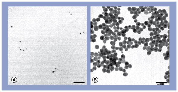
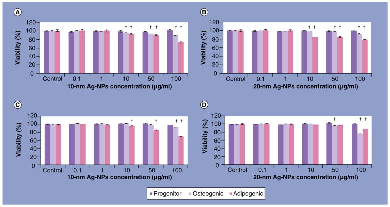
Materials & methods: hASC were exposed to 10- or 20-nm Ag-NPs at concentrations of 0.1, 1.0, 10.0, 50.0 and 100.0 µg/ml either before or after differentiation down the adipogenic or osteogenic pathways.

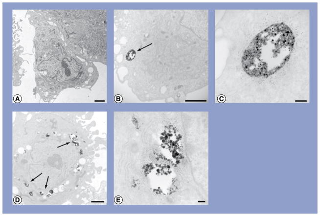
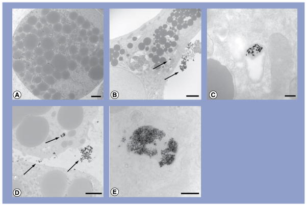
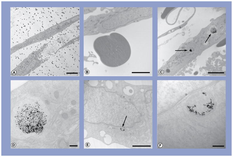
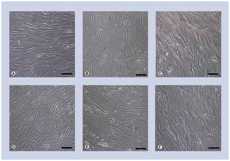
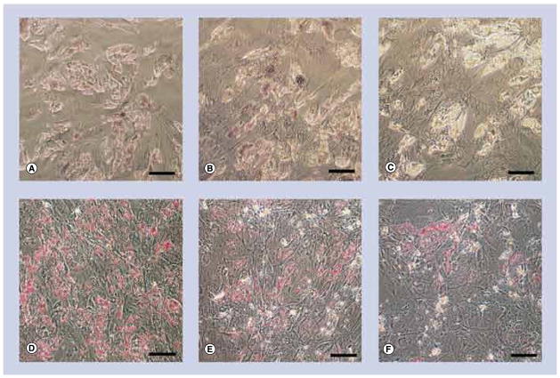
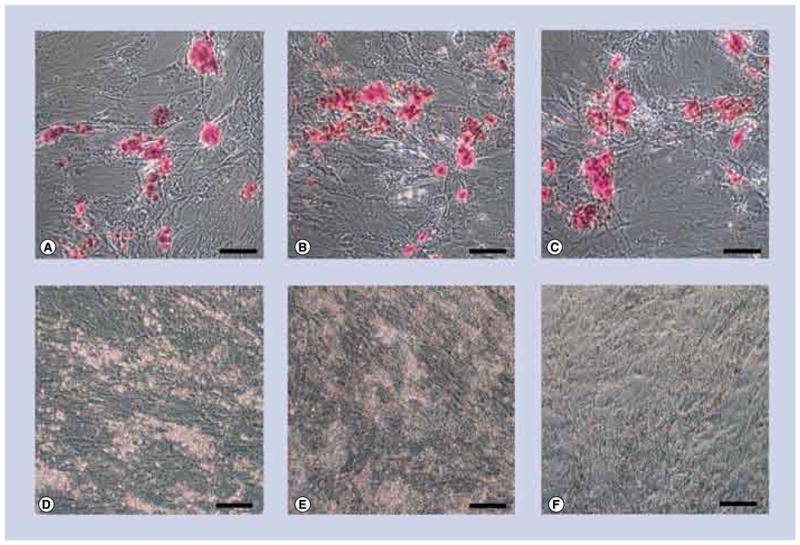
Results: Exposure of hASC to either 10- or 20-nm Ag-NPs resulted in no significant cytotoxicity to hASC, and minimal dose-dependent toxicity to adipogenic and osteogenic cells at 10 µg/ml. Each of the hASC, adipogenic and osteogenic cells showed cellular uptake of both 10- and 20-nm Ag-NPs, without causing significant ultrastructural alterations. Exposure to 10- or 20-nm Ag-NPs did not influence the differentiation of the cells, and at antimicrobial concentrations of Ag-NPs resulted in a minimal decrease in viability.

Conclusion: The biocompatibility of Ag-NPs with both undifferentiated and differentiated hASC establishes their suitability for incorporation into tissue-engineered graft scaffolds, for the prevention of bacterial contamination upon implantation.


Figures

Figure 1
Figure 1. Transmission electron micrographs of silver nanoparticles
(A) 10 nm and (B) 20 nm. Scale bars: 50 nm.
Figure 2
Figure 2. alamarBlue® viability of undifferentiated and differentiated human adipose-derived stem cells exposed to silver nanoparticles
alamarBlue viability of undifferentiated human adipose-derived stem cells, treated with either (A) 10 or (B) 20 nm Ag-NPs and then differentiated for 14 days; human adipose-derived stem cells differentiated for 14 days and then treated with (C) 10 or (D) 20-nm Ag-NPs. Significant differences (p < 0.05) in viability were assessed by multiple comparisons between concentrations, per cell pathway. Ag: Silver; NP: Nanoparticle.
Figure 3
Figure 3. Transmission electron micrographs of human adipose-derived stem cells exposed to either 10- or 20-nm silver nanoparticles at 10 μg/ml
(A) Human adipose-derived stem cells (hASCs); (B) hASC treated with 10-nm silver nanoparticles (Ag-NPs); (C) higher magnification of vacuole containing Ag-NPs from (B); (D) hASC treated with 20-nm Ag-NPs and (E) higher magnification of vacuoles containing Ag-NPs from (D). Scale bars: 2 μm (A, B & D); scale bars: 200 nm (C & E). Arrows denote agglomerates of Ag-NPs.
Figure 4
Figure 4. Transmission electron micrographs of adipogenic cells exposed to either 10- or 20-nm silver nanoparticles at 10 μg/ml
(A) Adipogenic cell showing spherical lipid accumulation; (B) adipogenic cell treated with 10-nm silver nanoparticles (Ag-NPs); (C) higher magnification of the vacuole containing Ag-NPs from (B); (D) adipogenic cell treated with 20-nm Ag-NPs and (E) higher magnification of the vacuole containing Ag-NPs from (D). Scale bars: 2 μm (A, B & D); scale bars: 200 nm (C & E). Arrows denote agglomerates of Ag-NPs.
Figure 5
Figure 5. Transmission electron micrographs of osteogenic cells exposed to either 10- or 20-nm silver nanoparticles at 10 μg/ml
(A) Osteogenic cell showing extracellular calcium accretion (dark dots); (B) higher magnification of accreted calcium; (C) osteogenic cell treated with 10-nm silver nanoparticles (Ag-NPs); (D) higher magnification of the vacuole containing Ag-NPs from (C); (E) adipogenic cell treated with 20-nm Ag-NPs and (F) higher magnification of the vacuole containing Ag-NPs from (E). Scale bar: 5 μm (A); scale bars: 2 μm (B, C & E); scale bars: 200 nm (D & F). Arrows denote agglomerates of Ag-NPs.
Figure 6
Figure 6. Light micrographs of undifferentiated human adipose-derived stem cells exposed to either 10- or 20-nm silver nanoparticles at 10 μg/ml
(A–C) No stain for either calcium accretion with Alizarin Red S or for (D–F) lipid accumulation with Oil Red O. Human adipose-derived stem cells (A & D) exposed to 10-nm silver nanoparticles (Ag-NPs) (B & E), or 20-nm Ag-NPs (C & F). Scale bars: 0.1 mm.
Figure 7
Figure 7. Light micrographs of adipogenic cells exposed to either 10- or 20-nm silver nanoparticles at 10 μg/ml. (A–C)
No stain for calcium accretion with Alizarin Red S and (D–F) strong staining for lipid accumulation with Oil Red O (cherry red spots). Control cells (A & D) exposed to 10-nm silver nanoparticles (Ag-NPs) (B & E), or 20-nm Ag-NPs (C & F). Scale bars: 0.1 mm.
Figure 8
Figure 8. Light micrographs of osteogenic cells exposed to either 10- or 20-nm silver nanoparticles at 10 μg/ml. (A–C)
exhibits strong staining for calcium accretion with Alizarin Red S (red spots) but none for (D–F) lipid accumulation with Oil Red O. Control cells (A & D) exposed to 10-nm silver nanoparticles (Ag-NPs) (B & E) or 20-nm Ag-NPs (C & F). Scale bars: 0.1 mm.

References

    1. Moucha C, Evans R, Clyburn T, et al. Orthopaedic infection prevention and control: an emerging new paradigm. Presented at: 77th Annual Meeting of the American Academy of Orthopaedic Surgeons; New Orleans, LA, USA. 9–12 March 2010.
    1. Samberg ME, Orndorff PE, Monteiro-Riviere NA. Antibacterial efficacy of silver nanoparticles of different sizes, surface conditions and synthesis methods. Nanotoxicology. 2011;5(2):244–253. Demonstrates the efficacy of silver nanoparticles with both Gram-negative and Gram-positive bacteria. - PubMed
    1. Morones JR, Elechiguerra JL, Camacho A, et al. The bactericidal effect of silver nanoparticles. Nanotechnology. 2005;16:2346–2353. - PubMed
    1. Sondi I, Salopek-Sondi B. Silver nanoparticles as antimicrobial agent: a case study on E. coli as a model for Gram-negative bacteria. J Colloid Interface Sci. 2004;275:177–182. - PubMed
    1. Wijnhoven SWP, Peijnenburg WJGM, Herberts CA, et al. Nano-silver – a review of available data and knowledge gaps in human and environmental risk assessment. Nanotoxicology. 2009;3:109–138.

Websites

    1. CDC. [Accessed 25 August 2011];Surgical Site Infection. www.cdc.gov/ncidod/dhqp/pdf/guidelines/SSI_tagged.pdf.
    1. The project on emerging nanotechnologies. Woodrow Wilson International Center for Scholars; Washington, DC, USA: [Accessed 15 November 2011]. www.nanotechproject.org/inventories/consumer/analysis_draft/
    1. Silver: Integrated Risk Information System. United States Environmental Protection Agency; [Accessed 14 November 2011]. www.epa.gov/iris/

Publication types