Review
. 2012 Aug;14(8):1183-92.
doi: 10.1111/j.1462-5822.2012.01812.x. Epub 2012 Jun 5.

The molecular basis of HIV entry

Affiliations
Review

The molecular basis of HIV entry

Per Johan Klasse. Cell Microbiol. 2012 Aug.

Abstract

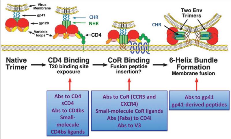
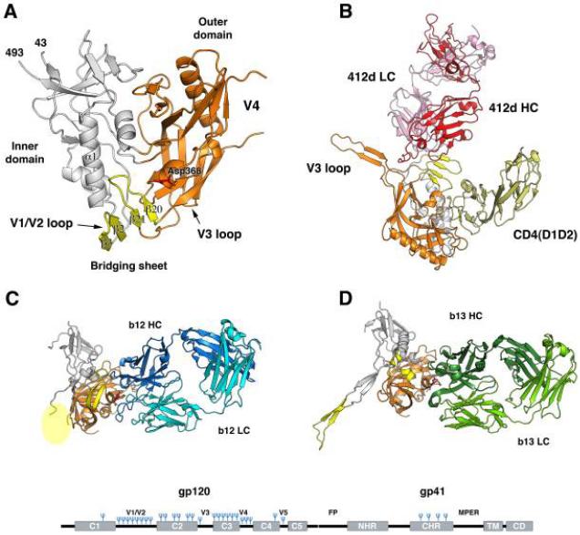
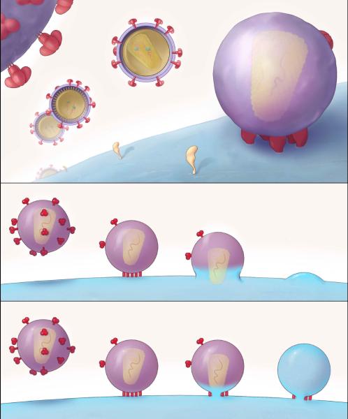
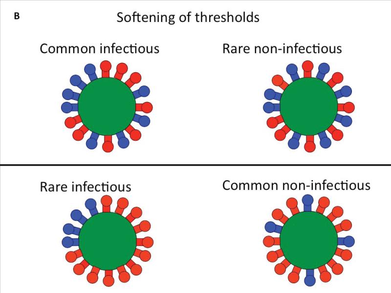
Infection by HIV starts when the virus attaches to a susceptible cell. For viral replication to continue, the viral envelope must fuse with a cellular membrane, thereby delivering the viral core to the cytoplasm, where the RNA genome is reverse-transcribed. The key players in this entry by fusion are the envelope glycoprotein, on the viral side, and CD4 and a co-receptor, CCR5 or CXCR4, on the cellular side. Here, the interplay of these molecules is reviewed from cell-biological, structural, mechanistic, and modelling-based perspectives. Hypotheses are evaluated regarding the cellular compartment for entry, the transfer of virus through direct cell-to-cell contact, the sequence of molecular events, and the number of molecules involved on each side of the virus-cell divide. An emerging theme is the heterogeneity among the entry mediators on both sides, a diversity that affects the efficacy of entry inhibitors, be they small-molecule ligands, peptides or neutralizing antibodies. These insights inform rational strategies for therapy as well as vaccination.


Figures

Figure 1
Figure 1. HIV enters susceptible cells through membrane fusion mediated by the viral Env protein
Top. Env mediates fusion A schematic of an Env trimer, anchored in the viral membrane, is shown in the first image to the left, and then a sequence of events is illustrated, from left to right, from receptor interactions to fusion of the viral envelope with the cell membrane. The second image of the trimer shows the binding of gp120 to the first domain of the CD4 receptor: NHR and CHR in gp41 become extended and the co-receptor-binding site is induced. In the third image, Env makes contact with a co-receptor, CCR5 or CXCR4. The bending of the hinge between domains 2 and 3 in CD4 and the interaction with the co-receptors pull the trimer to the target cell membrane. The co-receptor interaction also triggers the insertion of the gp41 “fusion peptide” (FP) into the cell membrane. Finally, in the fourth image, fusion has occurred, gp41 has refolded into a six-helix bundle, composed of the three copies of CHR slotting into the grooves of the trimer of NHRs. A number of principally different inhibitors of entry are listed in the boxes below the images of the steps they block. Amended and reproduced with permission from (Moore and Doms, 2003). Bottom. The three-dimensional structure of gp120, the receptor-binding subunit of Env Four different structures of gp120 are shown: gp120 consists of an inner domain (grey), an outer domain (orange), and a bridging sheet. (A) The tertiary structure of the core of a gp120 monomer was first obtained after several modifications: truncation of variable loops V1V2 and V3 as well as the N- and C-terminal segments, enzymatic trimming of glycans, and conformational stabilization by the binding of CD4 and a Fab to a CD4- induced epitope (these constituents of the complex are not shown). The bridging sheet is formed by the V1V2 stem and the hairpin of the β20-β21strands. Asp368 (red) contacts Arg59 in CD4. (B) Gp120 including V3 is complexed with CD4 (the D1D2 domains, pale yellow) and a Fab to a bridging-sheet epitope (412d light chain, LC, in pink, heavy chain, HC, in red). (C) Gp120 is bound by the neutralizing antibody b12 (HC in dark blue; LC in cyan), directed to the CD4-binding site. (D) gp120 is bound by the antibody b13 (HC in dark green; LC in light green); although b13, like b12, is directed to the CD4- binding site, it does not neutralize because its epitope is poorly antigenic on the trimeric form of Env, where the b12 epitope is well recognized. At the bottom of the figure is a schematic of the polypeptide chain of the Env precursor: the N-terminal subunit, gp120, contains five regions that are relatively conserved among viral strains (C1-C5). These form α helices and β strands in the folded protein. Gp120 is cleaved from gp41 at the C-terminal end of C5. Intercalated between the conserved regions of gp120 are variable regions, labeled V1V2-V5. In the more conserved gp41, the fusion peptide (FP), the N- and C-terminal helical regions (NHR and CHR), the membranee-proximal external region (MPER), the transmembrane segment (TM), and the cytoplasmic domain (CD) are marked. Glycans are shown as blue forks. Reproduced with permission from (Pejchal and Wilson, 2010); see references therein.
Figure 2
Figure 2
A. (Top) HIV virus particles (virions) are shown at different distances (they are all around 120 nm in diameter). The intersection in the middle shows the phospholipid bilayer of the viral envelope, surrounding a conical core. The viral envelope is studded with mushroom-like Env-protein trimers; the trimeric quaternary structure of Env is better discernable on the enlarged part of a virion in the upper left-hand corner. A cell surface is represented to the lower right, with some sparse receptors in yellow. (Middle) The virion docks onto the target cell and an entry claw forms by the lateral gathering of five Env trimers and a complement of receptors into a patch. When the juxtaposed areas of envelope and cell membrane have fused, the core passes into the cytoplasm. Fusion was thought to occur at the cell surface, with the plasma membrane, but cumulative evidence points to the endosome as the site of productive entry. (Bottom) A small fusion pore opens the communication between virion interior and cytoplasm. Expansion of the pore possibly involves more of the neighboring trimer-receptor rods in the entry claw. Reproduced from (Sougrat et al., 2007). B. A virion is postulated to be infectious only if it has more Env trimers than a certain threshold value. Heterogeneous distribution of Env trimers over the virion surface can soften such thresholds. Here the virions have 16 trimers each. Some are functional (blue; eight in top row; four in bottom row). If four contiguous trimers are needed for infection, most virions in the top row will be infectious, but few in the bottom row. In some studies virion infectivity has been postulated to be all-or-nothing, but the cartoon illustrates the possibility that a virion with more than a bare minimum number of trimers could have a greater propensity to infect than one that is just on the cusp of the threshold. Furthermore, different constellations of a minimum number of trimers might confer different propensities to infect. Whereas some virions are completely non-infectious, the infectious ones might not be equal but could display a spectrum of infectious propensity. C. The number of Env trimers per virion required for viral entry has been investigated by mixing defective and functional Env and by mathematically modeling the infectivity of the resulting virus. The relative infectivity (y axis) of such phenotypically mixed virus is a function of the proportion of functional Env protomers (x axis). A total of 9 potentially functional trimers per virion are postulated. The different curves represent degrees of blurring of the thresholds of absolute minimum numbers of functional trimers required for infectivity (as in B): high thresholds, around a minimum of 8 trimers, are shown in hard (red circles), intermediate (blue squares), and soft (green triangles) forms; low thresholds, around a minimum of 2 trimers, are also shown in hard (orange circles), intermediate (black squares), and soft (magenta triangles) forms. All the functions for these curves incorporate the premise that only trimers with three active protomers are fusogenic: thus at the level of the trimer the minimum or threshold for fusogenicity is three functional protomers. In general, threshold height at the trimer and virion levels compensate each other, so that theoretically distinct models become empirically indistinguishable. B and C are reproduced from (Klasse, 2007), where mathematical equations are given. D. Inhibition of CCR5-dependent HIV infection by a small-molecule CCR5 ligand (Vicriviroc, blue circles), a gp41-HR2-derived peptide (T-20, red squares), and their combination (green triangles). The synergy index calculated by non-linear regression was on average 0.22 (<1 indicates synergy) and the fold increase in apparent cooperativity or slope coefficient was 1.5. The synergy observed could be attributed partly to a prolongation of the HR2-peptide-sensitive intermediate by the CCR5 ligand, partly to heterogeneity in the target molecules on both sides. Reproduced from (Ketas et al., 2012).
Figure 2
Figure 2
A. (Top) HIV virus particles (virions) are shown at different distances (they are all around 120 nm in diameter). The intersection in the middle shows the phospholipid bilayer of the viral envelope, surrounding a conical core. The viral envelope is studded with mushroom-like Env-protein trimers; the trimeric quaternary structure of Env is better discernable on the enlarged part of a virion in the upper left-hand corner. A cell surface is represented to the lower right, with some sparse receptors in yellow. (Middle) The virion docks onto the target cell and an entry claw forms by the lateral gathering of five Env trimers and a complement of receptors into a patch. When the juxtaposed areas of envelope and cell membrane have fused, the core passes into the cytoplasm. Fusion was thought to occur at the cell surface, with the plasma membrane, but cumulative evidence points to the endosome as the site of productive entry. (Bottom) A small fusion pore opens the communication between virion interior and cytoplasm. Expansion of the pore possibly involves more of the neighboring trimer-receptor rods in the entry claw. Reproduced from (Sougrat et al., 2007). B. A virion is postulated to be infectious only if it has more Env trimers than a certain threshold value. Heterogeneous distribution of Env trimers over the virion surface can soften such thresholds. Here the virions have 16 trimers each. Some are functional (blue; eight in top row; four in bottom row). If four contiguous trimers are needed for infection, most virions in the top row will be infectious, but few in the bottom row. In some studies virion infectivity has been postulated to be all-or-nothing, but the cartoon illustrates the possibility that a virion with more than a bare minimum number of trimers could have a greater propensity to infect than one that is just on the cusp of the threshold. Furthermore, different constellations of a minimum number of trimers might confer different propensities to infect. Whereas some virions are completely non-infectious, the infectious ones might not be equal but could display a spectrum of infectious propensity. C. The number of Env trimers per virion required for viral entry has been investigated by mixing defective and functional Env and by mathematically modeling the infectivity of the resulting virus. The relative infectivity (y axis) of such phenotypically mixed virus is a function of the proportion of functional Env protomers (x axis). A total of 9 potentially functional trimers per virion are postulated. The different curves represent degrees of blurring of the thresholds of absolute minimum numbers of functional trimers required for infectivity (as in B): high thresholds, around a minimum of 8 trimers, are shown in hard (red circles), intermediate (blue squares), and soft (green triangles) forms; low thresholds, around a minimum of 2 trimers, are also shown in hard (orange circles), intermediate (black squares), and soft (magenta triangles) forms. All the functions for these curves incorporate the premise that only trimers with three active protomers are fusogenic: thus at the level of the trimer the minimum or threshold for fusogenicity is three functional protomers. In general, threshold height at the trimer and virion levels compensate each other, so that theoretically distinct models become empirically indistinguishable. B and C are reproduced from (Klasse, 2007), where mathematical equations are given. D. Inhibition of CCR5-dependent HIV infection by a small-molecule CCR5 ligand (Vicriviroc, blue circles), a gp41-HR2-derived peptide (T-20, red squares), and their combination (green triangles). The synergy index calculated by non-linear regression was on average 0.22 (<1 indicates synergy) and the fold increase in apparent cooperativity or slope coefficient was 1.5. The synergy observed could be attributed partly to a prolongation of the HR2-peptide-sensitive intermediate by the CCR5 ligand, partly to heterogeneity in the target molecules on both sides. Reproduced from (Ketas et al., 2012).

Similar articles

Cited by

References

    1. Abela IA, Berlinger L, Schanz M, Reynell L, Gunthard HF, Rusert P, Trkola A. Cell-Cell Transmission Enables HIV-1 to Evade Inhibition by Potent CD4bs Directed Antibodies. PLoS Pathog. 2012;8:e1002634. - PMC - PubMed
    1. Alkhatib G, Combadiere C, Broder CC, Feng Y, Kennedy PE, Murphy PM, Berger EA. CC CKR5: a RANTES, MIP-1alpha, MIP-1beta receptor as a fusion cofactor for macrophage-tropic HIV-1. Science. 1996;272:1955–1958. - PubMed
    1. Anastassopoulou CG, Ketas TJ, Klasse PJ, Moore JP. Resistance to CCR5 inhibitors caused by sequence changes in the fusion peptide of HIV-1 gp41. Proc Natl Acad Sci U S A. 2009;106:5318–5323. - PMC - PubMed
    1. Baribaud F, Edwards TG, Sharron M, Brelot A, Heveker N, Price K, Mortari F, Alizon M, Tsang M, Doms RW. Antigenically distinct conformations of CXCR4. J Virol. 2001;75:8957–8967. - PMC - PubMed
    1. Berro R, Klasse PJ, Lascano D, Flegler A, Nagashima KA, Sanders RW, Sakmar TP, Hope TJ, Moore JP. Multiple CCR5 conformations on the cell surface are used differentially by human immunodeficiency viruses resistant or sensitive to CCR5 inhibitors. J Virol. 2011;85:8227–8240. - PMC - PubMed

Publication types

Substances