. 2012 Jul 15;262(2):194-204.
doi: 10.1016/j.taap.2012.05.001. Epub 2012 May 11.

Neuregulin-1 is neuroprotective in a rat model of organophosphate-induced delayed neuronal injury

Affiliations

Neuregulin-1 is neuroprotective in a rat model of organophosphate-induced delayed neuronal injury

Yonggang Li et al. Toxicol Appl Pharmacol. .

Abstract

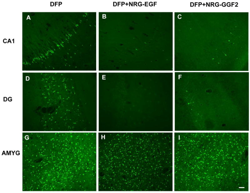
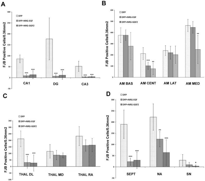
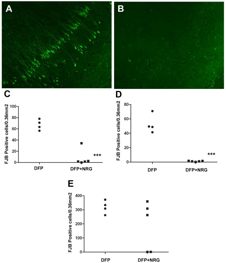
Current medical countermeasures against organophosphate (OP) nerve agents are effective in reducing mortality, but do not sufficiently protect the CNS from delayed brain damage and persistent neurological symptoms. In this study, we examined the efficacy of neuregulin-1 (NRG-1) in protecting against delayed neuronal cell death following acute intoxication with the OP diisopropylflurophosphate (DFP). Adult male Sprague-Dawley rats were pretreated with pyridostigmine (0.1 mg/kg BW, i.m.) and atropine methylnitrate (20 mg/kg BW, i.m.) prior to DFP (9 mg/kg BW, i.p.) intoxication to increase survival and reduce peripheral signs of cholinergic toxicity but not prevent DFP-induced seizures or delayed neuronal injury. Pretreatment with NRG-1 did not protect against seizures in rats exposed to DFP. However, neuronal injury was significantly reduced in most brain regions by pretreatment with NRG-1 isoforms NRG-EGF (3.2 μg/kg BW, i.a) or NRG-GGF2 (48 μg/kg BW, i.a.) as determined by FluroJade-B labeling in multiple brain regions at 24 h post-DFP injection. NRG-1 also blocked apoptosis and oxidative stress-mediated protein damage in the brains of DFP-intoxicated rats. Administration of NRG-1 at 1h after DFP injection similarly provided significant neuroprotection against delayed neuronal injury. These findings identify NRG-1 as a promising adjuvant therapy to current medical countermeasures for enhancing neuroprotection against acute OP intoxication.


Conflict of interest statement

Conflict of Interest Statement: Acorda Therapeutics, Inc. provided NRG-GGF2 and support for the studies involving NRG-GGF2. The sponsors were involved in the study design but not in the collection, analysis, and interpretation of data, in the writing of the report or in the decision to submit the paper for publication. Dr. Byron Ford has served as a paid consultant for Acorda Therapeutics.

Figures

Figure 1
Figure 1. NRG-1 pretreatment does not alter seizure activity in rats acutely intoxicated with DFP
Rats were pretreated with PB (0.1 mg/kg, i.m.) and AMN (20 mg/kg, i.m.) 30 or 10 min, respectively, prior to injection of DFP (9 mg/kg, i.p.) or an equal volume of vehicle (sterile water). NRG-1 (NRG-EGF, 3.2 μg/kg) or NRG-GGF2, 48 μg/kg) or vehicle (1% BSA in PBS) were administered via the carotid artery 5 min prior to DFP injection. Seizure severity was evaluated using a 5-point scale in which 0 indicates no response; 1, myoclonic jerks of the contralateral forelimb; 2, mouth and facial movements and head nodding with or without mild forelimb clonus; 3, severe forelimb clonus; 4, rearing and severe forelimb clonus; 5, rearing and falling.
Figure 2
Figure 2. NRG-1 pretreatment does not alter AChE activity in rats acutely intoxicated with DFP
Rats were pretreated with PB (0.1 mg/kg, i.m.) and AMN (20 mg/kg, i.m.) 30 or 10 min, respectively, prior to injection of DFP (9 mg/kg, i.p.) or an equal volume of vehicle (sterile water). NRG-GGF2 (48 μg/kg) or vehicle (1% BSA in PBS) was administered via the carotid artery 5 min prior to DFP injection. The Ellman assay was used to measure AChE activity which was normalized to protein concentration in brain samples and to volume for whole blood samples. Data in panels A and B are expressed as mean ± SEM (N = 5 per treatment group). Statistically significant differences were identified using one way ANOVA with post hoc Tukey's test; ***p < 0.001 compared to controls.
Figure 3
Figure 3. NRG-1 protects against DFP-induced delayed neurotoxicity
Rats were pretreated with PB and AMN prior to DFP (9 mg/kg, i.p.). NRG-1 (NRG-EGF, 3.2 μg/kg or NRG-GGF2, 48 μg/kg) or vehicle was administered via carotid artery 5 min prior to DFP exposure. Brains collected at 24 h post-DFP injection were labeled with Fluoro-Jade B (FJB). Representative photomicrographs of FJB labeling are shown from the CA1 region of the hippocampus (CA1; panels A,B,C), hippocampal dentate gyrus (DG; panels D,E,F) and amygdala basal nucleus (AMYG; panels G,H,I) of vehicle (A,D,G), NRG-EGF (B,E,H) and NRG-GGF2 (C,F,I) treated rats. Scale bar = 100 μm.
Figure 4
Figure 4. NRG-1 pretreatment protects against DFP-induced neuronal injury in the cerebral cortex
Rats were pretreated with PB and AMN prior to DFP (9 mg/kg, i.p.). NRG-1 (NRG-EGF, 3.2 μg/kg or NRG-GGF2, 48 μg/kg, i.a.) or vehicle was administered via carotid artery 5 min prior to DFP exposure. Brains collected at 24 h post-DFP injection were labeled with FJB. The number of FJB-positive cells was quantified from digital images of coronal sections (200× magnification) in specific brain regions at the same level across animals as determined using a brain atlas. Data are expressed as the mean ± SEM (N = 7 animals per treatment group). Statistically significant differences were identified using one way ANOVA with post hoc Tukey's test; *p <0.05, **p <0.01 and ***p <0.001 compared to controls. The brain regions analyzed were the auditory (AUD), cingulate (CING), motor (MOT), retrosplenial (RS), somatosensory (SS), visual (VIS), piriform (PIR), insular (INS), entorhinal (ENT) and ectorhinal (ECT) cortices.
Figure 5
Figure 5. Neuroprotection by NRG-1 against DFP neurotoxicity varies across brain regions
Rats were pretreated with PB and AMN prior to DFP (9 mg/kg, i.p.). NRG-1 (NRG-EGF, 3.2 μg/kg or NRG-GGF2, 48 μg/kg, i.a.) or vehicle was administered via carotid artery 5 min prior to DFP exposure. Brains collected at 24 h post-DFP injection were labeled with FJB. The number of FJB-positive neuronal cells was quantified from digital images of coronal sections (200× magnification) in specific brain regions at the same level across animals as determined using a brain atlas. Data are expressed as the mean ± SEM (N = 7 animals per treatment group). Statistically significant differences were identified using one way ANOVA with post hoc Tukey's test; *p <0.05, **p <0.01 and ***p <0.001 compared to controls. The brain areas examined were the CA1 (CA1), dentate gyrus (DG) and CA3 (CA3) regions of the hippocampus (A); the basal (AM BAS), central (AM CENT), medial (AM MED) and lateral (AM LAT) nuclei of the amygdala (B); the dorsolateral nucleus (THAL DL), mediodorsal (THAL MD) and reunion area (THAL RA) of the thalamus (C) and the septum (SEPT), nucleus accumbens (NA) and substantia nigra (SN) (D).
Figure 6
Figure 6. NRG-1 blocks DFP-induced apoptosis and rescues NeuN immunopositive cells
Rats were treated with PB, AMN and either vehicle or NRG-EGF (3.2 μg/kg, i.a.) before injection with DFP (9 mg/kg, i.p.). As illustrated in representative photomicrographs of the lateral dorsal thalamus, in control animals that did not receive NRG-1, at 24 h post-DFP injection there are many FJB-labeled cells (A), numerous TUNEL positive cells (B) and reduced NeuN immunoreactivity in injured neurons (C) with relatively higher levels of NeuN immunoreactivity in adjacent uninjured neurons (arrows). However, NRG-1 treatment significantly attenuated FJB labeling (B), abolished TUNEL staining (D) and rescued NeuN immunoreactivity (F). These figures are representative of brains from NRG-EGF treated animals, however similar results were seen when NRG-GGF2 was used. Scale bar = 100 μm.
Figure 7
Figure 7. Acute DFP intoxication causes oxidative stress that is reduced by NRG-1 pretreatment
Rats were treated with PB, AMN and either vehicle or NRG-EGF (3.2 μg/kg, i.a.) prior to injection with DFP (9 mg/kg, i.p.). Homogenates of cortical brain tissues collected at 24 (A) or 72 h (B) post-DFP injection were separated by SDS PAGE and probed with Ab that reacts with nitrotyrosine (nTyr) or the ATPase beta subunit. Densitometric analyses (C) indicate that DFP intoxication increased the normalized level of nitrated tyrosine and this effect was attenuated by pretreatment with NRG-EGF. ATPase activity was reduced by DFP and pretreatment with NRG-EGF protected ATPase activity (D). Experiments were run in duplicate or triplicate and repeated two times in independent experiments. Data were expressed as the mean ± SEM. Statistically significant differences were identified using the t-test (StatSimple v2.0.5; Nidus Technologies, Toronto, Canada); *p < 0.05 relative to control.
Figure 8
Figure 8. NRG-1 upregulates phospho-Akt immunoreactivity
Rats were treated with PB, AMN and either vehicle or NRG-EGF (3.2 μg/kg, i.a.) before injection with DFP (9 mg/kg, i.p.). Animals were euthanized 60 min after NRG-EGF administration, and brain sections from these animals were immunostained using antibodies that specifically recognized the phosphorylated form of Akt. As illustrated in representative photomicrographs from the somatosensory cortex, in control animals that did not receive NRG-EGF, there are few p-Akt -labeled cells (CTRL; A). Levels of phospho-Akt increase slightly at 60 min after DFP intoxication (DFP; B), but the percentage of phospho-Akt positive cells increases significantly 60 min after NRG-1 administration (DFP+NRG; C). The number of phospho-Akt positive neuronal cells in the somatosensory cortex increased 5-fold following NRG-EGF administration (D). Statistically significant differences were identified using one way ANOVA with post hoc Tukey's test (n = 3); ***p <0.001 compared to controls. Scale bar = 100 μm.
Figure 9
Figure 9. NRG-1 is neuroprotective when administered 1 h post-DFP injection
Rats were pretreated with PB and AMN prior to injection with DFP (9 mg/kg, i.p.) or vehicle (control). Between PB and DFP injections, catheters were placed into the external carotid artery. Animals were awake 2-3 min after isoflurane was discontinued and allowed to recover prior to NRG-1 administration. One h after DFP injection, animals were restrained and a single bolus dose of vehicle or NRG-1 (16 μg/kg) was administered via the catheter. Animals were euthanized 24 h post-DFP injection and neuronal injury quantified using FJB. Representative photomicrographs of FJB labeling in the CA1 region of the hippocampus from animals intoxicated with DFP in the absence (A; N = 4) or presence (B; N = 5) of NRG-1 treatment indicate that administration of NRG-1 1 h after DFP injection provides significant protection against DFP-induced delayed neuronal injury. These findings are confirmed by quantitative analyses of the number of FJB-labeled in cells the CA1 region of the hippocampus (C), cingulate cortex (D) and amygdala medial nucleus (E). Statistically significant differences were identified using a t- test; ***p<0.001. Scale bar = 100 μm.

References

    1. Carlsson T, Schindler FR, Hollerhage M, Depboylu C, Arias-Carrion O, Schnurrbusch S, Rosler TW, Wozny W, Schwall GP, Groebe K, Oertel WH, Brundin P, Schrattenholz A, Hoglinger GU. Systemic administration of neuregulin-1beta(1) protects dopaminergic neurons in a mouse model of Parkinson's disease. J Neurochem. 2011;117:1066–1074. - PubMed
    1. Collombet JM, Masqueliez C, Four E, Burckhart MF, Bernabe D, Baubichon D, Lallement G. Early reduction of NeuN antigenicity induced by soman poisoning in mice can be used to predict delayed neuronal degeneration in the hippocampus. Neurosci Lett. 2006;398:337–342. - PubMed
    1. Croslan DR, Schoell MC, Ford GD, Pulliam JV, Gates A, Clement CM, Harris AE, Ford BD. Neuroprotective effects of neuregulin-1 on B35 neuronal cells following ischemia. Brain Res. 2008;1210:39–47. - PMC - PubMed
    1. De Sarro G, Di Paola ED, De Sarro A, Vidal MJ. L-arginine potentiates excitatory amino acid-induced seizures elicited in the deep prepiriform cortex. Eur J Pharmacol. 1993;230:151–158. - PubMed
    1. Deshpande LS, Carter DS, Blair RE, DeLorenzo RJ. Development of a prolonged calcium plateau in hippocampal neurons in rats surviving status epilepticus induced by the organophosphate diisopropylfluorophosphate. Toxicol Sci. 2010;116:623–631. - PMC - PubMed

Publication types