

A review and synthesis of the first 20 years of PET and fMRI studies of heard speech, spoken language and reading
- PMID: 22584224
- PMCID: PMC3398395
- DOI: 10.1016/j.neuroimage.2012.04.062
A review and synthesis of the first 20 years of PET and fMRI studies of heard speech, spoken language and reading
Abstract
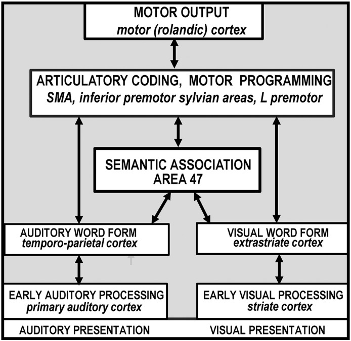
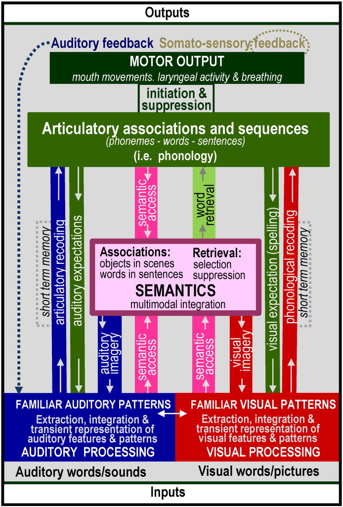
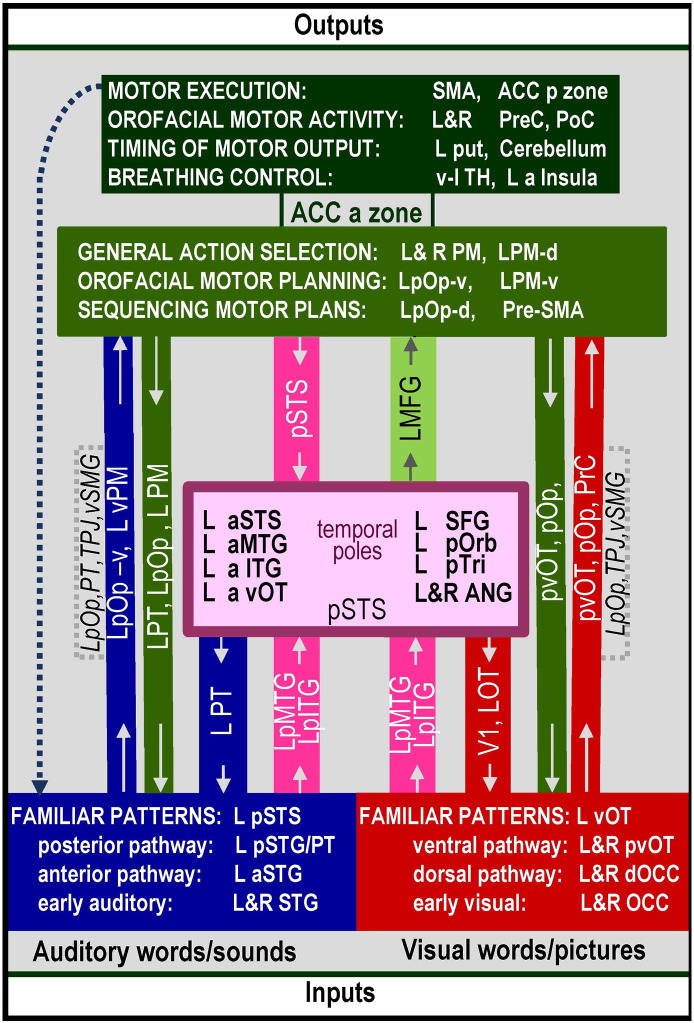
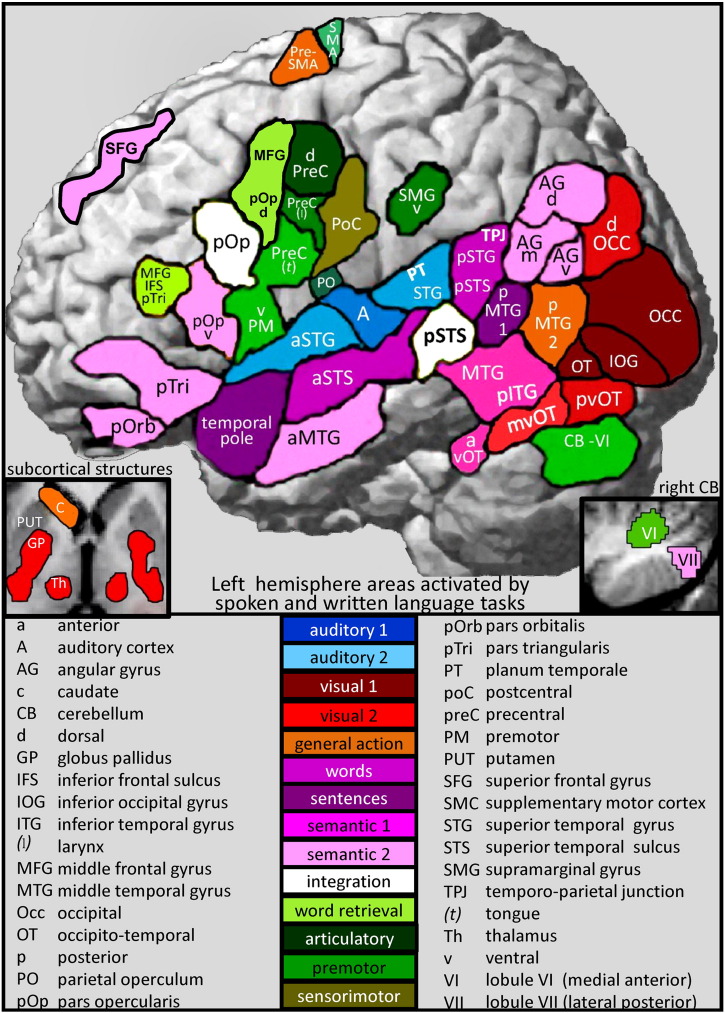
The anatomy of language has been investigated with PET or fMRI for more than 20 years. Here I attempt to provide an overview of the brain areas associated with heard speech, speech production and reading. The conclusions of many hundreds of studies were considered, grouped according to the type of processing, and reported in the order that they were published. Many findings have been replicated time and time again leading to some consistent and undisputable conclusions. These are summarised in an anatomical model that indicates the location of the language areas and the most consistent functions that have been assigned to them. The implications for cognitive models of language processing are also considered. In particular, a distinction can be made between processes that are localized to specific structures (e.g. sensory and motor processing) and processes where specialisation arises in the distributed pattern of activation over many different areas that each participate in multiple functions. For example, phonological processing of heard speech is supported by the functional integration of auditory processing and articulation; and orthographic processing is supported by the functional integration of visual processing, articulation and semantics. Future studies will undoubtedly be able to improve the spatial precision with which functional regions can be dissociated but the greatest challenge will be to understand how different brain regions interact with one another in their attempts to comprehend and produce language.
Copyright © 2012 Elsevier Inc. All rights reserved.
Figures
References
-
- Abel S., Dressel K., Bitzer R., Kummerer D., Mader I., Weiller C., Huber W. The separation of processing stages in a lexical interference fMRI-paradigm. Neuroimage. 2009;44:1113–1124. - PubMed
-
- Ackermann H. Cerebellar contributions to speech production and speech perception: psycholinguistic and neurobiological perspectives. Trends Neurosci. 2008;31:265–272. - PubMed
-
- Ackermann H., Riecker A. The contribution of the insula to motor aspects of speech production: a review and a hypothesis. Brain Lang. 2004;89:320–328. - PubMed
-
- Ackermann H., Riecker A. The contribution(s) of the insula to speech production: a review of the clinical and functional imaging literature. Brain Struct. Funct. 2010;214:419–433. - PubMed
-
- Ackermann H., Riecker A., Mathiak K., Erb M., Grodd W., Wildgruber D. Rate-dependent activation of a prefrontal-insular-cerebellar network during passive listening to trains of click stimuli: an fMRI study. Neuroreport. 2001;12:4087–4092. - PubMed
Publication types
MeSH terms
Grants and funding
LinkOut - more resources
Full Text Sources
Medical
