Comparative Study
. 2012 Sep;36(9):2093-7.
doi: 10.1007/s00268-012-1651-8.

Learning curve of thoracoscopic repair of esophageal atresia

Affiliations
Comparative Study

Learning curve of thoracoscopic repair of esophageal atresia

David C van der Zee et al. World J Surg. 2012 Sep.

Abstract

Background: Thoracoscopic repair of esophageal atresia is considered to be one of the more advanced pediatric surgical procedures, and it undoubtedly has a learning curve. This is a single-center study that was designed to determine the learning curve of thoracoscopic repair of esophageal atresia.

Methods: The study involved comparison of the first and second five-year outcomes of thoracoscopic esophageal atresia repair.

Results: The demographics of the two groups were comparable. There was a remarkable reduction of postoperative leakage or stenosis, and recurrence of fistulae, in spite of the fact that nowadays the procedure is mainly performed by young staff members and fellows.

Conclusions: There is a considerable learning curve for thoracoscopic repair of esophageal atresia. Centers with the ambition to start up a program for thoracoscopic repair of esophageal atresia should do so with the guidance of experienced centers.


Figures
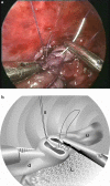
Fig. 1
Fig. 1
Running suture of posterior wall. Upper esophagus (u), distal esophagus (d), and lung (L). A stay suture (S) stabilizes the esophagus for suturing. b Drawing of the picture in a schematically displaying the posterior wall of the upper (u) and distal (d) esophagus, where a running suture has been laid. This running suture is tied to the stay suture (S) laid at the posterior wall. The collapsed lung (L) is at the lower end
Fig. 2
Fig. 2
Start of a running suture along the anterior side of the esophagus (this can be the same suture used for the posterior wall after it has been tied to the stay suture). Upper esophagus (u), distal esophagus (d), and lung (L), as well as the stay suture (S), are marked. b Drawing of the picture in a demonstrating the start of the anterior running suture

References

    1. Lobe TE, Rothenberg SS, Waldschmidt J. Thoracoscopic repair of esophageal atresia in an infant: a surgical first. Pediatr Endosurg Innov Tech. 1999;3:141–148. doi: 10.1089/pei.1999.3.141. - DOI
    1. Bax KM, van der Zee DC. Feasibility of thoracoscopic repair of esophageal atresia with distal fistula. J Pediatr Surg. 2002;37:192–196. doi: 10.1053/jpsu.2002.30253. - DOI - PubMed
    1. Rothenberg SS. Thoracoscopic repair of tracheoesophageal fistula in newborns. J Pediatr Surg. 2002;37:869–872. doi: 10.1053/jpsu.2002.32891. - DOI - PubMed
    1. Holcomb GW, 3rd, Rothenberg SS, Bax KM, et al. Thoracoscopic repair of esophageal atresia and tracheoesophageal fistula: a multi-institutional analysis. Ann Surg. 2005;242:422–428. - PMC - PubMed
    1. Allal H, Kalfa N, Lopez M, et al. Benefits of the thoracoscopic approach for short- or long-gap esophageal atresia. J Laparoendosc Adv Surg Tech A. 2005;15:673–677. doi: 10.1089/lap.2005.15.673. - DOI - PubMed

Publication types