. 2012 Feb;2(2):156-71.
doi: 10.1158/2159-8290.CD-11-0256.

The OPCML tumor suppressor functions as a cell surface repressor-adaptor, negatively regulating receptor tyrosine kinases in epithelial ovarian cancer

Affiliations

The OPCML tumor suppressor functions as a cell surface repressor-adaptor, negatively regulating receptor tyrosine kinases in epithelial ovarian cancer

Arthur B McKie et al. Cancer Discov. 2012 Feb.

Abstract

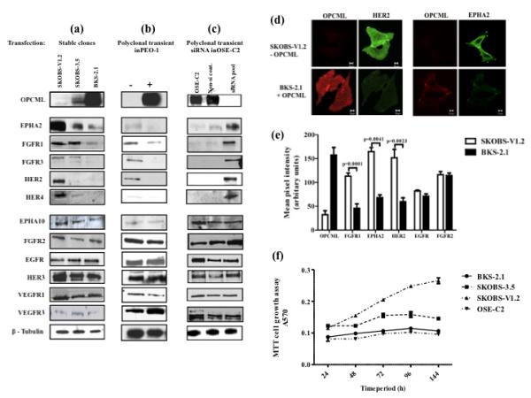
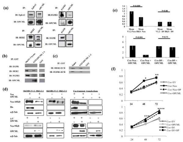
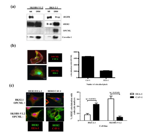
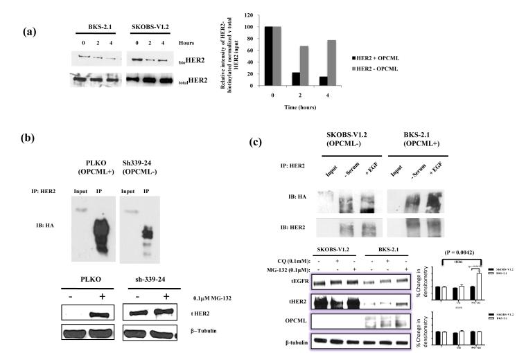
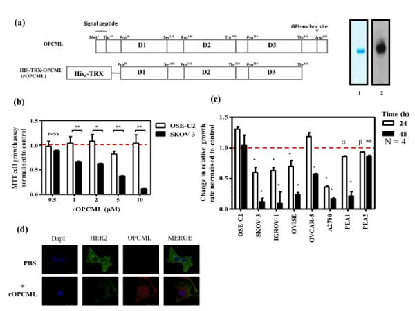
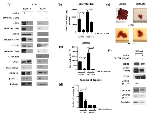
Epithelial ovarian cancer is the leading cause of death from gynecologic malignancy, and its molecular basis is poorly understood. We previously demonstrated that opioid binding protein cell adhesion molecule (OPCML) was frequently epigenetically inactivated in epithelial ovarian cancers, with tumor suppressor function in vitro and in vivo. Here, we further show the clinical relevance of OPCML and demonstrate that OPCML functions by a novel mechanism in epithelial ovarian cancer cell lines and normal ovarian surface epithelial cells by regulating a specific repertoire of receptor tyrosine kinases: EPHA2, FGFR1, FGFR3, HER2, and HER4. OPCML negatively regulates receptor tyrosine kinases by binding their extracellular domains, altering trafficking via nonclathrin-dependent endocytosis, and promoting their degradation via a polyubiquitination-associated proteasomal mechanism leading to signaling and growth inhibition. Exogenous recombinant OPCML domain 1-3 protein inhibited the cell growth of epithelial ovarian cancers cell in vitro and in vivo in 2 murine ovarian cancer intraperitoneal models that used an identical mechanism. These findings demonstrate a novel mechanism of OPCML-mediated tumor suppression and provide a proof-of-concept for recombinant OPCML protein therapy in epithelial ovarian cancers.

Significance: The OPCML tumor suppressor negatively regulates a specific spectrum of receptor tyrosine kinases in ovarian cancer cells by binding to their extracellular domain and altering trafficking to a nonclathrin, caveolin-1–associated endosomal pathway that results in receptor tyrosine kinase polyubiquitination and proteasomal degradation. Recombinant OPCML domain 1-3 recapitulates this mechanism and may allow for the implementation of an extracellular tumor-suppressor replacement strategy.


Figures

Figure 1
Figure 1. OPCML negatively regulates a specific repertoire of receptor tyrosine kinases
Western blots demonstrating that OPCML negatively regulates EPHA2, FGFR1, FGFR3, HER2, and HER4 but not EPHA10, FGFR2, EGFR, HER3, VEGFR1 and VEGFR3; shown in stably transfected SKOV-3 cells (a), containing pcDNA3.1vector only (−) and pcDNA3.1-OPCML with lower (SKOBS-3.5) and higher (BKS2.1) OPCML expression; polyclonal transiently transfected PEO1 cells (b) with vector only (−) and OPCML (+). OSE-C2 (normal ovarian surface epithelial) cells (c), physiologically expressing OPCML which were untreated (−), or transiently transfected with non-silencing duplex (Non-si), or OPCML-directed siRNA duplexes (si) demonstrated reciprocal upregulation of the same specific repertoire of RTKs. β-tubulin control for loading is shown in bottom panel. (d): Immunofluorescence microscopy showing HER2 and EPHA2 expression in vector control SKOBS-V1.2 and stable transfection OPCML expressing BKS2.1 cells. Abundant HER2 and EPHA2 expression seen in SKOBS-V1.2 (green) is abrogated in BKS2.1 associated with expression of OPCML (red). (e): Comparison of vector (white bars) and OPCML (black bars) transfected SKOV-3 cells showing quantification of the immunofluorescent pixel intensity for OPCML, FGFR1, EPHA2, HER2, EGFR and FGFR2 demonstrates the same OPCML-associated RTK spectrum of down-regulation by a second method. (f) A 144 h in vitro growth assay comparing vector control SKOBS V1.2 (OPCML deficient), BKS2.1 (strongly expressing OPCML), SKOBS-3.5 (lower OPCML expressor) and OSE-C2 (normal ovarian surface epithelial cell line). Cell proliferation was quantified by MTT assay showing OPCML expression is strongly growth suppressive.
Figure 2
Figure 2. OPCML associated RTK modulation affects downstream signalling
(a)Western blots of total and phospho HER2 and EGFR protein from SKOBS-V1.2 (vector control), BKS-2.1 cells and SKOBS-3.5 were subjected to a 60 min EGF (50 ng/ml) timecourse demonstrating profound abrogation of tHER2, HER2 and EGFR phosphorylation (Y-1248 and Y-1173 resp.) in BKS2.1 and SKOBS-3.5. OPCML transfection was also associated with abrogation of EGF phospho-activation of ERK 1 & 2 (T-202/T-204) and Akt (S-473), more profoundly in BKS-2.1 line. Representative western blots were run in parallel with equivalent loadings of the same lysate preparation for each of the 3 cell lines, and are defined by their beta tubulin bands. BKS2.1 tERK/pERK (left) is from the same blot as tERK/pERK for SKOBS-V1.2/SKOBS3.5 on the right, and therefore the same SKOBSV1.2 empty vector transfected control data is shown alongside for ease of interpretation (original blot shown in supplementary figure 9). BKS2.1 and SKOBS V1.2 tAkt/pAkt are from the same blot: removal of irrelevant additional sample lanes, for ease of interpretation, is indicated by black boxes. SKOBS V1.2 and SKOBS 3.5 tAkt/pAkt (bottom right) are a separate representative blot. (b) The same cells subjected to a 60 min FGF1 (10 ng/ml) timecourse again demonstrate loss of total and phospho (Y-766) FGFR1.(c) Analysis of shRNA clones by qRT-PCR for OPCML showing two clones exhibiting reduced OPCML mRNA levels relative to the PLKO-1 and PLKO-2 (empty vector clones) and shSCRAMbled controls. We focused on two clones; * sh464-23 which exhibited 60% KD and ** sh339-24 which exhibited 95% KD of OPCML. (e) Western analysis demonstrated that total HER2 and EphA2 are strongly up-regulated in the shRNA lines with 95% OPCML KD. (f) Western blots showing the upregulation of HER2 and EphA2 upon 95% KD of OPCML selectively on serum and on exposure to ligand stimulation of cells. (g) Cell proliferation assay shows that OSE-C2-sh339-24 line with 95% OPCML KD exhibits higher proliferation rates at 48, 72 and 96 hours compared to PLKO-2 control (P = * 0.002, **0.009, ***0.003 resp.) compared with shSCRAMBLED and empty vector PLKO-2 controls. No such growth rate difference was demonstrated in 0.5% FBS underscoring the RTK ligand dependency of OPCML effect (data not shown).
Figure 3
Figure 3. OPCML directly interacts with the ECD of EPHA2, FGFR1 and HER2 – a prerequisite for down-regulation
(a)IP for OPCML showing binding for EPHA2, FGFR1 and HER2. Similarly, IP for each of these proteins in turn showed reciprocal binding for OPCML. We were not able to demonstrate co-IP with EGFR and OPCML after numerous attempts. (b) GST pull-down assay with SKOV-3 cell lysate showing binding of HER2 and FGFR1to GST-OPCML1-3 and no binding of EGFR to GST-OPCML 1-3 protein. The presence of OPCML fusion proteins in these assays was verified by immunoblotting (bottom panel). (c) Confirmatory in vitro interaction studies were undertaken verifying binding of HER2 and FGFR1 extracellular domain protein, generated from in vitro translation kit (TnT) (HER2 ECD) and in E.coli (FGFR-ECD) to GST-OPCML 1-3 fusion protein. Both HER2 and FGFR1 ECDs were seen to bind to GST-OPCML 1-3. (d) To demonstrate a mechanistic link between the RTK ECD and OPCML, we undertook transient transfection of His-tagged full length Neu/HER2 ECD or ECD-less Neu (Δ5-P95) demonstrating down-regulation of the transfected full length 185kD ECD containing Neu in either the OPCML BKS2.1 or OPCML transiently co-transfected Cos-1 (Cos-Neu+OPCML); in contrast there was no change in expression of the transiently transfected ECD-less Neu (Δ5-P95) in the presence of OPCML. (e) The relative mean band intensity from 5 separate Neu construct transfections in BKS2.1/SKOBS-V1.2 and 3 separate co-transfections in Cos1 cells shows that in both cell systems, the level of full length Neu protein is significantly reduced by 50-80% when co-expressed with OPCML, (p = 0.009 & 0.041 respectively) while the level of the ECD-less 95kD Neu species is unchanged in both systems regardless of OPCML status. (f) Growth assays in Neu and OPCML non-expresing Cos-1 cells conclusively demonstrate that the significant growth acceleration (p=0.003) induced by transiently transfected p185 Neu (Cos+ Neu) is abrogated by transient co-transfection (dotted line) with OPCML (Cos+Neu+OP, p= 0.004at 48hr*, p=0.016 at 72hr**), reducing the growth rate to that of the empty vector (Cos+EV) and OPCML (Cos+OPCML) controls. In contrast the similarly significantly growth accelerated ECD-less Δ5-P95 (p=0.015) was not inhibited by OPCML co-transfection(Cos+Neu+OP, p= 0.56at 48hr***, p=0.092 at 72hr****), and this demonstrates the functional necessity of the ECD for regulation of the RTK by OPCML.
Figure 4
Figure 4. Location and co-localisation of OPCML with HER2 to a detergent-resistant/ Cav-1 specific compartment
(a) OPCML sequesters RTKs into detergent-resistant membrane fraction; SKOBS-V1.2 and BKS2.1 cells were subjected to membrane fractionation to separate detergent resistant membrane fraction (DRMF)/lipid raft fraction from the detergent soluble/bulk membrane phase. An equal volume from each fraction was analysed by SDS-PAGE followed by Western blotting with anti-EGFR, anti-HER2, anti-OPCML and anti-Cav-1 antibodies. OPCML was found to be strictly located in the DRMF with Cav-1. Transfection of OPCML was associated with shift of HER2 and EGFR to the DRMF. (b) Confocal co-immunofluorescence demonstrates increased co-localisation of Cav-1with OPCML and concurrently shows decreased co-localisation with EEA-1 suggesting a redistribution of HER2 to lipid-raft domains. The bar chart (35) shows quantification of the total number of OPCML pixels colocalised with Cav-1 and EEA-1, implying that OPCML localises mainly with lipid-raft domains. (c) Confocal co-immunofluorescence of HER2 co-localisation with EEA-1 and Cav-1(left) with quantification (35) as a percentage of the total number of HER2 pixels detected within the cells. These studies demonstrate that OPCML expressing cells are associated with a switch of HER2 trafficking from clathrin-mediated internalisation of early-endosomes to lipid-raft domain mechanisms of internalisation.
Figure 5
Figure 5. Mechanism of OPCML mediated RTK down-regulation
(a) Pulse-chase biotinylation followed by western blotting demonstrates more rapid loss of HER2 protein in OPCML transfected BKS2.1 than OPCML deficient SKOBS V1.2 (left). Quantitation of relative signal intensity of biotinylated HER2 normalized against input total HER2 expressed as a percentage of the initial pulse (35). (b) Normal OSE-C2 cells PLKO (OPCML+) and sh339-24(OPCML−) (above) were transiently transfected with an HA-tagged ubiquitin, serum starved then treated with MG-132 for 2hours. Ubiquitinated proteins were detected by immunoprecipitating (IP) with HER2 then immunoblotting (IB) with an anti-HA antibody and show that OPCML KD dramatically reduces polyubiquitination of HER2 . Western blot of PLKO (OPCML+) and sh339-24 (OPCML KD) treated with the proteasomal inhibitor MG-132 (0.1 μM) for 2 h demonstrate clear upregulation of tHER2 by MG132 in OPCML containing but not OPCML deficient OSE-C2 (below). (c) IP of HER2 followed by anti-HA antibody in SKOBS-V1.2 (OPCML−) and BKS-2.1(OPCML+) (above), showing that OPCML expression in BKS2.1 is associated with polyubiquitination of HER2 and conversely this polyubiquitination was diminished in OPCML deficient SKOBS-V1.2. BKS-2.1(OPCML+) and SKOBS-V1.2 were treated with the lysosomal inhibitor chloroquine (CQ; 0.1 mM) or the proteasomal inhibitor MG-132 (0.1 μM) for 2 h (below). Immunoblotting for EGFR, HER2, OPCML and β-tubulin is shown. Densitometric analysis of these WB data for HER2 (upper chart) and EGFR (lower chart) using β-tubulin as a control demonstrate that MG132 proteasomal inhibition selectively up-regulates of HER2 but not EGFR in OPCML expressing BKS2.1 but not OPCML deficient SKOBS-V1.2.
Figure 6
Figure 6. Recombinant OPCML (rOPCML) protein effect in vitro
(a) (Left) schematic diagram of OPCML and rOPCML. (35) SDS–PAGE analysis of rOPCML expressed in E.coli. Lane 1, Coomassie Blue, lane 2, rOPCML Western blot. (b) rOPCML targets cancer but not normal cells (left panel); OSE-C2 and SKOV-3 cells were subjected to varying concentrations of rOPCML ( 0.5, 1, 2, 5 and 10 μM). The MTT proliferation assay is shown relative to control vehicle-treated (dotted line) at 48hrs. A dose dependent inhibition of SKOV-3 cell growth demonstrates the specificity of rOPCML for OPCML deficient cells (NS = non significant, * P=0.004, **P<0.0001). (c) The OSE-C2 line and a panel of ovarian cancer lines (SKOV-3, IGROV, OVISE, OVCAR-5, A2780, PEA1 and PEA2) were exposed to 10 μM of rOPCML for 24hr (white bar) and 48 hr (black bar) with MTT cell growth was normalised to vehicle only controls (*P<0.0001, aP=0.0284 and bP=0.0079). This data demonstrates that 6/7 (86%) of the ovarian cancer lines (but not OSE-C2) were significantly and profoundly growth suppressed after exposure to rOPCML. (d) Confocal co-immunofluorescence demonstrates the relative abundance of HER2 protein in SKOV-3 cells after either PBS application (above) or rOPCML application (below) with punctate co-localisation of OPCML with HER2 in the latter.
Figure 7
Figure 7. Recombinant OPCML (rOPCML) abrogates an identical spectrum of RTKs and their downstream signalling in SKOV3 and A2780 in vitro and after Intra-peritoneal (IP) administration of rOPCML in vivo
(a) rOPCML specifically abrogates total/phospho-HER2, total/ phospho-FGFR1 and phospho- but not total EGFR (*n.b. A2780 does not express EGFR, although VEGFR1 was expressed and not altered). rOPCML abrogates downstream substrates pAkt (S-473) and pErk 1 & 2 in both SKOV-3 and A2780 (35). (b) Quantitation of mean tumour weight per mouse (n=4) (c) rOPCML significantly abrogates ascites both IP tumor models (n=4) and (d) significantly reduces mean number of tumour deposits in the A2780 IP model. (e) Comparison is shown between IP tumours collected from BSA and rOPCML-IP treated mice and demonstrates the tumor suppressor effect of rOPCML in-vivo. (f) Western blot analysis of recovered IP tumour lysate demonstrating the impact of rOPCML protein treatment on cell signalling: rOPCML specifically abrogates total/phospho-HER2, total/phospho-FGFR1 and phospho- but not total EGFR.

Comment in

  • New roles opined for OPCML.
    Wu SY, Sood AK. Wu SY, et al. Cancer Discov. 2012 Feb;2(2):115-6. doi: 10.1158/2159-8290.CD-11-0356. Cancer Discov. 2012. PMID: 22585855 Free PMC article.

Similar articles

Cited by

References

    1. Statistics UKofN . Cancer trends in England and Wales 1950-1999. Office of NationalStatistics; 2002.
    1. Hall J, Paul J, Brown R. Critical evaluation of p53 as a prognostic marker in ovarian cancer. Expert Reviews in Molecular Medicine. 2004;6:1–20. - PubMed
    1. Radice P. Mutations of BRCA genes in hereditary breast and ovarian cancer. Journal of Experimental & Clinical Cancer Research. 2002;21:9–12. - PubMed
    1. Meng Q, Xia C, Fang J, Rojanasakul Y, Jiang B. Role of PI3K and AKT specific isoforms in ovarian cancer cell migration, invasion and proliferation through the p70S6K1 pathway. Cellular Signalling. 2006;18:2262–71. - PubMed
    1. Maihle NJ, Baron AT, Barrette BA, Boardman CH, Christensen TA, Cora EM, et al. EGF/ErbB receptor family in ovarian cancer. Cancer Treat Res. 2002;107:247–58. - PubMed

Publication types

MeSH terms

Substances