

Brain responses to high-protein diets
- PMID: 22585905
- PMCID: PMC3649463
- DOI: 10.3945/an.112.002071
Brain responses to high-protein diets
Abstract
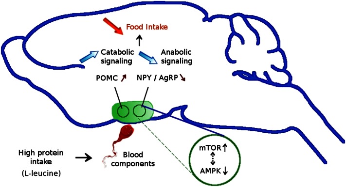
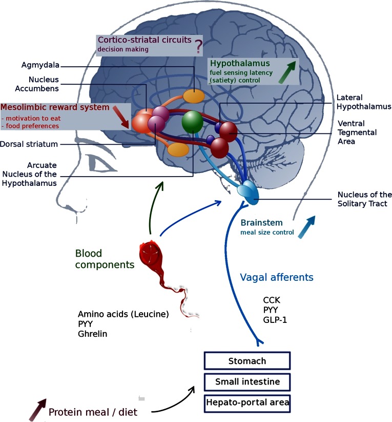
Proteins are suspected to have a greater satiating effect than the other 2 macronutrients. After protein consumption, peptide hormones released from the gastrointestinal tract (mainly anorexigenic gut peptides such as cholecystokinin, glucagon peptide 1, and peptide YY) communicate information about the energy status to the brain. These hormones and vagal afferents control food intake by acting on brain regions involved in energy homeostasis such as the brainstem and the hypothalamus. In fact, a high-protein diet leads to greater activation than a normal-protein diet in the nucleus tractus solitarius and in the arcuate nucleus. More specifically, neural mechanisms triggered particularly by leucine consumption involve 2 cellular energy sensors: the mammalian target of rapamycin and AMP-activated protein kinase. In addition, reward and motivation aspects of eating behavior, controlled mainly by neurons present in limbic regions, play an important role in the reduced hedonic response of a high-protein diet. This review examines how metabolic signals emanating from the gastrointestinal tract after protein ingestion target the brain to control feeding, energy expenditure, and hormones. Understanding the functional roles of brain areas involved in the satiating effect of proteins and their interactions will demonstrate how homeostasis and reward are integrated with the signals from peripheral organs after protein consumption.
Conflict of interest statement
Author disclosures: M. Journel, C. Chaumontet, N. Darcel, G. Fromentin and D. Tomé, no conflicts of interest.
Figures
Similar articles
-
Peripheral and central mechanisms involved in the control of food intake by dietary amino acids and proteins.Nutr Res Rev. 2012 Jun;25(1):29-39. doi: 10.1017/S0954422411000175. Epub 2012 May 29. Nutr Res Rev. 2012. PMID: 22643031 Review.
-
The interaction of amylin with other hormones in the control of eating.Diabetes Obes Metab. 2013 Feb;15(2):99-111. doi: 10.1111/j.1463-1326.2012.01670.x. Epub 2012 Aug 29. Diabetes Obes Metab. 2013. PMID: 22862822 Review.
-
How Satiating Are the 'Satiety' Peptides: A Problem of Pharmacology versus Physiology in the Development of Novel Foods for Regulation of Food Intake.Nutrients. 2019 Jul 4;11(7):1517. doi: 10.3390/nu11071517. Nutrients. 2019. PMID: 31277416 Free PMC article. Review.
-
Peptide signals regulating food intake and energy homeostasis.Can J Physiol Pharmacol. 2002 May;80(5):396-406. doi: 10.1139/y02-035. Can J Physiol Pharmacol. 2002. PMID: 12056545 Review.
-
The Importance of the Gastrointestinal Tract in Controlling Food Intake and Regulating Energy Balance.Gastroenterology. 2017 May;152(7):1707-1717.e2. doi: 10.1053/j.gastro.2017.01.053. Epub 2017 Feb 11. Gastroenterology. 2017. PMID: 28193513
Cited by
-
Neuronal Mechanisms that Drive Organismal Aging Through the Lens of Perception.Annu Rev Physiol. 2020 Feb 10;82:227-249. doi: 10.1146/annurev-physiol-021119-034440. Epub 2019 Oct 21. Annu Rev Physiol. 2020. PMID: 31635526 Free PMC article. Review.
-
High Protein Diet Induces Oxidative Stress in Rat Cerebral Cortex and Hypothalamus.Int J Mol Sci. 2019 Mar 28;20(7):1547. doi: 10.3390/ijms20071547. Int J Mol Sci. 2019. PMID: 30925663 Free PMC article.
-
Diet-induced changes in the Lean Brain: Hypercaloric high-fat-high-sugar snacking decreases serotonin transporters in the human hypothalamic region.Mol Metab. 2013 Aug 7;2(4):417-22. doi: 10.1016/j.molmet.2013.07.006. eCollection 2013. Mol Metab. 2013. PMID: 24327957 Free PMC article.
-
Mice lacking neutral amino acid transporter B(0)AT1 (Slc6a19) have elevated levels of FGF21 and GLP-1 and improved glycaemic control.Mol Metab. 2015 Feb 16;4(5):406-17. doi: 10.1016/j.molmet.2015.02.003. eCollection 2015 May. Mol Metab. 2015. PMID: 25973388 Free PMC article.
-
FGF21 as a mediator of adaptive changes in food intake and macronutrient preference in response to protein restriction.Neuropharmacology. 2024 Sep 1;255:110010. doi: 10.1016/j.neuropharm.2024.110010. Epub 2024 May 24. Neuropharmacology. 2024. PMID: 38797244 Free PMC article. Review.
References
-
- Fromentin G, Darcel N, Chaumontet C, Marsset-Baglieri A, Nadkarni N, Tome D. Peripheral and central mechanisms involved in the control of food intake by dietary amino acids and proteins. NRR. 2012; in press. - PubMed
-
- Koda S, Date Y, Murakami N, Shimbara T, Hanada T, Toshinai K, Niijima A, Furuya M, Inomata N, Osuye K, et al. The role of the vagal nerve in peripheral PYY3–36-induced feeding reduction in rats. Endocrinology. 2005;146:2369–75 - PubMed
-
- Veldhorst M, Smeets A, Soenen S, Hochstenbach-Waelen A, Hursel R, Diepvens K, Lejeune M, Luscombe-Marsh N, Westerterp-Plantenga M. Protein-induced satiety: effects and mechanisms of different proteins. Physiol Behav. 2008;94:300–7 - PubMed
-
- Feurte S, Nicolaidis S, Even PC, Tome D, Mahe S, Fromentin G. Rapid fall in plasma threonine followed by increased intermeal interval in response to first ingestion of a threonine-devoid diet in rats. Appetite. 1999;33:329–41 - PubMed
Publication types
MeSH terms
Substances
LinkOut - more resources
Full Text Sources
Other Literature Sources
Medical
