. 2012 Jun 12;125(23):2844-53.
doi: 10.1161/CIRCULATIONAHA.111.060889. Epub 2012 May 14.

Ventricular assist device implantation corrects myocardial lipotoxicity, reverses insulin resistance, and normalizes cardiac metabolism in patients with advanced heart failure

Affiliations

Ventricular assist device implantation corrects myocardial lipotoxicity, reverses insulin resistance, and normalizes cardiac metabolism in patients with advanced heart failure

Aalap Chokshi et al. Circulation. .

Abstract

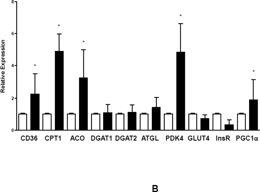
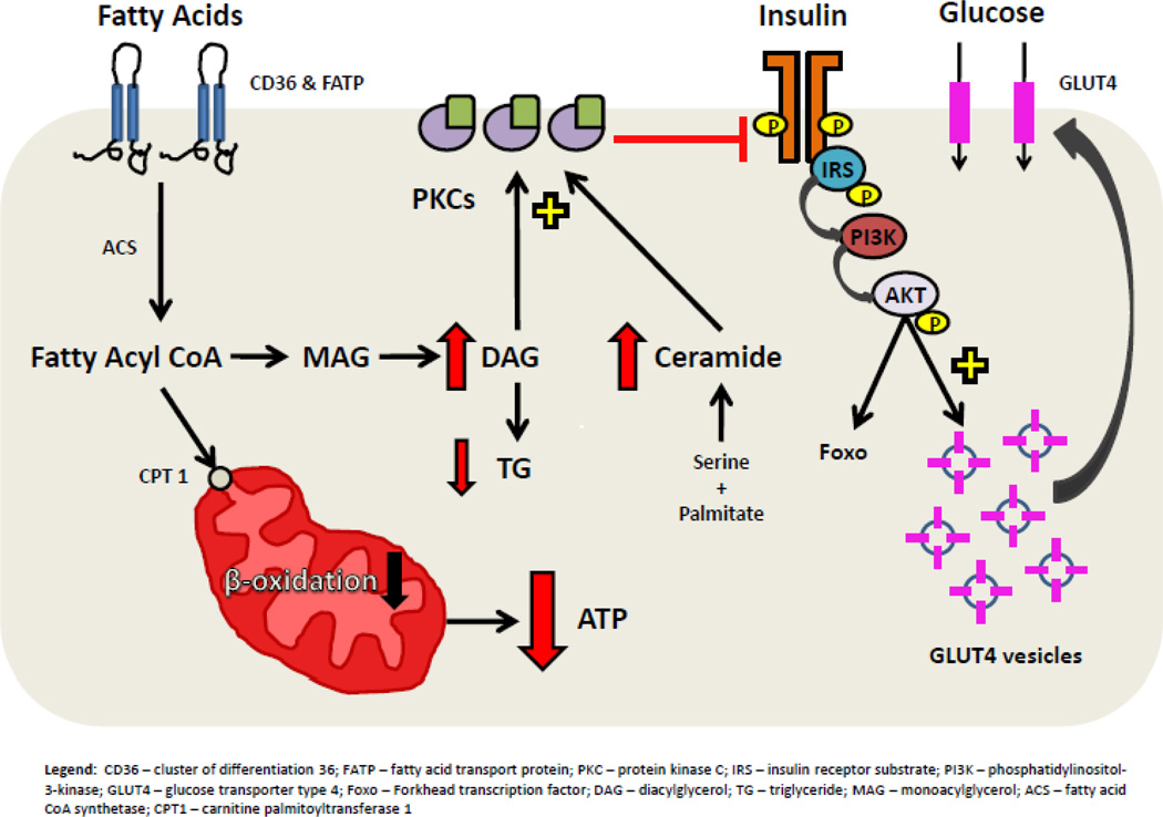
Background: Heart failure is associated with impaired myocardial metabolism with a shift from fatty acids to glucose use for ATP generation. We hypothesized that cardiac accumulation of toxic lipid intermediates inhibits insulin signaling in advanced heart failure and that mechanical unloading of the failing myocardium corrects impaired cardiac metabolism.

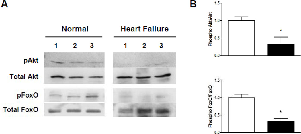
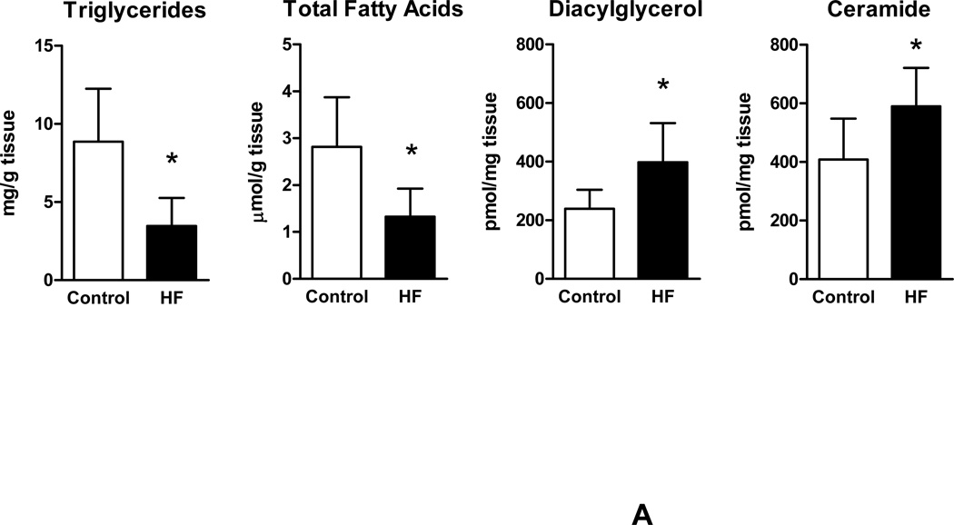
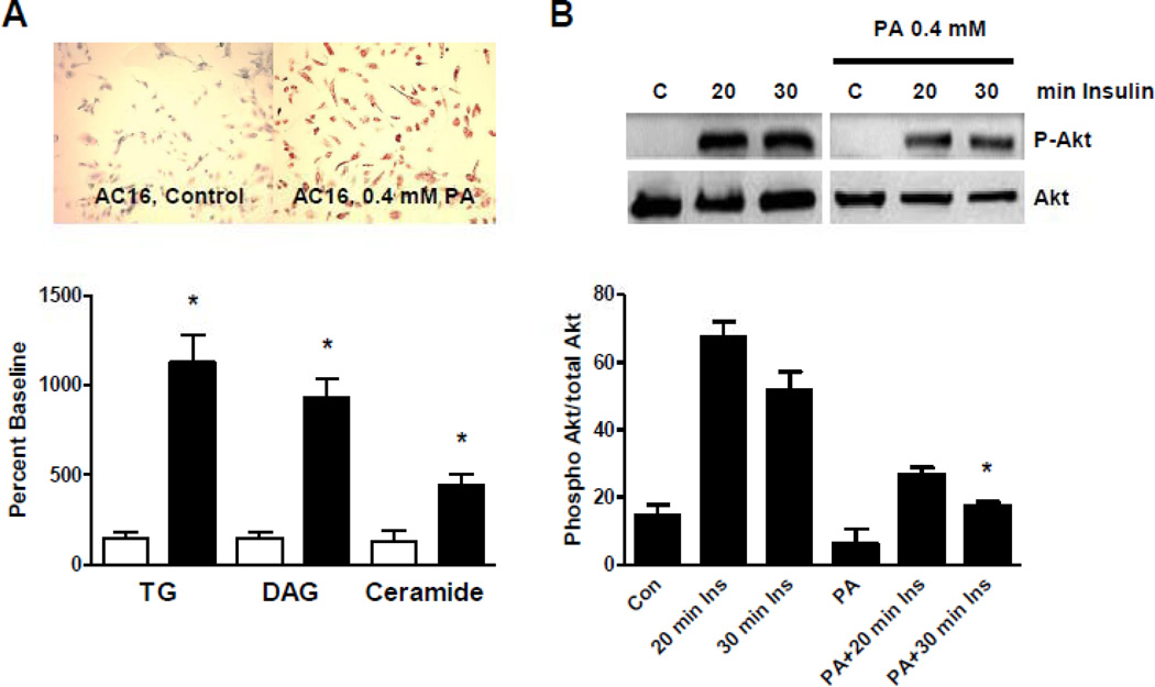
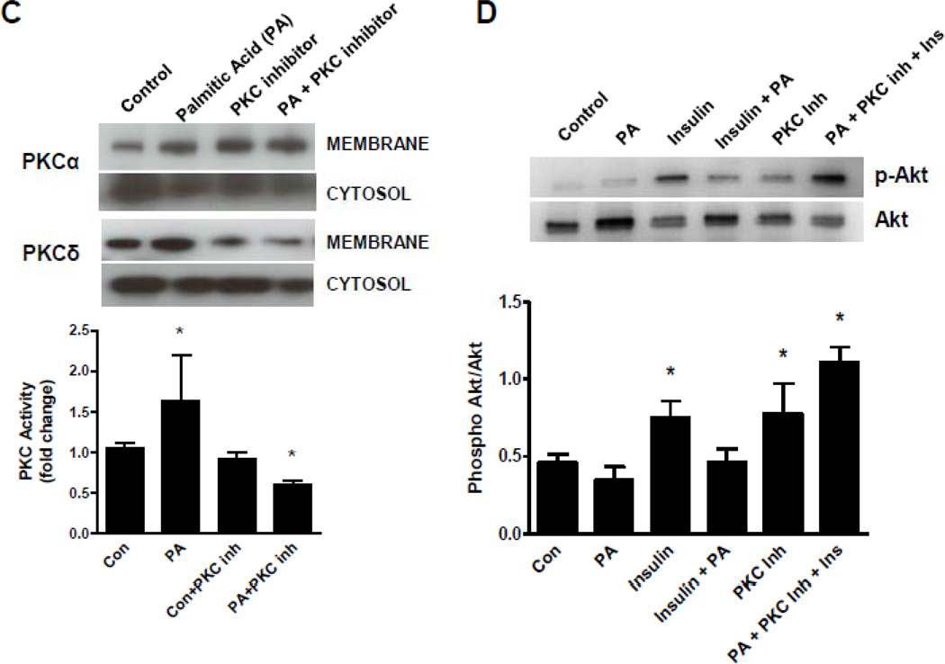
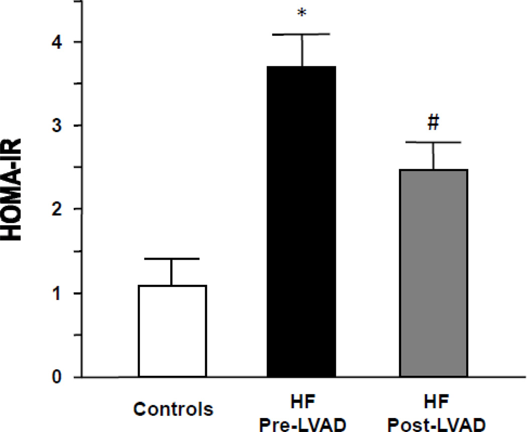
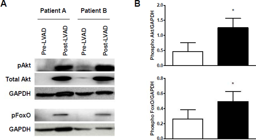
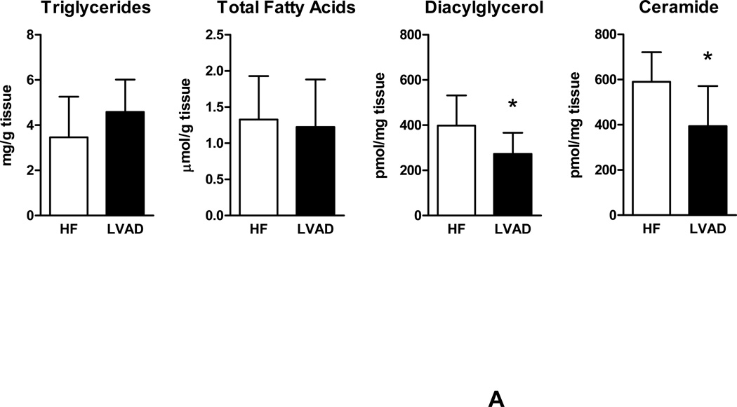
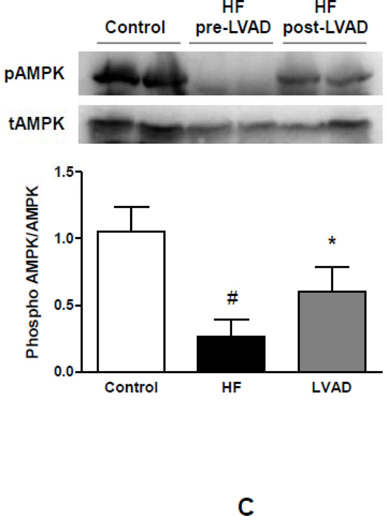
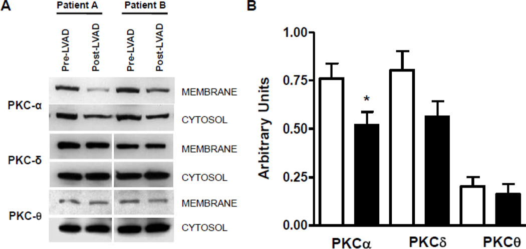
Methods and results: We analyzed the myocardium and serum of 61 patients with heart failure (body mass index, 26.5±5.1 kg/m(2); age, 51±12 years) obtained during left ventricular assist device implantation and at explantation (mean duration, 185±156 days) and from 9 control subjects. Systemic insulin resistance in heart failure was accompanied by decreased myocardial triglyceride and overall fatty acid content but increased toxic lipid intermediates, diacylglycerol, and ceramide. Increased membrane localization of protein kinase C isoforms, inhibitors of insulin signaling, and decreased activity of insulin signaling molecules Akt and Foxo were detectable in heart failure compared with control subjects. Left ventricular assist device implantation improved whole-body insulin resistance (homeostatic model of analysis-insulin resistance, 4.5±0.6-3.2±0.5; P<0.05) and decreased myocardial levels of diacylglycerol and ceramide, whereas triglyceride and fatty acid content remained unchanged. Improved activation of the insulin/phosphatidylinositol-3 kinase/Akt signaling cascade after left ventricular assist device implantation was confirmed by increased phosphorylation of Akt and Foxo, which was accompanied by decreased membrane localization of protein kinase C isoforms after left ventricular assist device implantation.

Conclusions: Mechanical unloading after left ventricular assist device implantation corrects systemic and local metabolic derangements in advanced heart failure, leading to reduced myocardial levels of toxic lipid intermediates and improved cardiac insulin signaling.


Conflict of interest statement

Conflict of Interest Disclosures: None

Figures

Figure 1
Figure 1
Impairment of PI3Kinase/Akt signaling in human advanced HF. (A) Reduced activation indicated by decreased phosphorylation status of Akt and FOXO in samples from patients with advanced HF compared to control subjects (three representative samples per group). (B) Decreased myocardial phosphorylation levels of Akt and FOXO in patients with advanced HF compared to controls (*p<0.05, n=6–8 samples per group).
Figure 2
Figure 2
Myocardial lipid content and transcriptional changes of glucose and lipid metabolism genes. (A) Myocardial lipid content in patients with advanced HF and controls. (B) Relative changes in gene expression of metabolic genes controlling fatty acid and glucose uptake and oxidation (empty bars – controls; filled bars – HF; n=6–8 individual patients in each group, *p<0.05 vs. controls).
Figure 3
Figure 3
Inhibition of insulin signaling by increased cardiomyocyte lipid content. (A) Incubation of AC16 cells with 0.4 mM palmitic acid (PA) resulted in increased cellular lipid content measured by oil red O staining (top) and by quantitative lipid analysis (bottom, *p<0.05 vs. unstimulated cells). (B) Overnight stimulation of AC16 cells resulted in inhibition of insulin-mediated phosphorylation of Akt (*p<0.05 vs. 30 min insulin). (C) PKC activity in control and PA-treated cells. (D) Inhibition of PKC activity normalized insulin-mediated Akt phosphorylation in AC16 under high PA stimulation (n=3–4 individual samples per group, *p<0.05 vs. control).
Figure 3
Figure 3
Inhibition of insulin signaling by increased cardiomyocyte lipid content. (A) Incubation of AC16 cells with 0.4 mM palmitic acid (PA) resulted in increased cellular lipid content measured by oil red O staining (top) and by quantitative lipid analysis (bottom, *p<0.05 vs. unstimulated cells). (B) Overnight stimulation of AC16 cells resulted in inhibition of insulin-mediated phosphorylation of Akt (*p<0.05 vs. 30 min insulin). (C) PKC activity in control and PA-treated cells. (D) Inhibition of PKC activity normalized insulin-mediated Akt phosphorylation in AC16 under high PA stimulation (n=3–4 individual samples per group, *p<0.05 vs. control).
Figure 4
Figure 4
Insulin resistance in patients with advanced HF corrects following LVAD placement (white bars – controls, n=10; black bars – HF pre-LVAD, n=36; grey bars – HF post-LVAD, n=30; *p<0.01 vs. controls; #p<0.05 vs. HF pre-LVAD).
Figure 5
Figure 5
Mechanical unloading of the failing myocardium increased cardiac PI3K/Akt/Foxo signaling. (A) Increased myocardial activation of Akt and Foxo following LVAD placement. (B) Quantitative analysis of Akt and Foxo activation. (empty bars – pre-LVAD; filled bars – post-LVAD; n=6 individual patients before and after LVAD implantation; *p<0.05 vs. pre-LVAD).
Figure 6
Figure 6
Impact of mechanical unloading on myocardial lipid content, metabolic gene expression and AMPK activation in patients with advanced HF. (A) No changes are detectable in cardiac levels of triglycerides and free fatty acids. Increased levels of DAG and ceramide in advanced HF correct after mechanical unloading of the failing myocardium. (B) Changes in myocardial metabolic gene expression in response to mechanical unloading (empty bars – pre-LVAD; filled bars – post-LVAD; *p<0.05 vs. pre-LVAD). (C) Reduced myocardial AMPK phosphorylation in patients with advanced HF compared to controls partially corrects after mechanical unloading of the failing myocardium (empty bars – controls; filled bars - pre-LVAD; grey bars – post-LVAD; *p<0.05 vs. pre-LVAD; #p<0.01 vs. controls; n=6–8 individual patients before and after LVAD implantation).
Figure 6
Figure 6
Impact of mechanical unloading on myocardial lipid content, metabolic gene expression and AMPK activation in patients with advanced HF. (A) No changes are detectable in cardiac levels of triglycerides and free fatty acids. Increased levels of DAG and ceramide in advanced HF correct after mechanical unloading of the failing myocardium. (B) Changes in myocardial metabolic gene expression in response to mechanical unloading (empty bars – pre-LVAD; filled bars – post-LVAD; *p<0.05 vs. pre-LVAD). (C) Reduced myocardial AMPK phosphorylation in patients with advanced HF compared to controls partially corrects after mechanical unloading of the failing myocardium (empty bars – controls; filled bars - pre-LVAD; grey bars – post-LVAD; *p<0.05 vs. pre-LVAD; #p<0.01 vs. controls; n=6–8 individual patients before and after LVAD implantation).
Figure 7
Figure 7
Myocardial activation of PKC isoforms in patients with advanced HF before and after mechanical unloading. (A) Decreased membrane localization of PKC isoforms following mechanical unloading through ventricular assist device placement. (B) Semiquantitative assessment of PKC isoform localization (empty bars – pre-LVAD; filled bars – post-LVAD; n=8 individual patients before and after LVAD implantation; *p<0.05 vs. pre-LVAD).
Figure 8
Figure 8
Lipotoxic inhibition of insulin signaling in advanced HF. CD36 – cluster of differentiation 36; FATP – fatty acid transport protein; PKC – protein kinase C; IRS – insulin receptor substrate; PI3K – phosphatidylinositol-3-kinase; GLUT4 – glucose transporter type 4; Foxo – forkhead transcription factor; GSK3 – glycogen synthase kinase 3; p70s6k – 70-kDa ribosomal protein S6 kinase; DAG – diacylglycerol; TG – triglyceride; MAG – monoacylglycerol; ACS – fatty acid CoA synthase; CPT1 – carnitine palmitoyltransferase 1.

Comment in

References

    1. Hunter JJ, Chien KR. Signaling pathways for cardiac hypertrophy and failure. N Engl J Med. 1999;341:1276–1283. - PubMed
    1. Neubauer S. The failing heart--an engine out of fuel. N Engl J Med. 2007;356:1140–1151. - PubMed
    1. Tian R. Transcriptional regulation of energy substrate metabolism in normal and hypertrophied heart. Curr Hypertens Rep. 2003;5:454–458. - PubMed
    1. Ingwall JS. On substrate selection for atp synthesis in the failing human myocardium. Am J Physiol Heart Circ Physiol. 2007;293:H3225–H3226. - PubMed
    1. Razeghi P, Young ME, Alcorn JL, Moravec CS, Frazier OH, Taegtmeyer H. Metabolic gene expression in fetal and failing human heart. Circulation. 2001;104:2923–2931. - PubMed

Publication types

MeSH terms