

Vascular inflammatory cells in hypertension
- PMID: 22586409
- PMCID: PMC3345946
- DOI: 10.3389/fphys.2012.00128
Vascular inflammatory cells in hypertension
Abstract
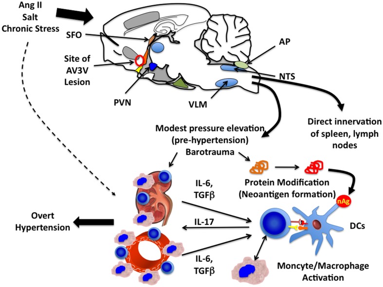
Hypertension is a common disorder with uncertain etiology. In the last several years, it has become evident that components of both the innate and adaptive immune system play an essential role in hypertension. Macrophages and T cells accumulate in the perivascular fat, the heart and the kidney of hypertensive patients, and in animals with experimental hypertension. Various immunosuppressive agents lower blood pressure and prevent end-organ damage. Mice lacking lymphocytes are protected against hypertension, and adoptive transfer of T cells, but not B cells in the animals restores their blood pressure response to stimuli such as angiotensin II or high salt. Recent studies have shown that mice lacking macrophages have blunted hypertension in response to angiotensin II and that genetic deletion of macrophages markedly reduces experimental hypertension. Dendritic cells have also been implicated in this disease. Many hypertensive stimuli have triggering effects on the central nervous system and signals arising from the circumventricular organ seem to promote inflammation. Studies have suggested that central signals activate macrophages and T cells, which home to the kidney and vasculature and release cytokines, including IL-6 and IL-17, which in turn cause renal and vascular dysfunction and lead to blood pressure elevation. These recent discoveries provide a new understanding of hypertension and provide novel therapeutic opportunities for treatment of this serious disease.
Keywords: T cells; blood pressure; dendritic cells; interleukin 17; interleukin 6; macrophages; superoxide; sympathetic nerves.
Figures
References
-
- Ba D., Takeichi N., Kodama T., Kobayashi H. (1982). Restoration of T cell depression and suppression of blood pressure in spontaneously hypertensive rats (SHR) by thymus grafts or thymus extracts. J. Immunol. 128, 1211–1216 - PubMed
-
- Brands M. W., Banes-Berceli A. K., Inscho E. W., Al-Azawi H., Allen A. J., Labazi H. (2010). Interleukin 6 knockout prevents angiotensin II hypertension: role of renal vasoconstriction and janus kinase 2/signal transducer and activator of transcription 3 activation. Hypertension 56, 879–88410.1161/HYPERTENSIONAHA.110.158071 - DOI - PMC - PubMed
-
- Chae C. U., Lee R. T., Rifai N., Ridker P. M. (2001). Blood pressure and inflammation in apparently healthy men. Hypertension 38, 399–403 - PubMed
Grants and funding
LinkOut - more resources
Full Text Sources