

Periodontal tissue reaction to customized nano-hydroxyapatite block scaffold in one-wall intrabony defect: a histologic study in dogs
- PMID: 22586523
- PMCID: PMC3349047
- DOI: 10.5051/jpis.2012.42.2.50
Periodontal tissue reaction to customized nano-hydroxyapatite block scaffold in one-wall intrabony defect: a histologic study in dogs
Abstract
Purpose: This study evaluated histologically the tissue responses to and the effects of a customized nano-hydroxyapatite (n-HA) block bone graft on periodontal regeneration in a one-wall periodontal-defect model.
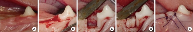
Methods: A customized block bone for filling in the standardized periodontal defect was fabricated from prefabricated n-HA powders and a polymeric sponge. Bilateral 4×4×5 mm (buccolingual width×mesiodistal width×depth), one-wall, critical-size intrabony periodontal defects were surgically created at the mandibular second and fourth premolars of five Beagle dogs. In each dog, one defect was filled with block-type HA and the other served as a sham-surgery control. The animals were sacrificed following an 8-week healing interval for clinical and histological evaluations.
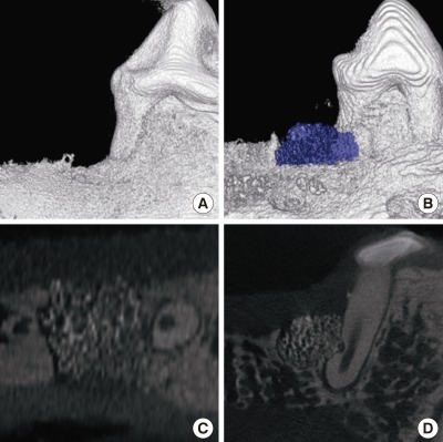
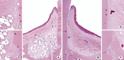
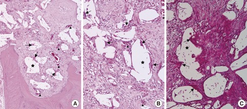
Results: Although the sites that received an n-HA block showed minimal bone formation, the n-HA block was maintained within the defect with its original hexahedral shape. In addition, only a limited inflammatory reaction was observed at sites that received an n-HA block, which might have been due to the high stability of the customized block bone.
Conclusions: In the limitation of this study, customized n-HA block could provide a space for periodontal tissue engineering, with minimal inflammation.
Keywords: Bone substitutes; Guided tissue regeneration; Periodontal disease; Tissue engineering; Tissue scaffolds.
Conflict of interest statement
No potential conflict of interest relevant to this article was reported.
Figures
References
-
- Bartold PM, McCulloch CA, Narayanan AS, Pitaru S. Tissue engineering: a new paradigm for periodontal regeneration based on molecular and cell biology. Periodontol 2000. 2000;24:253–269. - PubMed
-
- Trombelli L. Which reconstructive procedures are effective for treating the periodontal intraosseous defect? Periodontol 2000. 2005;37:88–105. - PubMed
-
- Bender SA, Rogalski JB, Mills MP, Arnold RM, Cochran DL, Mellonig JT. Evaluation of demineralized bone matrix paste and putty in periodontal intraosseous defects. J Periodontol. 2005;76:768–777. - PubMed
-
- Caton J, Nyman S, Zander H. Histometric evaluation of periodontal surgery. II. Connective tissue attachment levels after four regenerative procedures. J Clin Periodontol. 1980;7:224–231. - PubMed
-
- Heitz-Mayfield L, Tonetti MS, Cortellini P, Lang NP European Research Group on Periodontology (ERGOPERIO) Microbial colonization patterns predict the outcomes of surgical treatment of intrabony defects. J Clin Periodontol. 2006;33:62–68. - PubMed
LinkOut - more resources
Full Text Sources
