. 2012 Aug;61(8):2004-15.
doi: 10.2337/db11-0802. Epub 2012 May 14.

ChREBP mediates glucose-stimulated pancreatic β-cell proliferation

Affiliations

ChREBP mediates glucose-stimulated pancreatic β-cell proliferation

Mallikarjuna R Metukuri et al. Diabetes. 2012 Aug.

Abstract

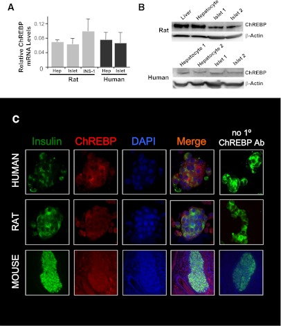
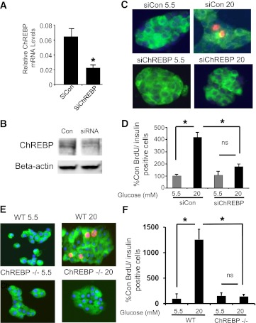
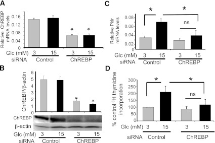
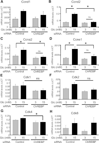
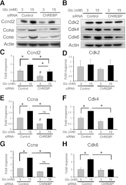
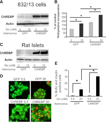
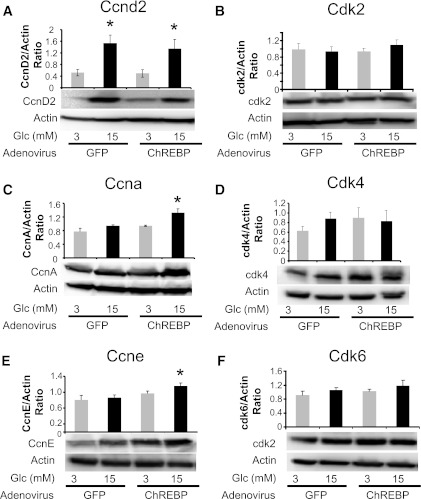
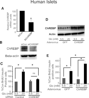
Glucose stimulates rodent and human β-cell replication, but the intracellular signaling mechanisms are poorly understood. Carbohydrate response element-binding protein (ChREBP) is a lipogenic glucose-sensing transcription factor with unknown functions in pancreatic β-cells. We tested the hypothesis that ChREBP is required for glucose-stimulated β-cell proliferation. The relative expression of ChREBP was determined in liver and β-cells using quantitative RT-PCR (qRT-PCR), immunoblotting, and immunohistochemistry. Loss- and gain-of-function studies were performed using small interfering RNA and genetic deletion of ChREBP and adenoviral overexpression of ChREBP in rodent and human β-cells. Proliferation was measured by 5-bromo-2'-deoxyuridine incorporation, [(3)H]thymidine incorporation, and fluorescence-activated cell sorter analysis. In addition, the expression of cell cycle regulatory genes was measured by qRT-PCR and immunoblotting. ChREBP expression was comparable with liver in mouse pancreata and in rat and human islets. Depletion of ChREBP decreased glucose-stimulated proliferation in β-cells isolated from ChREBP(-/-) mice, in INS-1-derived 832/13 cells, and in primary rat and human β-cells. Furthermore, depletion of ChREBP decreased the glucose-stimulated expression of cell cycle accelerators. Overexpression of ChREBP amplified glucose-stimulated proliferation in rat and human β-cells, with concomitant increases in cyclin gene expression. In conclusion, ChREBP mediates glucose-stimulated proliferation in pancreatic β-cells.


Figures

FIG. 1.
FIG. 1.
ChREBP abundance in rodent and human β-cells is comparable with liver. A: Relative mRNA abundance from rat or human liver or pancreatic β-cells, using quantitative RT-PCR and species-specific primers. Results were normalized to β-actin and are the means of three independent experiments ± SEM. B: Relative abundance of ChREBP protein in immunoblots from whole protein extracts from liver, primary hepatocytes, or isolated islets from rats or humans. β-Actin is used as a loading control, and results are representative of three independent experiments. C: Immunofluorescence using species-specific antibodies against ChREBP in dispersed rat and human islets cells and in a section of a mouse pancreas using antibodies against ChREBP (red), insulin (green), and with a nuclear stain (DAPI). The far right column confirms that red ChREBP-dependent fluorescence is not a result of the nonspecific binding of secondary antibody. Results shown are representative of three independent experiments. (A high-quality digital representation of this figure is available in the online issue.)
FIG. 2.
FIG. 2.
ChREBP is required for glucose-stimulated rat β-cell proliferation. Rat islets were isolated and dispersed by trypsinization and treated for 72 h in 5.5 mmol/L glucose with control or ChREBP-specific Accell siRNA. RNA was isolated and subjected to quantitative RT-PCR with primers specific for ChREBP and β-actin (A), or protein was isolated and subjected to immunoblotting with antibodies directed against either ChREBP or β-actin (B). Results in A are the means ± SE; n = 3; *P < 0.05; results in B are representative of two independent experiments with identical results. C: Dispersed rat islets cells treated with Accell siRNA were cultured on coverslips in either 5.5 or 20 mmol/L glucose for 72 h. BrdU was added for the last 36 h. Cells were fixed and stained with anti-insulin (green) and BrdU (red) antibodies, and nuclei were stained with DAPI (blue). D: Shown are the means ± SE of the percentage of BrdU-positive:insulin-positive cells, counted in a blinded fashion, and normalized to the low glucose control (average = 0.7%), from four independent experiments (*P < 0.05). At least 1,300 insulin-positive cells were counted for each treatment group. E: Dispersed islets from control (C57BL/6J) or ChREBP−/− mice on the same genetic background were cultured for 72 h in either 5.5 or 20 mmol/L glucose, and BrdU was added for the last 48 h. Cells were fixed and stained with anti-insulin (green) and BrdU (red) antibodies, and nuclei were stained with DAPI (blue). F: Results are the means ± SE of the percentage of BrdU-positive:insulin-positive cells, counted in a blinded fashion, and normalized to the low glucose control (average = 0.3%), from four control and four ChREBP−/− animals (*P < 0.05). An average of 1,988 ± 265 insulin-positive cells were counted for each treatment group. WT, wild type; n.s., not significant; Con, control; siCon, siControl. (A high-quality digital representation of this figure is available in the online issue.)
FIG. 3.
FIG. 3.
Depletion of ChREBP in INS-1–derived 832/13 cells inhibits glucose-stimulated proliferation. INS-1–derived 832/13 cells were transfected with either ChREBP-specific or control siRNA in 11 mmol/L glucose. After 48 h, media were switched to 3 mmol/L glucose for 6 h and then incubated in 3 or 15 mmol/L glucose for an additional 16 h. Total RNA or protein was extracted and used to determine relative levels of ChREBP by quantitative RT-PCR after normalization to β-actin (A) or by immunoblotting relative to β-actin (B), respectively. Pklr mRNA expression was determined in C. In addition, proliferation rates were determined by [3H]thymidine incorporation (D). Results are expressed as the means ± SE (n = 3 to 5; *P < 0.05; n.s., not significant).
FIG. 4.
FIG. 4.
Depletion of ChREBP decreases mRNA expression of cell cycle regulators. INS-1–derived 832/13 cells were transfected for 48 h with either ChREBP-specific or scrambled siRNAs. Cells were pretreated in 3 mmol/L glucose for 6 h and then cultured in 3 or 15 mmol/L glucose for 16 h. Relative mRNA levels of the indicated genes were determined by quantitative RT-PCR and normalized to actin. Shown are the means ± SE (n = 3 to 4; *P < 0.05; n.s., not significant).
FIG. 5.
FIG. 5.
Depletion of ChREBP leads to decreased protein expression of cell cycle regulators. INS-1–derived 832/13 cells were transfected for 48 h with either ChREBP-specific or scrambled siRNAs. Cells were pretreated in 3 mmol/L glucose for 6 h and then cultured in 3 or 15 mmol/L glucose for 16 h. Immunoblot analysis was used to determine the relative protein expression of the indicated cell cycle regulators. Shown are representative autoradiograms for cyclins (A) and cyclin-dependent kinases (B). CH: The results of densitometry from immunoblots as in A and B are presented as means from three to four separate experiments (± SE; *P < 0.05; #P < 0.05 compared with 3 mmol/L glucose control; n.s., not significant).
FIG. 6.
FIG. 6.
Overexpression of ChREBP amplifies glucose-stimulated β-cell proliferation. Transduction of adenovirus expressing ChREBP in INS-1–derived 832/13 cells (A) or isolated rat islets (C) led to robust expression of ChREBP as determined by immunoblotting. Shown are blots representative of two independent experiments with identical results. B: INS-1–derived 832/13 cells were treated with adenovirus expressing ChREBP, or GFP as a control, for 2 h and cultured for an additional 22 h in 11 mmol/L glucose. Cells were then pretreated for 6 h in 3 mmol/L glucose, and after 16 h of culture in either 3 or 15 mmol/L glucose, the relative amount [3H]thymidine incorporation was determined. Results are expressed as means ± SE (n = 3 to 5; *P < 0.05). CE: Dispersed rat islet cells were transduced with adenovirus expressing ChREBP or GFP for 2 h. Cells were cultured on coverslips in medium containing 5.5 mmol/L glucose for 24 h, at which time the high glucose group was adjusted to 20 mmol/L glucose and the cells were cultured for an additional 60 h. D: BrdU was added for the last 36 h, and cells were fixed and stained for insulin (green) or BrdU (red) and DAPI (blue). E: Cells were counted in a blinded fashion, and the results are expressed as a percentage of insulin:BrdU-positive cells (± SE; n = 4; *P < 0.05). At least 1,500 cells were counted for each treatment group. siCon, siControl. (A high-quality digital representation of this figure is available in the online issue.)
FIG. 7.
FIG. 7.
Overexpression of ChREBP increases expression of cyclins A and E. INS-1–derived 832/13 were transduced with adenoviruses expressing either GFP or ChREBP as in Fig. 6 and were cultured for 18 h in 3 or 15 mmol/L glucose and processed for immunoblotting. AF: Representative immunoblots of the indicated proteins are shown, with results of densitometry, normalized to β-actin shown above (means ± SE; n = 3 to 4; *P < 0.05).
FIG. 8.
FIG. 8.
ChREBP regulates glucose-stimulated proliferation in human β-cells. Isolated human islets were dispersed by trypsinization and treated with control or ChREBP-specific Accell siRNA in 5.5 mmol/L glucose, and after 72 h, the relative abundance of ChREBP mRNA was determined by quantitative RT-PCR (A) or protein was isolated and subjected to immunoblotting with antibodies directed against either ChREBP or β-actin (B). Results in A are the means ± SE (n = 3; *P < 0.05); results in B are representative of two independent experiments with identical results. C: Dispersed human islets cells treated with Accell siRNA were cultured on coverslips in either 5.5 or 20 mmol/L glucose for 72 h. BrdU was added for the last 48 h. Cells were fixed and stained with anti-insulin (green) and BrdU (red) antibodies, and nuclei were stained with DAPI (blue) (see Supplementary Fig. 1). At least 1,500 cells were counted for each treatment group. Shown are the means ± SE of the percentage of BrdU-positive:insulin-positive cells, counted in a blinded fashion, normalized to the low glucose control (average = 0.3%), from four independent experiments (*P < 0.05). D and E: Dispersed human islet cells were transduced with adenovirus expressing ChREBP or GFP and were treated exactly as described for isolated rat islet cells in Fig. 6, except BrdU was added 48 h before the end of the experiment. D: A representative immunoblot is shown with antibodies specific for either ChREBP or β-actin as indicated. Cells were fixed and stained with antibodies against insulin (green) and BrdU (red), with nuclei stained with DAPI (blue) (see Supplementary Fig. 1). E: Cells were counted in a blinded fashion, and the results are expressed as a percentage of insulin:BrdU-positive cells, normalized to the low glucose control, which averaged 0.3% (means ± SE; n = 4; *P < 0.05). At least 1,500 β-cells were counted for each condition in each of four independent experiments. Con, control; n.s., not significant.

References

    1. Muoio DM, Newgard CB. Mechanisms of disease: molecular and metabolic mechanisms of insulin resistance and beta-cell failure in type 2 diabetes. Nat Rev Mol Cell Biol 2008;9:193–205 - PubMed
    1. Cozar-Castellano I, Fiaschi-Taesch N, Bigatel TA, et al. . Molecular control of cell cycle progression in the pancreatic beta-cell. Endocr Rev 2006;27:356–370 - PubMed
    1. Cozar-Castellano I, Harb G, Selk K, et al. . Lessons from the first comprehensive molecular characterization of cell cycle control in rodent insulinoma cell lines. Diabetes 2008;57:3056–3068 - PMC - PubMed
    1. Fiaschi-Taesch NM, Salim F, Kleinberger J, et al. . Induction of human β-cell proliferation and engraftment using a single G1/S regulatory molecule, cdk6. Diabetes 2010;59:1926–1936 - PMC - PubMed
    1. Fiaschi-Taesch N, Bigatel TA, Sicari B, et al. . Survey of the human pancreatic β-cell G1/S proteome reveals a potential therapeutic role for cdk-6 and cyclin D1 in enhancing human β-cell replication and function in vivo. Diabetes 2009;58:882–893 - PMC - PubMed

Publication types

MeSH terms