Review
. 2012 Jun 1;4(6):a012401.
doi: 10.1101/cshperspect.a012401.

Deconstructing pancreas developmental biology

Affiliations
Review

Deconstructing pancreas developmental biology

Cecil M Benitez et al. Cold Spring Harb Perspect Biol. .

Abstract

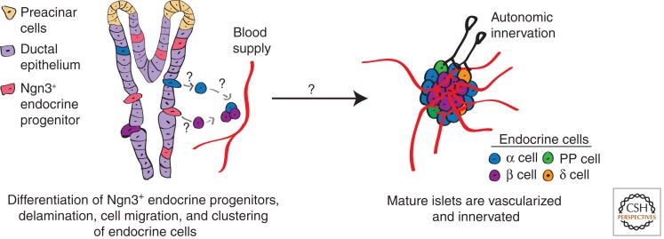
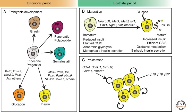
The relentless nature and increasing prevalence of human pancreatic diseases, in particular, diabetes mellitus and adenocarcinoma, has motivated further understanding of pancreas organogenesis. The pancreas is a multifunctional organ whose epithelial cells govern a diversity of physiologically vital endocrine and exocrine functions. The mechanisms governing the birth, differentiation, morphogenesis, growth, maturation, and maintenance of the endocrine and exocrine components in the pancreas have been discovered recently with increasing tempo. This includes recent studies unveiling mechanisms permitting unexpected flexibility in the developmental potential of immature and mature pancreatic cell subsets, including the ability to interconvert fates. In this article, we describe how classical cell biology, genetic analysis, lineage tracing, and embryological investigations are being complemented by powerful modern methods including epigenetic analysis, time-lapse imaging, and flow cytometry-based cell purification to dissect fundamental processes of pancreas development.


Figures

Figure 1.
Figure 1.
Pancreatic morphogenesis and developmental regulation. Mouse pancreatic development is characterized by a “primary transition” from embryonic day (E) 9.5 to E12.5 and a “secondary transition” from E13 to birth. (Panel 1) Pancreatic budding and pancreatic proliferation occur at approximately E9.5 and a subset of epithelial cells at that stage express pancreas-specific transcription factor 1a (Ptf1a), pancreatic and duodenal homeobox 1 (Pdx1), sry-box 9 (Sox9), cMyc, and other transcription factors. (Panel 2) Proliferation of epithelial cells results in the formation of microlumens (empty white spaces) at E11. Mesenchymal cells (brown crosshatch) overlie the developing pancreatic bud and secrete a variety of growth and differentiation factors (see text). Before E13, Cdc42 influences microtubule coalescence and formation of a continuous branched tube. (Panel 3) Coincident with tubulogenesis, multipotent “tip” and bipotent “trunk” domains establish. The multipotent Ptf1a+ tip progenitors derive acinar, duct, and endocrine cells, whereas the bipotent Nkx6 homeobox (Nkx6+) trunk progenitors produce duct and endocrine cells. (Panel 4) After E13 and during the secondary transition, pancreatic branching, cell differentiation, acinar cell expansion, and islet formation drive pancreatic morphogenesis. Islets represent clusters of endocrine cells. At this stage, the “tip” domain will derive acinar cells, whereas the “trunk” domain will derive duct and endocrine cells.
Figure 2.
Figure 2.
Pancreatic acinar and ductal cell differentiation. Apolar multipotent progenitors develop into three distinct progeny: acinar cells (top, green), duct cells (bottom, brown), and endocrine islet cells (not shown). (A) During acinar development expression of Ptf1a represses Nkx6, thereby suppressing alternative duct and endocrine cell fates. The commitment of early acinar cells requires the formation of a Ptf1a-Rbpj trimeric complex (PTF1-J). Acinar maturation requires formation of a Ptf1a-Rbpjl trimeric complex (PTF1-L) and is dependent on muscle, intestine, and stomach expression 1 (Mist1) and recombination signal-binding protein for immunoglobulin kappa J region-like (Rbpjl) and possibly other unknown factors. Maturation produces pyramidal polarized acinar cells with specialized organelles and high-secretory capacity. Cholecystokinin (CCK) and NFAT signaling influence adaptive growth of acinar cells. (B) Duct cells derive from apolar progenitors that become polarized through unknown mechanisms and form primary cilia, an organelle whose development requires both hepatocyte nuclear factor 6 (Hnf6) and Hnf1β. Duct cell heterogeneity within pancreatic branches is depicted (see text).
Figure 3.
Figure 3.
Events culminating in islet morphogenesis. Endocrine cells derive from unipotent Neurogenin3 (Ngn3+) endocrine progenitors (pink). On differentiation, endocrine cells delaminate from the ductal epithelia (light purple), migrate toward the mesenchyme (not shown), and aggregate into clusters called islets. For simplicity only α cells (blue) and β cells (purple) are depicted delaminating from the ductal epithelia and migrating toward blood vessels (red). Coincident with islet morphogenesis, vascularization, and innervation of islets by the autonomic nervous system occurs (black).
Figure 4.
Figure 4.
Embryonic and postnatal development of pancreatic endocrine cells. (A) During embryonic development, pancreatic endocrine cells are formed by differentiation from progenitor cells expressing the bHLH transcription factor Neurogenin3 (Ngn3). Differentiation into distinct lineages requires the expression of a cascade of different transcriptional factors (TFs). Key α-cell TFs include Forkhead box A2 (Foxa2), NK2 homeobox 2 (Nkx2.2), Paired box 6 (Pax6), and aristaless (Arx), whereas β-cell differentiation requires expression of musculoaponeurotic fibrosarcoma oncogene homolog B (MafB), Pancreatic and duodenal homeobox 1 (Pdx1), Homeobox protein HB9 (Hlxb9), Pax4, Pax6, Islet1 (Isl1), Nkx2.2, and Nkx6.1 among others (B,C). During the “postnatal period” (loosely defined here as “birth until weaning” in mice and “birth until adolescence” in humans), β cells undergo two critical events that enable the establishment of a normal, functional β-cell mass. (B) First, β cells undergo functional maturation by increasing insulin production and enhancing glucose-stimulated insulin secretion (GSIS). Known transcriptional regulators of the maturation process include Neurogenic differentiation 1 (NeuroD1), MafA, MafB, Isl1, Pdx1, Ngn3, and Von Hippel–Lindau (Vhl). (C) Second, β cells undergo a transient burst of β-cell proliferation that coincides with a significant increase in β-cell mass expansion. Cell-cycle regulators of this process include cyclin-dependent kinases (Cdk4), D-type cyclins (CcnD1 and CcnD2), CDK inhibitors (CKIs—p16INK4a, p19Arf, and p27Kip1), and the transcription factor FoxM1. Refer to text, Table 1, and cited references for further information.

Similar articles

Cited by

References

    1. Aguayo-Mazzucato C, Koh A, El Khattabi I, Li W-C, Toschi E, Jermendy A, Juhl K, Mao K, Weir GC, Sharma A, et al. 2011. Mafa expression enhances glucose-responsive insulin secretion in neonatal rat β cells. Diabetologia 54: 583–593 - PMC - PubMed
    1. Ahlgren U, Jonsson J, Edlund H. 1996. The morphogenesis of the pancreatic mesenchyme is uncoupled from that of the pancreatic epithelium in IPF/PDX1-deficient mice. Development 122: 1409–1416 - PubMed
    1. Ahlgren U, Jonsson J, Jonsson L, Simu K, Edlund H 1998. β-cell-specific inactivation of the mouse Ipf1/Pdx1 gene results in loss of the β-cell phenotype and maturity onset diabetes. Genes Dev 12: 1763–1768 - PMC - PubMed
    1. Akirav EM, Lebastchi J, Galvan EM, Henegariu O, Akirav M, Ablamunitis V, Lizardi PM, Herold KC 2011. Detection of β cell death in diabetes using differentially methylated circulating DNA. Proc Natl Acad Sci 108: 19018–19023 - PMC - PubMed
    1. Apelqvist A, Li H, Sommer L, Beatus P, Anderson DJ, Honjo T, Hrabe de Angelis M, Lendahl U, Edlund H 1999. Notch signalling controls pancreatic cell differentiation. Nature 400: 877–881 - PubMed

Publication types

LinkOut - more resources