

Temporal expression of hypoxia-regulated genes is associated with early changes in redox status in irradiated lung
- PMID: 22588005
- PMCID: PMC3649014
- DOI: 10.1016/j.freeradbiomed.2012.04.014
Temporal expression of hypoxia-regulated genes is associated with early changes in redox status in irradiated lung
Abstract
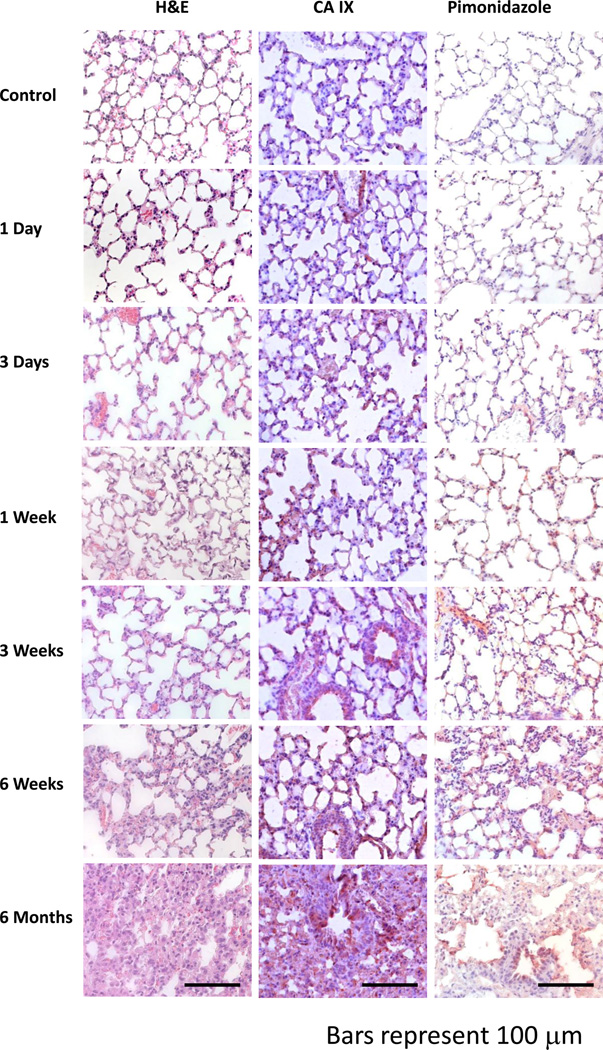
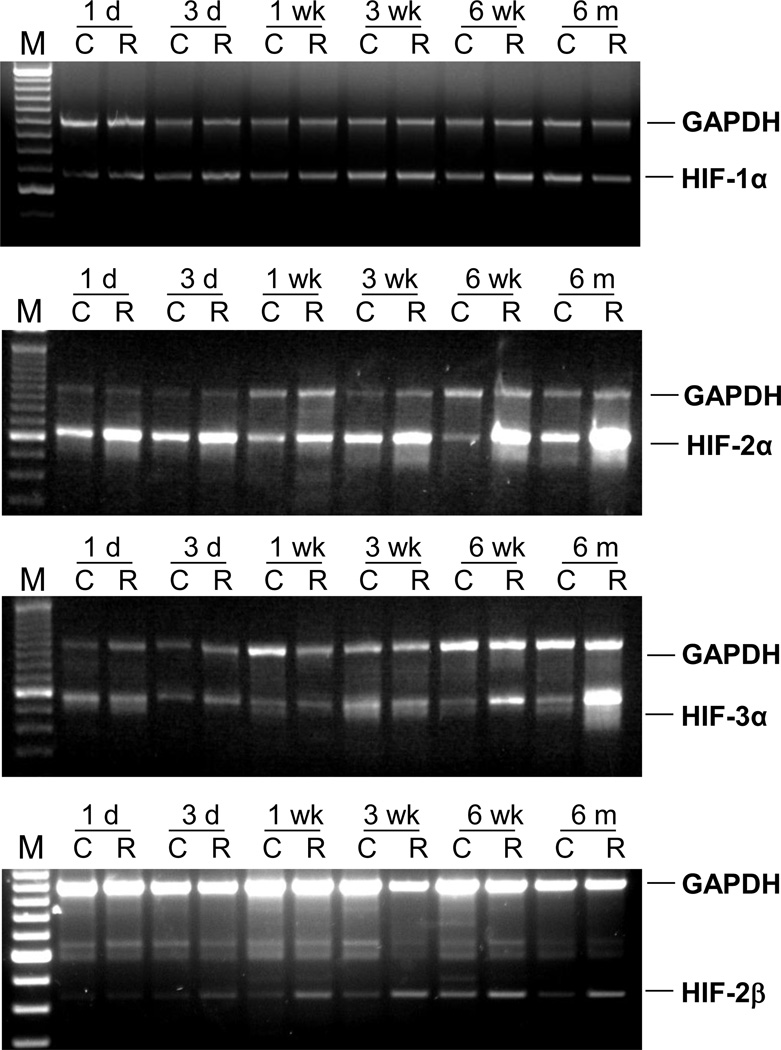
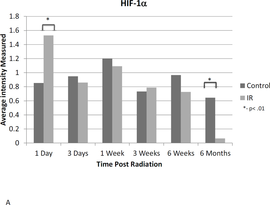
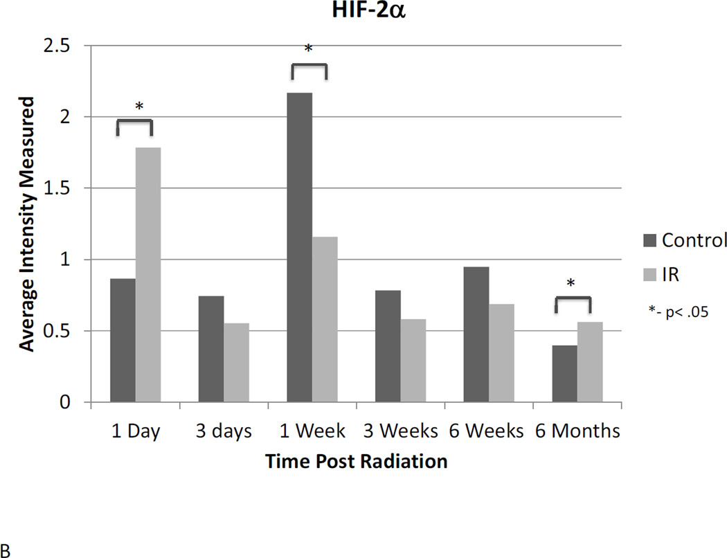
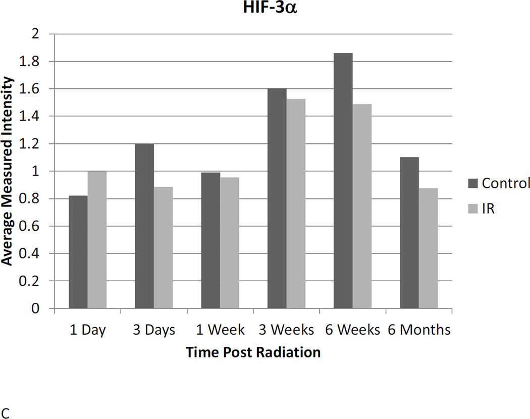
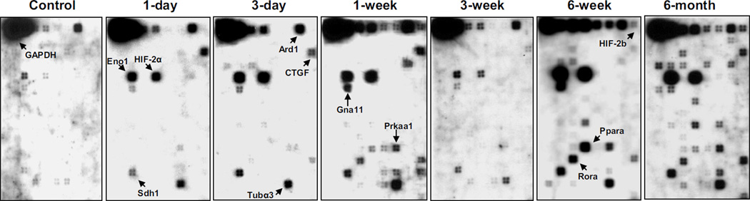
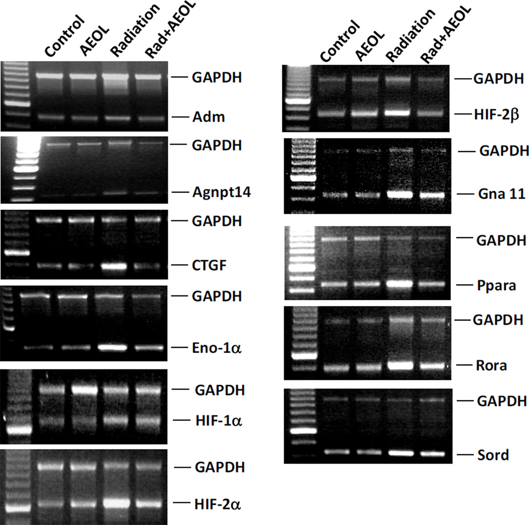
The development of normal lung tissue toxicity after radiation exposure results from multiple changes in cell signaling and communication initiated at the time of the ionizing event. The onset of gross pulmonary injury is preceded by tissue hypoxia and chronic oxidative stress. We have previously shown that development of debilitating lung injury can be mitigated or prevented by administration of AEOL10150, a potent catalytic antioxidant, 24h after radiation. This suggests that hypoxia-mediated signaling pathways may play a role in late radiation injury, but the exact mechanism remains unclear. The purpose of this study was to evaluate changes in the temporal expression of hypoxia-associated genes in irradiated mouse lung and determine whether AEOL10150 alters expression of these genes. A focused oligo array was used to establish a hypoxia-associated gene expression signature for lung tissue from sham-irradiated or irradiated mice treated with or without AEOL10150. Results were further verified by RT-PCR. Forty-four genes associated with metabolism, cell growth, apoptosis, inflammation, oxidative stress, and extracellular matrix synthesis were upregulated after radiation. Elevated expression of 31 of these genes was attenuated in animals treated with AEOL10150, suggesting that expression of a number of hypoxia-associated genes is regulated by early development of oxidative stress after radiation. Genes identified herein could provide insight into the role of hypoxic signaling in radiation lung injury, suggesting novel therapeutic targets, as well as clues to the mechanism by which AEOL10150 confers pulmonary radioprotection.
Copyright © 2012 Elsevier Inc. All rights reserved.
Conflict of interest statement
Isabel L. Jackson is a consultant for AEOLUS Pharmaceuticals, Inc. All other authors declare no conflict of interest.
Figures
References
-
- Barcellos-Hoff MH, Dix TA. Redox-mediated activation of latent transforming growth factor-beta 1. Molecular endocrinology (Baltimore, Md. 1996;10(9):1077–1083. - PubMed
-
- Cucoranu I, Clempus R, Dikalova A, Phelan PJ, Ariyan S, Dikalov S, et al. NAD(P)H oxidase 4 mediates transforming growth factor-beta1-induced differentiation of cardiac fibroblasts into myofibroblasts. Circ Res. 2005;97(9):900–907. - PubMed
-
- Sturrock A, Cahill B, Norman K, Huecksteadt TP, Hill K, Sanders K, et al. Transforming growth factor-beta1 induces Nox4 NAD(P)H oxidase and reactive oxygen species-dependent proliferation in human pulmonary artery smooth muscle cells. American journal of physiology. 2006;290(4):L661–L673. - PubMed
-
- Ward WF, Solliday NH, Molteni A, Port CD. Radiation injury in rat lung. II. Angiotensin-converting enzyme activity. Radiation research. 1983;96(2):294–300. - PubMed
Publication types
MeSH terms
Substances
Grants and funding
LinkOut - more resources
Full Text Sources
Medical
