Review
. 2012:114:21-57.
doi: 10.1016/B978-0-12-386503-8.00002-8.

Essential roles of EphB receptors and EphrinB ligands in endothelial cell function and angiogenesis

Affiliations
Review

Essential roles of EphB receptors and EphrinB ligands in endothelial cell function and angiogenesis

Ombretta Salvucci et al. Adv Cancer Res. 2012.

Abstract

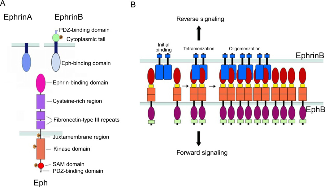
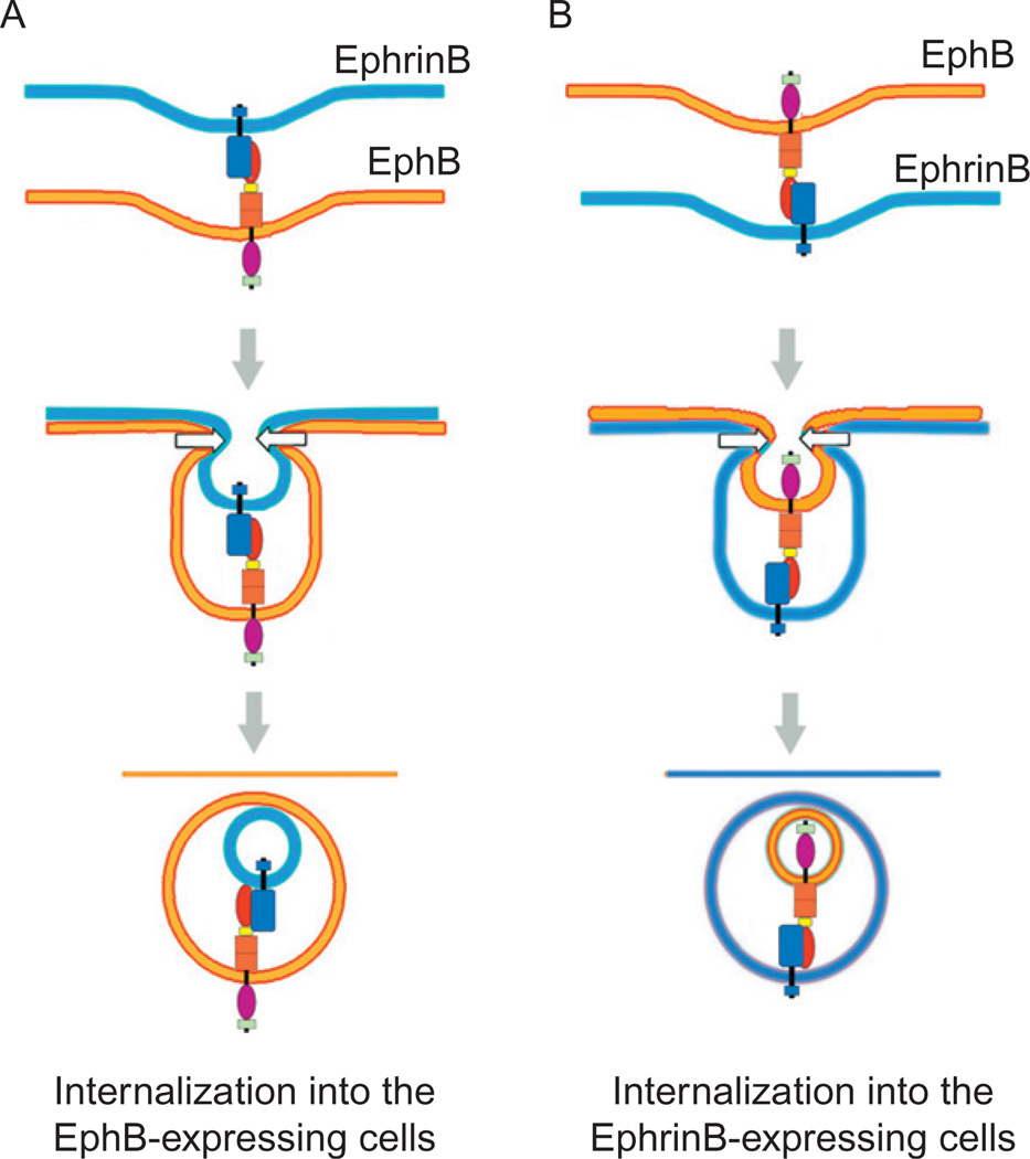
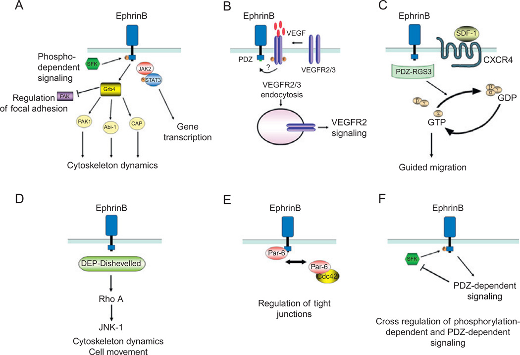
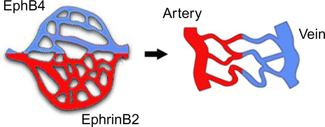
Eph receptor tyrosine kinases and their Ephrin ligands represent an important signaling system with widespread roles in cell physiology and disease. Receptors and ligands in this family are anchored to the cell surface; thus Eph/Ephrin interactions mainly occur at sites of cell-to-cell contact. EphB4 and EphrinB2 are the Eph/Ephrin molecules that play essential roles in vascular development and postnatal angiogenesis. Analysis of expression patterns and function has linked EphB4/EphrinB2 to endothelial cell growth, survival, migration, assembly, and angiogenesis. Signaling from these molecules is complex, with the potential for being bidirectional, emanating both from the Eph receptors (forward signaling) and from the Ephrin ligands (reverse signaling). In this review, we describe recent advances on the roles of EphB/EphrinB protein family in endothelial cell function and outline potential approaches to inhibit pathological angiogenesis based on this understanding.


Figures

Fig. 1
Fig. 1
(A) Schematic representation of the domain structure and binding interfaces of Ephrins and Eph receptors. EphrinA ligands are attached to the cell surface through a glycosylphosphatidylinositol (GPI)-anchor; the extracellular domain contains an Eph receptor-binding domain that is connected to the transmembrane segment. EphrinB ligands are transmembrane proteins with an extracellular Eph receptor-binding domain connected to a transmembrane segment, which is followed by a short intracellular domain. The Eph receptors include an extracellular domain composed of an Ephrin-binding domain, a cysteine-rich segment that contains an epidermal growth factor (EGF)-like motif, and two fibronectin-type III domains; and a cytoplasmic region that contains a juxtamembrane region, the kinase domain, a sterile a-motif (SAM), and a binding site for PDZ-containing proteins. (B) Representation of initial binding of cell surface Eph and Ephrin molecules to form heterotetramers, which initiate signaling, and subsequent oligomerization to form large receptor/ligand clusters that expand laterally through hemophilic interactions between Eph receptors.
Fig. 2
Fig. 2
Interaction of EphrinB with EphB can lead to (A) trans-endocytosis in the direction of the receptor (forward endocytosis) or (B) trans-endocytosis in the direction of the ligand (reverse endocytosis). Both processes lead to the internalization of full-length ligand and receptor.
Fig. 3
Fig. 3
Mechanisms of EphrinB reverse signaling. (A) EphrinB activation by EphB receptor leads to the recruitment of Src family kinases (SFKs) that phosphorylate EphrinB intracellular domain at tyrosine residues. The adaptor molecule Grb4, which contains a Src-homology-2 (SH2) domain, is recruited to the phosphorylated EphrinB and initiates a number of signaling events that regulate cytoskeleton dynamics and focal adhesions. The phosphorylated EphrinB can also recruit the Jak2/STAT3 complex; the phosphorylated STAT3 translocates to the nucleus regulating expression of target genes. (B) Activated EphrinB regulates the internalization of VEGFR2 and VEGFR3 through PDZ-mediated binding of yet unknown protein(s). After VEGF binding, VEGFR is phosphorylated; signaling from activated VEGFR requires internalization, which is positively regulated by EphrinB signaling. (C) RGS3 is a PDZ-containing protein that constitutively binds to EphrinB and links signaling from the G-protein-coupled receptor CXCR4 to EphrinB. CXCR4 signaling in response to the ligand SDF1 is induced by the dissociation of Gβγ and GTP-Gα subunits. PDZ-RGS3 can inhibit CXCR4 signaling by enhancing the GTPase activity of the Gα subunit, resulting in the reformation of the inactive heterodimeric CXCR4 receptor. (D) The scaffold protein Dishevelled binds EphrinB through its DEP domain and mediates signaling via the Rho small GTPase pathway. (E) The scaffold protein Par6 associates with EphrinB resulting in the loss of tight junctions. PAR6 forms a complex with activated apical protein kinase C (aPKC) and Cdc42-GTP; the complex localizes to the apical cell junctions where it regulates tight junctions. EphrinB1 can compete with Cdc42 for binding to Par6 and thus reduce tight junctions. (F) Cross-regulation between EphrinB phosphorylation- and PDZ-dependent signaling pathways. The phosphatase PTB-BL, recruited through its PDZ domain to EphrinB cytoplasmic domain, inactivates Src and dephosphorylates EphrinB.
Fig. 4
Fig. 4
A role for EphB4 and EphrinB2 in arterial and venous fate determination. In the primitive vascular plexus, presumptive arterial and venous territories are marked by the distinctive expression of EphrinB2 or EphB4, prior to the formation of EphrinB2+ arteries and EphB4+ veins.
Fig. 5
Fig. 5
Angiogenesis is often initiated by the sprouting of endothelial cells from existing vessels. The leading cell in the sprout, the so-called tip cell, is characterized by filopodia extensions; the cells at the base of the sprout, the so-called stalk cells, are proliferating. VEGF/VEGFR2, the Notch ligands Delta4 and Notch signaling participate in orchestrating sprouting angiogenesis.
Fig. 6
Fig. 6
EphrinB is phosphorylated in remodeling but not in fully developed retinal vessels. (A) EphrinB phosphorylation is not detected in fully developed mouse retinal vessels, including the CD31+ endothelial cells and theNG2+ pericytes. (B) EphrinB2 is widely phosphorylated in the remodeling retinal vessels from 6-day-old mice, both in the endothelial cells and in the pericytes.
Fig. 7
Fig. 7
Primary endothelial cells assemble into cord-like structures on extracellular matrix, a process that requires EphB/EphrinB interaction. (A) Time-dependent assembly of primary endothelial cells on Matrigel. Note the appearance after 1–2h of needle-like endothelial cell extensions that eventually form the initial bridging of endothelial cells with each other. (B) EphrinB2 is broadly and time-dependently phosphorylated in endothelial cells as they assemble into cord-like structures. (C) The EphB receptor inhibitors SNEW and TNYL-RAW peptides prevent the assembly of endothelial cells into cord-like structures.
Fig. 8
Fig. 8
Endothelial cells and mesenchymal stem cells/pericytes assemble in cord-like structures on Matrigel, with mesenchymal stem cells (green) in the nodes anchoring the cord structures. (B) EphrinB is phosphorylated (white) at points of contact between endothelial cells (HUVEC green) and mesenchymal stem cells (MSC red) forming cord-like structures. (C) The silencing of EphrinB2 in mesenchymal stem cells (green) prevents their assembly with endothelial cells (unstained). (D) The silencing of EphrinB2 in endothelial cells (unstained) prevents their assembly with MSC (green).
Fig. 9
Fig. 9
(A) EphrinB is phosphorylated in angiogenic vessels associated with skin wound healing. (B) EphrinB is phosphorylated in angiogenic retinal vessels induced by hypoxia (areas of hypoxia visualized in green; ROP model). (C) EphrinB is phosphorylated in angiogenic tumor vessels. CD31 identifies endothelial cells; NG2 pericytes/mural cells.

References

    1. Adams RH, Wilkinson GA, Weiss C, Diella F, Gale NW, Deutsch U, Risau W, Klein R. Roles of ephrinB ligands EphB receptors in cardiovascular development: demarcation of arterial/venous domains, vascular morphogenesis, sprouting angiogenesis. Genes Dev. 1999;13:295–306. - PMC - PubMed
    1. Adams RH, Diella F, Hennig S, Helmbacher F, Deutsch U, Klein R. The cytoplasmic domain of the ligand ephrinB2 is required for vascular morphogenesis but not cranial neural crest migration. Cell. 2001;104:57–69. - PubMed
    1. Alon T, Hemo I, Itin A, Pe’er J, Stone J, Keshet E. Vascular endothelial growth factor acts as a survival factor for newly formed retinal vessels has implications for retinopathy of prematurity. Nat. Med. 1995;1:1024–1028. - PubMed
    1. Armulik A, Abramsson A, Betsholtz C. Endothelial/pericyte interactions. Circ. Res. 2005;97:512–523. - PubMed
    1. Armulik A, Genove G, Mae M, Nisancioglu MH, Wallgard E, Niaudet C, He L, Norlin J, Lindblom P, Strittmatter K, Johansson BR, Betsholtz C. Pericytes regulate the blood–brain barrier. Nature. 2010;468:557–561. - PubMed

Publication types

MeSH terms