. 2012 May;2(5):414-24.
doi: 10.1158/2159-8290.CD-12-0022. Epub 2012 Apr 1.

Preexisting MEK1 exon 3 mutations in V600E/KBRAF melanomas do not confer resistance to BRAF inhibitors

Affiliations

Preexisting MEK1 exon 3 mutations in V600E/KBRAF melanomas do not confer resistance to BRAF inhibitors

Hubing Shi et al. Cancer Discov. 2012 May.

Abstract

BRAF inhibitors (BRAFi) induce antitumor responses in nearly 60% of patients with advanced V600E/KBRAF melanomas. Somatic activating MEK1 mutations are thought to be rare in melanomas, but their potential concurrence with V600E/KBRAF may be selected for by BRAFi. We sequenced MEK1/2 exon 3 in melanomas at baseline and upon disease progression. Of 31 baseline V600E/KBRAF melanomas, 5 (16%) carried concurrent somatic BRAF/MEK1 activating mutations. Three of 5 patients with BRAF/MEK1 double-mutant baseline melanomas showed objective tumor responses, consistent with the overall 60% frequency. No MEK1 mutation was found in disease progression melanomas, except when it was already identified at baseline. MEK1-mutant expression in V600E/KBRAF melanoma cell lines resulted in no significant alterations in p-ERK1/2 levels or growth-inhibitory sensitivities to BRAFi, MEK1/2 inhibitor (MEKi), or their combination. Thus, activating MEK1 exon 3 mutations identified herein and concurrent with V600E/KBRAF do not cause BRAFi resistance in melanoma.

Significance: As BRAF inhibitors gain widespread use for treatment of advanced melanoma, biomarkers for drug sensitivity or resistance are urgently needed. We identify here concurrent activating mutations in BRAF and MEK1 in melanomas and show that the presence of a downstream mutation in MEK1 does not necessarily make BRAF–mutant melanomas resistant to BRAF inhibitors.


Conflict of interest statement

Disclosure of Potential Conflicts of Interest

A. Ribas and R.S. Lo declare patent application under PCT application serial no. PCT/US11/061552 (Compositions and Methods for Detection and Treatment of BRAF Inhibitor-Resistant Melanomas). K.B. Dahlman, B. Chmielowski, J.A. Sosman, R.F. Kefford, G.V. Long, and A. Ribas have received honoraria from or served as consultants to pharmaceutical firms (Roche-Genentech, GlaxoSmithKline, Illumina). No potential conflicts of interest were disclosed by the other authors.

Figures
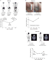
Figure 1
Figure 1
Spectrum of clinical responses of V600E/KBRAF/P124SMEK1 double-mutant melanomas to BRAF inhibitors. A, in patient 2, a right axillary nodal melanoma clearly responded to vemurafenib at day 15 [positron emission tomography (PET) scan], at which time a “baseline” biopsy of the same tumor revealed mutations in both BRAF and MEK1. Tumor tracking shows timing of baseline and DP tumor biopsies. Best overall response demonstrates a partial response, and biopsy locations are indicated. B, in patient 12, a 2-cm right axillary tumor (which was not a chosen target tumor tracked below), from which a 3-mm punch biopsy prior to dabrafenib initiation revealed both BRAF and MEK1 mutations, became nonpalpable within 14 days of starting on dabrafenib. Despite tumor tracking not reaching partial response before DP, initial tumor response in this patient was associated with a dramatic increase in right shoulder mobility as well as decreased right breast/axillary swelling (Supplementary Fig. S2). C, in patient 28, a baseline tumor biopsy in the right side of the neck taken 4 months and 11 days prior to dabrafenib initiation was shown to harbor both BRAF and MEK1 mutations. PET scans before and after dabrafenib initiation clearly showed a rapid metabolic response at day 15 on treatment. Best overall response indicates a PR, and the same neck lesion that responded on day 15 was biopsied when it progressed.
Figure 2
Figure 2
Regulated P124SMEK1 expression cannot alter p-ERK levels or sensitivity to BRAF or MEK inhibition in V600EBRAF melanoma cell lines. A, doxycycline-repressible expression of vector (control), FLAG-MEK1 WT, versus FLAG-MEK1 P124S in V600EBRAF melanoma cell lines (M229, M238). Protein lysates (48 hours post seeding at 0, 0.1, and 10 ng/mL doxycycline) were probed by Western blotting for the indicated phospho- and total protein levels. Tubulin, loading control. B, dose-dependent suppression of p-ERK levels by vemurafenib (PLX4032) in a V600EBRAF background with or without concurrent P124SMEK1. Indicated M238 stable cell lines were washed free of doxycycline for 48 hours, inducing exogenous FLAG-MEK1 WT or FLAG-MEK1 P124S expression, and treated for 1 hour with increasing doses (μM) of vemurafenib: 0 (DMSO), 0.01, 0.1, 1.0, and 10. Cell lysates were then probed for the indicated protein levels. C, stable MEK1 knockdown in the naturally occurring V600EBRAF/P124SMEK1 double-mutant melanoma short-term culture (YUKSI). Protein lysates were probed for the indicated protein levels. D, impact of P124SMEK1 on cellular p-ERK levels in BRAF WT versus V600E backgrounds. Indicated FLAG-tagged expression constructs were transiently transfected into HEK293T cells (BRAF WT) with either pBABE-PURO (empty vector) or pBABE-PURO-V600EBRAF. After 72 hours, cell lysates were probed for the indicated protein levels by Western blotting. E, stable cell lines (M229, top; M238, bottom) were either maintained with doxycycline (10 ng/mL) or washed and released incrementally (0.1 or 0 ng/mL) from doxycycline-mediated suppression of FLAG-MEK1 WT or FLAG-MEK1 P124S gene expression for 24 hours prior to treatment with increasing concentrations of vemurafenib/PLX4032 (black) or selumetinib/AZD6244 (red). Survival curves are shown after 72 hours of drug treatments, and data represent percent surviving cells relative to DMSO-treated controls (mean ± SEM, n = 5). The dashed line corresponds to 50% cell killing. F, the BRAF/MEK1 double-mutant melanoma short-term culture, YUKSI, was infected with either a control or shMEK1 virus and subjected to PLX4032 or AZD6244 treatments for 72 hours. G, indicated M238 stable cell lines maintained with doxycycline (100 ng/mL) or washed free of doxycycline, and seeded at single-cell density. At 24 hours after seeding, cells were treated with indicated concentrations of PLX4032. Cellular colonies were visualized by staining with crystal violet at 12 days after drug treatments. Photographs are representative of 2 independent experiments and time points.
Figure 3
Figure 3
I111SMEK1 expression fails to modulate p-ERK levels or melanoma sensitivity to BRAF or MEK inhibitors in the presence of V600EBRAF. A, doxycycline-repressible expression vector, FLAG-MEK1 I111S, or FLAG-MEK1 C121S in the V600EBRAF melanoma cell lines M229. Protein lysates (48 hours post seeding at 0, 0.1, and 10 ng/mL doxycycline) were probed by Western blotting for the indicated phospho- and total protein levels. Tubulin, loading control. B, M229 stable cell lines were either maintained with doxycycline (10 ng/mL) or washed and released incrementally (0.1 or 0 ng/mL) from doxycycline-mediated suppression of gene expression for 24 hours prior to treatment with increasing concentrations of vemurafenib/PLX4032 (black) or selumetinib/AZD6244 (red). Survival curves are shown after 72 hours of drug treatments, and data represent percent surviving cells relative to DMSO-treated controls (mean ± SEM, n = 5). The dashed line corresponds to 50% cell killing. C, dose-dependent suppression of p-ERK levels by vemurafenib/PLX4032 in a V600EBRAF background with or without concurrent WTMEK1, C121SMEK1, or I111SMEK1 expression. Indicated M229 stable cell lines were treated for 1 hour with increasing doses (μM) of PLX4032: 0 (DMSO), 0.01, 0.1, 1.0, and 10. Cell lysates were then probed for the indicated protein levels. D, impact of indicated MEK1 mutants on cellular p-ERK levels in BRAF WT versus V600E backgrounds. Indicated FLAG-tagged expression constructs were transiently transfected into HEK293T cells (BRAF WT) with either pBABE-PURO (empty vector) or pBABE-PURO-V600EBRAF. After 72 hours, cell lysates were probed for the indicated protein levels by Western blotting.

Comment in

References

    1. Long GV, Menzies AM, Nagrial AM, Haydu LE, Hamilton AL, Mann GJ, et al. Prognostic and clinicopathologic associations of oncogenic BRAF in metastatic melanoma. J Clin Oncol. 2011;29:1239–46. - PubMed
    1. Bollag G, Hirth P, Tsai J, Zhang J, Ibrahim PN, Cho H, et al. Clinical efficacy of a RAF inhibitor needs broad target blockade in BRAF-mutant melanoma. Nature. 2010;467:596–9. - PMC - PubMed
    1. Chapman PB, Hauschild A, Robert C, Haanen JB, Ascierto P, Larkin J, et al. Improved survival with vemurafenib in melanoma with BRAF V600E mutation. N Engl J Med. 2011;364:2507–16. - PMC - PubMed
    1. Flaherty KT, Puzanov I, Kim KB, Ribas A, McArthur GA, Sosman JA, et al. Inhibition of mutated, activated BRAF in metastatic melanoma. N Engl J Med. 2010;363:809–19. - PMC - PubMed
    1. Kefford R, Arkenau H, Brown MP, Millward M, Infante JR, Long GV, et al. Phase I/II study of GSK2118436, a selective inhibitor of oncogenic mutant BRAF kinase, in patients with metastatic melanoma and other solid tumors. J Clin Oncol. 2010;28(15s) suppl;abstr 8503.

Publication types

MeSH terms