. 2012 May;2(5):450-7.
doi: 10.1158/2159-8290.CD-11-0287. Epub 2012 Mar 31.

Kinetics of inhibitor cycling underlie therapeutic disparities between EGFR-driven lung and brain cancers

Affiliations

Kinetics of inhibitor cycling underlie therapeutic disparities between EGFR-driven lung and brain cancers

Krister J Barkovich et al. Cancer Discov. 2012 May.

Abstract

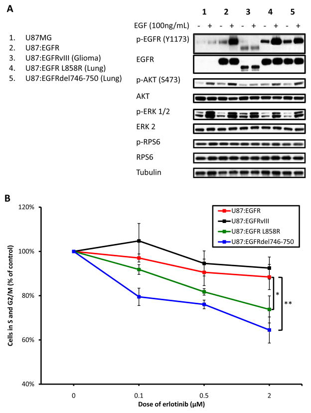
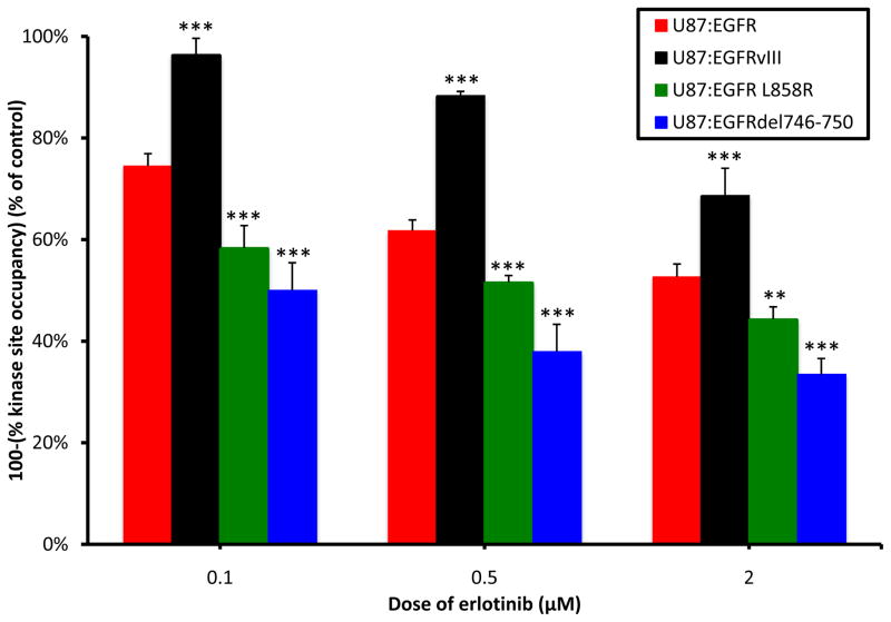
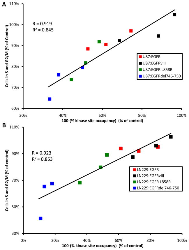
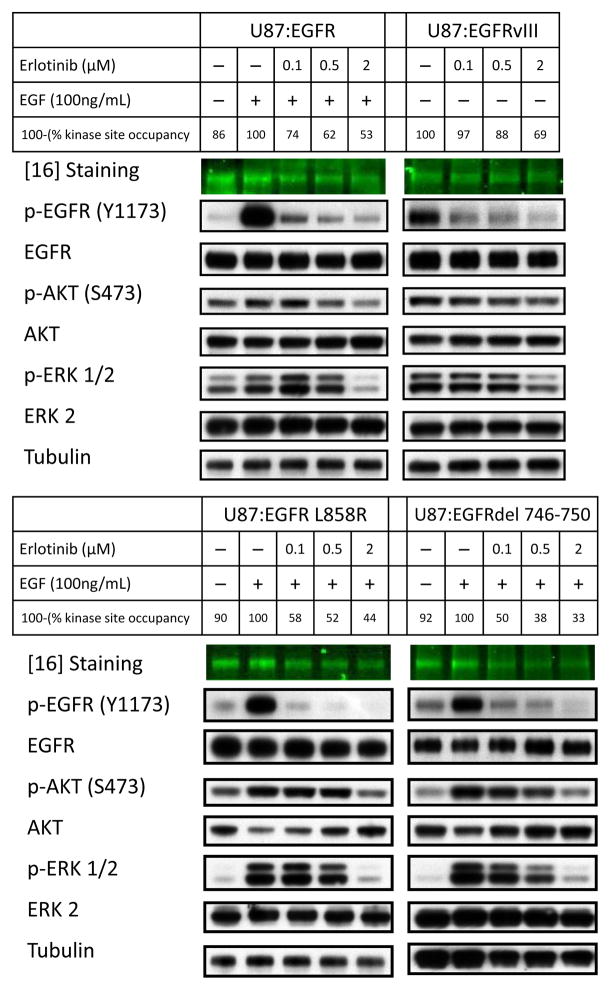
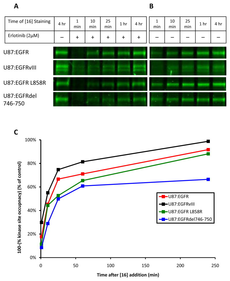
Although mutational activation of the epidermal growth factor receptor (EGFR) features prominently in glioma and non-small cell lung cancer (NSCLC), inhibitors of EGFR improve survival only in patients with NCSLC. To understand how mutations in EGFR influence response to therapy, we generated glioma cells expressing either glioma- or NSCLC-derived alleles and quantified kinase-site occupancy by clinical inhibitors with the use of a novel affinity probe and kinetic methodology. At equivalent doses, erlotinib achieved lower kinase-site occupancy in glioma-derived EGFRvIII compared with NSCLC-derived EGFR mutants. Kinase-site occupancy correlated directly with cell-cycle arrest. EGFRvIII released erlotinib rapidly compared with wild-type EGFR, whereas NSCLC-derived mutants released erlotinib slowly.

Significance: These data suggest that kinase-site occupancy is a biomarker for efficacy of EGFR inhibitors, that rapid binding and release of erlotinib in glioma-derived EGFRvIII opposes the blockade of downstream signaling, and that slower cycling of erlotinib within the active site of NSCLC-derived mutants underlies their improved clinical response.


Conflict of interest statement

DISCLOSURE OF POTENTIAL CONFLICTS OF INTEREST

We have no potential conflicts of interest to disclose.

Figures

Figure 1
Figure 1
EGFR mutants exhibit differential growth inhibition in response to erlotinib. A, U87MG cells were transduced with wild type-EGFR, EGFRvIII, EGFR L858R, or EGFRdel746-750. Cells were treated or untreated with EGF 30 min prior to harvest, as shown. Cells were then lysed and analyzed by immunoblot. Signaling downstream of EGFR was analyzed using antisera to total and phospho-specific proteins shown. B, The U87MG panel was treated for 24 hours with three doses of erlotinib, as shown. Cells were analyzed for their DNA content, measured by propidium iodide staining. For each sample, the proportion of cells in S and G2/M phases was combined and graphed. Treatments were completed in triplicate with error bars representing one standard deviation (* p<0.05, ** p<0.01).
Figure 2
Figure 2
EGFR alleles differ in levels of kinase site occupancy achieved after erlotinib treatment. The U87MG panel was treated overnight with erlotinib at doses shown, and stimulated for 30 minutes with 100ng/mL EGF. Cells were subjected to a 25-minute pulse-chase with 60μM [16], then lysed and separated by SDS-PAGE. Gels were scanned on a Typhoon fluorescence imager using a 488nm excitation laser. Levels of fluorescence correspond to the amount of kinase active site that is unbound by erlotinib (100-(% kinase site occupancy)), and thus is available for probe binding. The fluorescence intensity for each treatment was quantified by densitometry and scaled as a percent of the EGF-stimulated control lane. Results were completed in triplicate with error bars representing one standard deviation (** p<0.01, *** p<0.005).
Figure 3
Figure 3
Kinase site occupancy is a better biomarker than abundance of p-EGFR, for efficacy of erlotinib. A, For each EGFR-allele and at each dose of erlotinib, 100-(% kinase site occupancy) was assigned the x-coordinate. The proportion of cells in S and G2/M phases was combined and graphed on the y-coordinate. We found a strong (R=0.916) correlation between kinase site occupancy and efficacy for the U87 panel. B, The same analysis was applied to the LN229 panel, where a correlation of R=0.923 was found between kinase site occupancy and efficacy.
Figure 4
Figure 4
Antiproliferative effects of erlotinib correlate poorly with abundance of p-EGFR. The U87MG panel was treated with three doses of erlotinib, as shown, then pulsed with 100 ng/mL EGF prior to harvesting. Phospho- and total protein levels were visualized by western blotting. While low dosages of erlotinib efficiently block p-EGFR (Y1173) in all cell lines, levels of p-ERK 1/2 (T202/Y204) were decreased in NSCLC-derived alleles, compared with brain-cancer derived EGFRvIII, paralleling the antiproliferative response in Figure 1B. U87MG cells are mutant for PTEN, disconnecting signaling between EGFR and PI3K. Thus, the differential abundance of p-AKT (S473) was less apparent in this experiment, as compared with PTENWT LN229 cells (Supplemental Figure 10). Levels of kinase site occupancy more closely follow abundance of phosphorylated downstream molecules.
Figure 5
Figure 5
Kinetics of erlotinib binding/unbinding differ across EGFR-alleles. A and B, The U87 panel was treated with 2μM erlotinib (A) or DMSO (B) for 24 hours and then pulsed with 60μM [16] for 1 minute, 10 minutes, 25 minutes, 1 hour, or 4 hours. In the drug treated experiment (A), the control was untreated and pulsed with [16] for 4 hours. As erlotinib cycles out of EGFR, the active site is irreversibly bound by [16], preventing erlotinib re-binding. Over time, this occurs with all EGFR-bound erlotinib. The rate with which this replacement occurs is related to the speed with which erlotinib is unbound by EGFR. The control (B) established the rate at which [16] alone binds each EGFR-allele (k 2 in Equation (2)). C, Analysis of (A) and (B) by densitometry allowed for the quantification of [16] binding over time in the presence or absence of erlotinib. The level of [16] staining of a single EGFR-allele (as a % of the 4hr control lane) determined in (A) was divided by the level of [16] staining of that same EGFR-allele (as a % of the 4hr control lane) determined in (B), allowing for the tracking of kinase site occupancy for each EGFR-allele over time. These data determine that erlotinib replacement occurs more quickly in glioma-derived EGFRvIII than in NSCLC-derived EGFR-mutants.

Comment in

  • Occupy EGFR.
    Park JH, Lemmon MA. Park JH, et al. Cancer Discov. 2012 May;2(5):398-400. doi: 10.1158/2159-8290.CD-12-0144. Cancer Discov. 2012. PMID: 22588876 Free PMC article.

References

    1. Holbro T, Civenni G, Hynes NE. The ErbB receptors and their role in cancer progression. Exp Cell Res. 2003;284:99–110. - PubMed
    1. Zandi R, Larsen AB, Andersen P, Stockhausen M-R, Poulsen HS. Mechanisms for oncogenic activation of the epidermal growth factor receptor. Cell Signal. 2007;19:2013–23. - PubMed
    1. Frederick L, Wang X-Y, Eley G, James CD. Diversity and frequency of epidermal growth factor receptor mutations in human glioblastomas. Cancer Res. 2000;60:1383–7. - PubMed
    1. Sagawa N, Ekstrand AJ, James CD, Collins VP. Identical splicing of aberrant epidermal growth factor receptor transcripts from amplified rearranged genes in human glioblastomas. Proc Natl Acad Sci. 1990;87:8602–6. - PMC - PubMed
    1. Huang H-JS, Nagane M, Klingbeil CK, Lin H, Nishikawa R, Ji X-D, et al. The enhanced tumorigenic activity of a mutant epidermal growth factor receptor common in human cancers is mediated by threshold levels of constitutive tyrosine phosphorylation and unattenuated signaling. J Bio Chem. 1997;272:2927–35. - PubMed

Publication types

MeSH terms