. 2012 May;2(5):458-71.
doi: 10.1158/2159-8290.CD-11-0284. Epub 2012 Mar 31.

Differential sensitivity of glioma- versus lung cancer-specific EGFR mutations to EGFR kinase inhibitors

Affiliations

Differential sensitivity of glioma- versus lung cancer-specific EGFR mutations to EGFR kinase inhibitors

Igor Vivanco et al. Cancer Discov. 2012 May.

Abstract

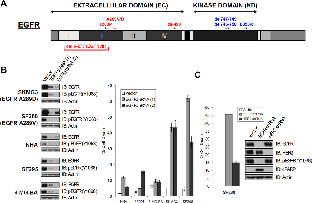
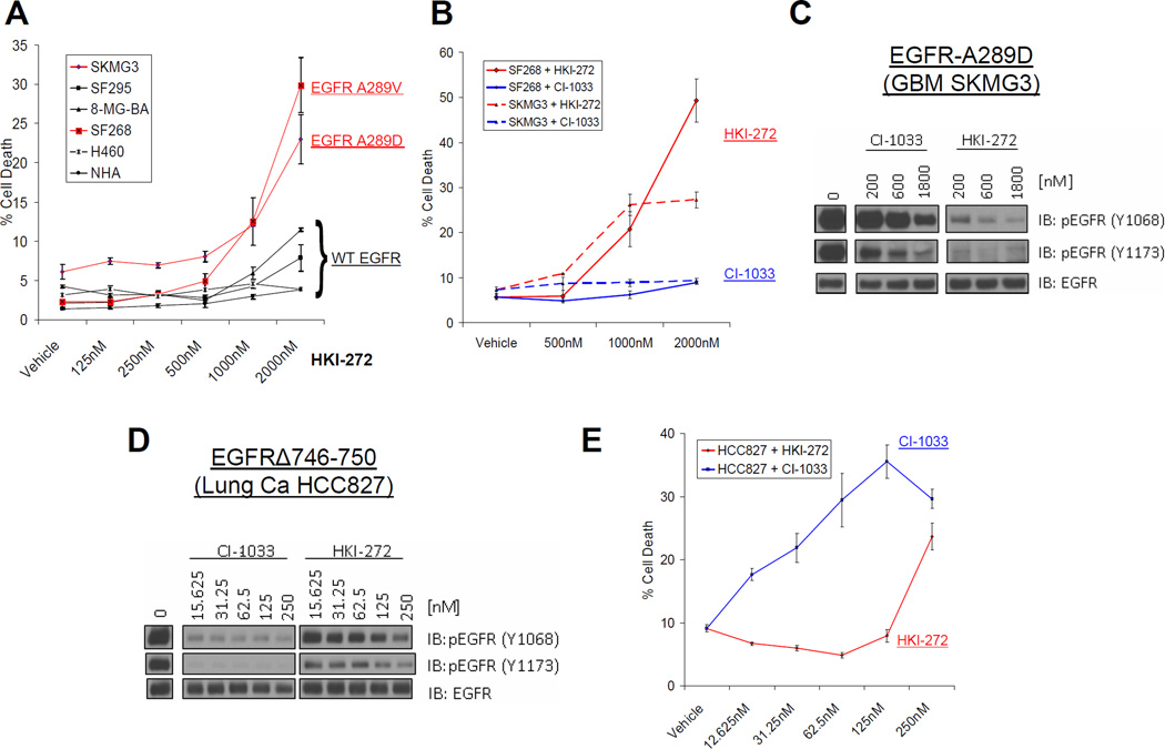
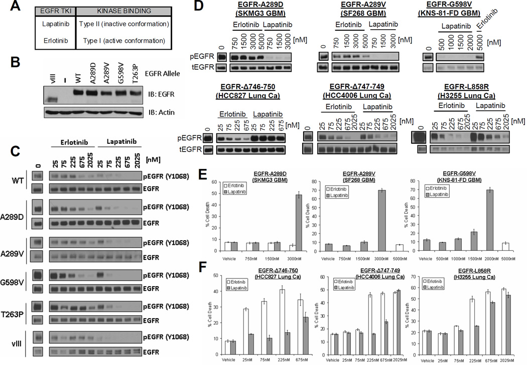
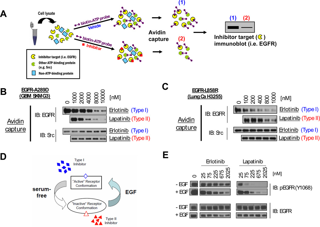
Activation of the epidermal growth factor receptor (EGFR) in glioblastoma (GBM) occurs through mutations or deletions in the extracellular (EC) domain. Unlike lung cancers with EGFR kinase domain (KD) mutations, GBMs respond poorly to the EGFR inhibitor erlotinib. Using RNAi, we show that GBM cells carrying EGFR EC mutations display EGFR addiction. In contrast to KD mutants found in lung cancer, glioma-specific EGFR EC mutants are poorly inhibited by EGFR inhibitors that target the active kinase conformation (e.g., erlotinib). Inhibitors that bind to the inactive EGFR conformation, however, potently inhibit EGFR EC mutants and induce cell death in EGFR-mutant GBM cells. Our results provide first evidence for single kinase addiction in GBM and suggest that the disappointing clinical activity of first-generation EGFR inhibitors in GBM versus lung cancer may be attributed to the different conformational requirements of mutant EGFR in these 2 cancer types.

Significance: Approximately 40% of human glioblastomas harbor oncogenic EGFR alterations, but attempts to therapeutically target EGFR with first-generation EGFR kinase inhibitors have failed. Here, we demonstrate selective sensitivity of glioma-specific EGFR mutants to ATP-site competitive EGFR kinase inhibitors that target the inactive conformation of the catalytic domain.


Figures

FIGURE 1
FIGURE 1. EGFR-knockdown induces cell death in GBM cells with EGFR EC mutations
A. EGFR domain structure. Mutations indicated in red have been documented in glioblastoma (GBM) but not in lung cancer, while those indicated in blue are seen in non-small cell lung cancer (NSCLC) and not in GBM. Roman numerals indicate subdomains within the EC domain. B. EGFR-mutant GBM lines are sensitive to EGFR knockdown. The indicated cell lines were acutely transduced with control or two different EGFR-targeted shRNAs. The extent of EGFR knockdown was assessed by immunoblot (left panel). The effects of the hairpins on cell death was assessed by trypan blue exclusion 5 days post-infection (right panel). (NHA, normal human astrocytes). C. HER2 knockdown only induces minimal cell death in EGFR mutant SF268 GBM cells. Cells were acutely transduced with control, EGFR-targeted, or HER2-targeted shRNAs. The extent of EGFR and HER2 knockdown was evaluated by immunoblot (inset). Cell death was assessed as in B. Confer Suppl. Figure 2 for results of HER2 knockdown in EGFR mutant SKMG3 GBM cells.
FIGURE 2
FIGURE 2. Differential sensitivity of EGFR mutant glioma and lung cancer cell lines to the irreversible EGFR inhibitors HKI-272 and CI-1033
A. HKI-272 induces cell death in GBM cells with EGFR EC mutation (SKMG3, SF268), but not EGFR wildtype cancer cell lines or astrocytes (NHA). Cell death was assessed by trypan blue exclusion following five days of inhibitor treatment. Cells lines in black express wild type EGFR, while those in red contain EGFR EC mutations. B. CI-1033, unlike HKI-272, does not induce cell death in GBM cells with EGFR EC mutation. SF268 (solid) or SKMG3 (dashed) cells were treated with the indicated doses of HKI-272 (red) or CI-1033 (blue) for 5 days. Cell death was evaluated at day 5 by trypan blue exclusion. C. HKI-272 is more potent than CI-1033 in blocking EGFR phosphorylation in SKMG3 cells with EGFR EC mutation. SKMG3 cells were treated with the indicated doses of CI-1033 or HKI-272 and whole lysates were analyzed by immunoblot with the indicated antibodies. D. CI-1033 is more potent than HKI-272 in blocking EGFR phosphorylation in HCC827 lung cancer cells harboring the (Δ746-750) EGFR kinase domain mutant. HCC827 were treated with the indicated doses of HKI-272 or CI-1033. Lysates of these cells were made and analyzed by immunoblot as indicated. E. CI-1033 is more potent than HKI-272 in inducing cell death in HCC827 lung cancer cells. Cell death was also assessed five days after treatment as before.
FIGURE 3
FIGURE 3. Differential sensitivity of glioma- versus lung cancer-specific EGFR mutants to the reversible EGFR inhibitors lapatinib and erlotinib
A. Classification of reversible ATP-competitive EGFR kinase inhibitors based on crystal structures. B. Ectopic expression of glioma-specific EGFR EC mutants in EGFR-deficient (EGFR-NEG) NR6 fibroblasts. Shown are immunoblots of whole cell lystates. C. Enhanced sensitivity of EGFR EC mutants to lapatinib in NR6 cells. NR6 cells expressing the indicated alleles of EGFR were treated with increasing doses of erlotinib or lapatinib as shown. Cell lysates were made and analyzed by immunoblot with the indicated antibodies. D. Differential sensitivity of endogenously expressed glioma- versus lung cancer-specific EGFR mutants to lapatinib versus erlotinib. The indicated GBM (upper panels) and lung cancer (Lung Ca, lower panels) cell lines were treated with various doses of lapatinib or erlotinib. Cells were harvested and analyzed by western blot using antibodies for tyrosine-phosphorylated EGFR (pEGFR, top) or total EGFR (tEGFR, bottom). E and F. Differential cell death response of GBM (E) and lung cancer (F) cell lines to lapatinib versus erlotinib. Cell death was assessed by trypan blue exclusion assay after 5 days of incubation with the indicated drug.
FIGURE 4
FIGURE 4. Type II EGFR kinase inhibitors more effectively displace ATP from the EGFR kinase domain of EGFR ectodomain mutants than type I inhibitors
A. Schematic of ATP competition-binding assessment. Lysates of cells expressing the ATP-binding protein under study are simultaneously treated with a targeted agent (e.g. EGFR TKI) or vehicle, and a biotinylating ATP probe. All ATP-binding proteins will be biotinylated unless the ATP-binding pocket is occupied (e.g. with EGFR TKI). Lysates are subjected to avidin pulldown. The ability of the test compound to compete with ATP for binding to the target protein is assessed by immunoblot of the pulldown using antibodies against the target protein (e.g. EGFR). B. and C. Results of ATP competition assay in lysates from (B) EGFR EC mutant GBM cells and (C) EGFR KD mutant lung cancer cells. (B) Lapatinib more effectively competes with ATP for binding to the EGFR-TK in SKMG3 (EGFR A289D) cell lysates than erlotinib. (C.) Erlotinib more effectively competes with ATP for binding to the EGFR-TK in H3255 cell lysates (EGFR L858R KD mutation) than lapatinib. Cell lysates were carried through the assay described in A. The ATP probe was competed with the indicated doses of erlotinib or lapatinib. Following the avidin pulldown, samples were analyzed by immunoblot with antibodies for EGFR. Immunoblots were also probed with antibodies for Src as a control. D. A model of ligand-induced changes in EGFR conformation. In the absence of ligand (serum-free), the conformational equilibrium of EGFR-EC mutants is shifted towards to “inactive” conformation which is preferentially bound by type II inhibitors. In ligand-occupied receptor (EGF), the conformational equilibrium shifts towards the “active” conformation, which is the preferred substrate of type I inhibitors. E. EGF “desensitizes” the A289D EGFR EC mutant from lapatinib and “sensitizes” it to erlotinib. SKMG3 GBM cells were serum-starved, stimulated with EGF or vehicle, and subsequently treated with the indicated doses of erlotinib and lapatinib (while still under EGF treatment). Cell were lysed 30 minutes of drug treatment and analyzed by immunoblot with the indicated antibodies.
FIGURE 5
FIGURE 5. Lapatinib fails to achieve sufficient intratumoral concentrations in GBM patients
A. Design of the multicenter NABTC 04-01 biomarker trial for patients with recurrent GBM. GBM patients requiring tumor resection for recurrent disease received preoperative lapatinib (750mg p.o. BID). Lapatinib concentrations in tumor tissue and EGFR phosphorylation were assessed in surgical specimens. See Suppl. Information for details. B. Intratumoral lapatinib concentrations in GBM are below lapatinib concentrations required to induce cell death in EGFR mutant GBM cells (red line, 1.5µM). C. Incomplete inhibition of EGFR phosphorylation in GBM tumor tissue by lapatinib. Shown are levels of total EGFR (x-axis) and phosphorylated EGFR (y-axis) in tumors from GBM patients who received preoperative lapatinib (red diamonds) versus tumors from GBM patients who did not receive any EGFR kinase inhibitor prior to surgery (empty circles). Levels of tEGFR phosphorylation and total levels were measured concurrently with a multi-array immunoassay using electrochemiluminescence detection (see top panel). D. EGFR expression in GBMs from patients enrolled in the NABTC 04-01 study. Shown are immunoblots of tumor lysates probed with EGFR or a loading control. Full length (FL) and truncated EGFRvIII (vIII) mutant forms of EGFR are indicated by arrows. E. Incomplete EGFR inhibition in GBM tumor tissue by lapatinib. Shown are immunoblots of EGFR overexpressing GBMs on the NABTC 04-01 study (labeled “LAPATINIB”) versus tumors from GBM patients who did not receive an EGFR kinase inhibitor prior to surgery (“CONTROL”). Total levels of EGFR and EGFR tyrosine phosphorylation were analyzed by immunoblot. Total Erk levels were also examined as a loading control.
FIGURE 6
FIGURE 6. Level of EGFR inhibition determines cell death response in EGFR mutant GBM cells
A. Lapatinib induces cell death in SF268 GBM cells (A289V EGFR) at concentrations above 1.5µM. Cell death was assessed 5 days after treatment by trypan blue exclusion. Similar results are shown in Suppl. Figure 7A for SKMG3 cells (A289D-EGFR) and in KNS-81-FD cells (G598V-EGFR)(Suppl. Figure 7B). B. Cell death induction in SF268 GBM cells (A289V EGFR) requires near complete EGFR inactivation. SF268 cells were acutely transduced with a dilution series of a lentiviral EGFR-targeted shRNA (dilution factor is indicated in parenthesis) or with a control shRNA (empty vector). A fraction of the cells from each infection was used for immunoblot analysis with the indicated antibodies (left panel). The remaining cells were re-seeded and allowed to grow for 5 days post-infection. The fold number of viable cells (relative to day 1) and fraction of dead cells are shown in the middle and right panel, respectively. Similar results were obtained in SKMG3 GBM cells (Suppl. Figure 8). C. Lapatinib inhibits anchorage-independent growth of EGFR amplified (GS676, GS596, GS600), but not PDGFRA amplified, GBM tumor sphere cultures. Anchorage independent growth of four freshly derived GBM tumor sphere cultures was assessed in soft agar in the presence of the indicated concentrations of lapatinib. See also Suppl. Figure 9. D. Pulsatile lapatinib dosing (1000 mpk every 5 days) is superior to daily lapatinib dosing (200 mpk qd) in inducing growth inhibition and cell death induction in EGFR amplified GS676 GBM tumor sphere lines. GS676 cells were injected subcutaneously into SCID mice and treatment initiated at the indicated lapatinib dosing schedules after tumors were established. Tumor volumes (left panel) were evaluated at the end of treatment by caliper measurements. The right panel shows quantification of cleaved Caspase3 staining in tumor sections from the indicated cohorts, i.e. vehicle, 200mg/kg lapatinib, and 1000mg/kg lapatinib. IHC images are shown in Suppl. Figure 10.

Comment in

  • Targeted therapies: Glioma--it's all in the site occupancy.
    Hutchinson L. Hutchinson L. Nat Rev Clin Oncol. 2012 Apr 17;9(6):308. doi: 10.1038/nrclinonc.2012.65. Nat Rev Clin Oncol. 2012. PMID: 22508031 No abstract available.
  • Occupy EGFR.
    Park JH, Lemmon MA. Park JH, et al. Cancer Discov. 2012 May;2(5):398-400. doi: 10.1158/2159-8290.CD-12-0144. Cancer Discov. 2012. PMID: 22588876 Free PMC article.

References

    1. Wen PY, Kesari S. Malignant gliomas in adults. N Engl J Med. 2008 Jul 31;359(5):492–507. - PubMed
    1. Sawyers CL. Shifting paradigms: the seeds of oncogene addiction. Nat Med. 2009 Oct;15(10):1158–1161. - PubMed
    1. Sellers WR. A blueprint for advancing genetics-based cancer therapy. Cell. 2011 Sep 30;147(1):26–31. - PubMed
    1. McLendon R, Friedman A, Bigner D, Van Meir EG, Brat DJ, Mastrogianakis M, et al. Comprehensive genomic characterization defines human glioblastoma genes and core pathways. Nature. 2008 Oct 23;455(7216):1061–1068. - PMC - PubMed
    1. Parsons DW, Jones S, Zhang X, Lin JC, Leary RJ, Angenendt P, et al. An integrated genomic analysis of human glioblastoma multiforme. Science. 2008 Sep 26;321(5897):1807–1812. - PMC - PubMed

Publication types

MeSH terms