. 2012 May 15;72(10):2457-67.
doi: 10.1158/0008-5472.CAN-11-2612.

A comprehensive survey of Ras mutations in cancer

Affiliations

A comprehensive survey of Ras mutations in cancer

Ian A Prior et al. Cancer Res. .

Abstract

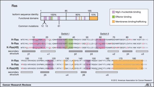
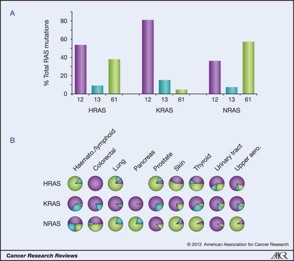
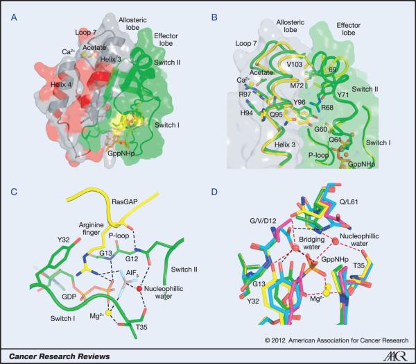
All mammalian cells express 3 closely related Ras proteins, termed H-Ras, K-Ras, and N-Ras, that promote oncogenesis when they are mutationally activated at codon 12, 13, or 61. Although there is a high degree of similarity among the isoforms, K-Ras mutations are far more frequently observed in cancer, and each isoform displays preferential coupling to particular cancer types. We examined the mutational spectra of Ras isoforms curated from large-scale tumor profiling and found that each isoform exhibits surprisingly distinctive codon mutation and amino-acid substitution biases. These findings were unexpected given that these mutations occur in regions that share 100% amino-acid sequence identity among the 3 isoforms. Of importance, many of these mutational biases were not due to differences in exposure to mutagens, because the patterns were still evident when compared within specific cancer types. We discuss potential genetic and epigenetic mechanisms, as well as isoform-specific differences in protein structure and signaling, that may promote these distinct mutation patterns and differential coupling to specific cancers.


Figures

Figure 1
Figure 1. Oncogenic mutations of Ras isoforms
The key oncogenic mutations are in the region that is identical between the three isoforms. 44 separate point mutations have been characterised in Ras isoforms with 99.2% of all mutations occurring at codons 12, 13 and 61. Mutations cluster in and around loops 1, 2 and 4 responsible for nucleotide binding and result in enhanced GTP binding. Residues mutated in cancer are highlighted in red, those mutated in developmental disorders are underlined and residues that are variable between isoforms are in grey (26, 65, 66).
Figure 2
Figure 2. Ras isoform-specific codon mutation bias
A. K-Ras is typically mutated at codon 12 whereas N-Ras favours codon 61. H-Ras displays intermediate behaviour. Data are averages of percentages collated from all cancers with at least 20 tumours scored. B. Analysis of individual cancer types reveals isoform-specific patterns of codon mutation even within the same tissue. Pie chart colours - black: codon 12; grey: codon 13; white: codon 61.
Figure 3
Figure 3. Structural features of Ras and its oncogenic mutations
A. The catalytic domain of Ras. The effector lobe is depicted in green and the allosteric lobe in grey. Residues that are not identical in all three isoforms are in red and are found entirely in the allosteric lobe. Residues 12, 13 and 61 are shown in yellow and GppNHp is in orange. Calcium acetate is shown bound at the allosteric site. B. The allosteric switch showing some of the key residues involved. The allosteric state in which switch II is disordered is shown in yellow (PDB code 2RGE). Residues 61-68 are disordered and therefore were removed from the model. The structure with calcium acetate in the allosteric site interacting with R97 is shown in green (PDB code 3K8Y). Switch II is ordered in this structure. The nucleotide is depicted in orange. C. The active site in the Ras/RasGAP complex (PDB code 1WQ1). GAP residues are shown in yellow with the arginine finger shown in stick. Ras residues are in green, including the GDP with phosphate groups in orange. AlF3 is shown with the aluminum atom in grey and fluorine atoms in cyan. G12, G13, Y32 T35 and Q61 are shown in stick. The Mg2+ ion is depicted as a yellow sphere and the nucleophilic water molecule as a red sphere. D. The active site for intrinsic hydrolysis in Ras. The wild type is shown in yellow (PDB code 3K8Y), G12V in magenta (PDB code 3OIW), G12D in cyan (PDB code 1AGP) and Q61L in green (PDB code 3OIU). The wild type structure contains both the nucleophilic and bridging water molecules. Note that in G12D the side chain of D12 replaces the bridging water molecule, whereas in G12V and Q61L there is a direct H-bond between Y32 and the γ-phosphate of the nucleotide. Mg2+ is shown in yellow sphere and water molecules in red spheres in each structure. Hydrogen bonds in the wild type structure are depicted in red dashed lines. Those in the G12D mutant are in black dashed line. H-bonds in the G12V and Q61L structures are not shown. Note the proximity of G13 to the Y32 side chain in the wild type structure.

References

    1. Quinlan MP, Settleman J. Isoform-specific ras functions in development and cancer. Future Oncol. 2009;5(1):105–16. - PubMed
    1. Omerovic J, Laude AJ, Prior IA. Ras proteins: paradigms for compartmentalised and isoform-specific signalling. Cell Mol Life Sci. 2007;64(19-20):2575–89. - PMC - PubMed
    1. Henis YI, Hancock JF, Prior IA. Ras acylation, compartmentalization and signaling nanoclusters (Review) Mol Membr Biol. 2009;26(1):80–92. - PMC - PubMed
    1. Bos JL. ras oncogenes in human cancer: a review. Cancer Res. 1989;49(17):4682–9. - PubMed
    1. Forbes SA, Bindal N, Bamford S, Cole C, Kok CY, Beare D, et al. COSMIC: mining complete cancer genomes in the Catalogue of Somatic Mutations in Cancer. Nucleic Acids Res. 2011;39(Database issue):D945–50. - PMC - PubMed

Publication types