. 2012 May 29;109(22):8699-704.
doi: 10.1073/pnas.1017909109. Epub 2012 May 15.

Suppression of heat shock protein 27 induces long-term dormancy in human breast cancer

Affiliations

Suppression of heat shock protein 27 induces long-term dormancy in human breast cancer

Oddbjørn Straume et al. Proc Natl Acad Sci U S A. .

Abstract

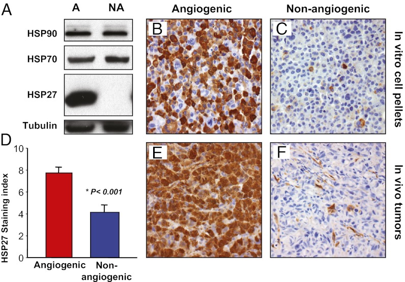
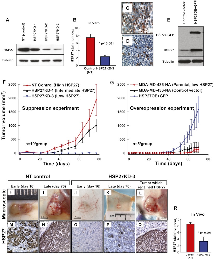
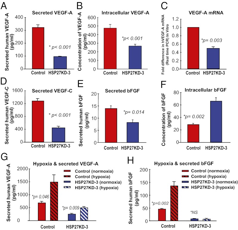
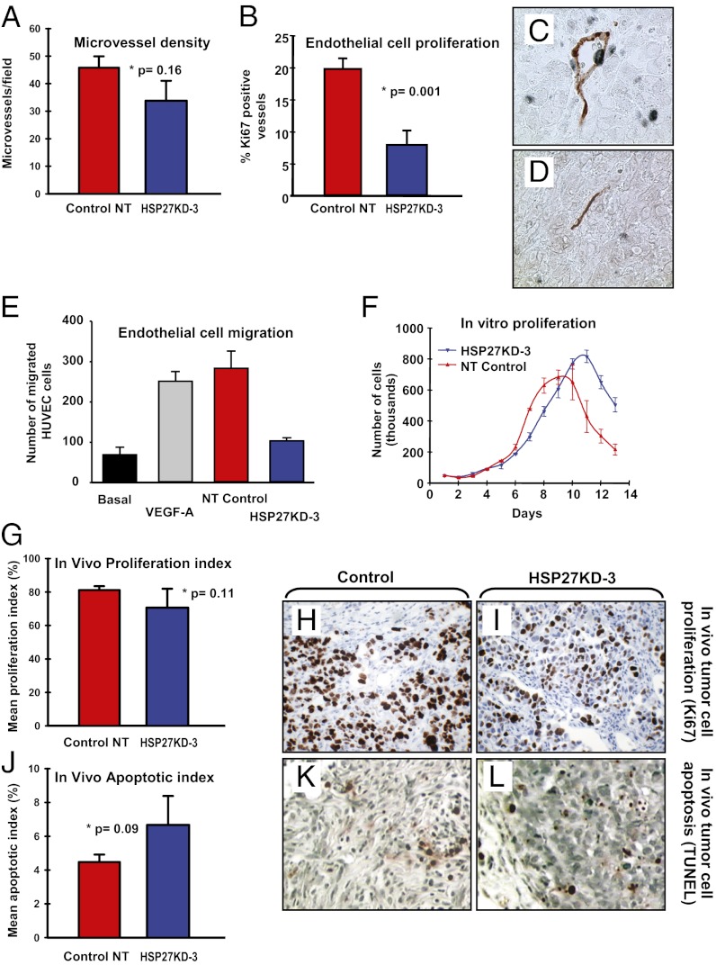
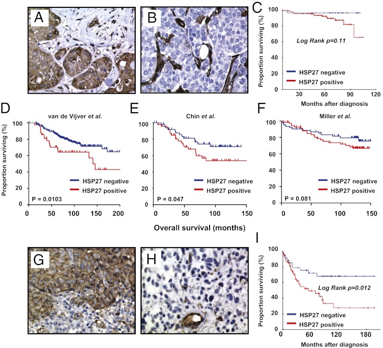
The mechanisms underlying tumor dormancy have been elusive and not well characterized. We recently published an experimental model for the study of human tumor dormancy and the role of angiogenesis, and reported that the angiogenic switch was preceded by a local increase in VEGF-A and basic fibroblast growth factor. In this breast cancer xenograft model (MDA-MB-436 cells), analysis of differentially expressed genes revealed that heat shock protein 27 (HSP27) was significantly up-regulated in angiogenic cells compared with nonangiogenic cells. The effect of HSP27 down-regulation was further evaluated in cell lines, mouse models, and clinical datasets of human patients with breast cancer and melanoma. Stable down-regulation of HSP27 in angiogenic tumor cells was followed by long-term tumor dormancy in vivo. Strikingly, only 4 of 30 HSP27 knockdown xenograft tumors initiated rapid growth after day 70, in correlation with a regain of HSP27 protein expression. Significantly, no tumors escaped from dormancy without HSP27 expression. Down-regulation of HSP27 was associated with reduced endothelial cell proliferation and decreased secretion of VEGF-A, VEGF-C, and basic fibroblast growth factor. Conversely, overexpression of HSP27 in nonangiogenic cells resulted in expansive tumor growth in vivo. By clinical validation, strong HSP27 protein expression was associated with markers of aggressive tumors and decreased survival in patients with breast cancer and melanoma. An HSP27-associated gene expression signature was related to molecular subgroups and survival in breast cancer. Our findings suggest a role for HSP27 in the balance between tumor dormancy and tumor progression, mediated by tumor-vascular interactions. Targeting HSP27 might offer a useful strategy in cancer treatment.


Conflict of interest statement

The authors declare no conflict of interest.

Figures

Fig. 1.
Fig. 1.
HSP27 is overexpressed in angiogenic human breast cancer cells compared with nonangiogenic human breast cancer cells. Differences in HSP27 expression between the angiogenic and nonangiogenic variants of the MDA-MB-436 cell line were validated. (A) Western blot analysis of HSP90, HSP70, and HSP27 protein expression in angiogenic (A) and nonangiogenic (NA) variants of the human breast cancer cell line. (B and C) Immunohistochemical staining for HSP27 protein in cell pellets containing angiogenic (B) or nonangiogenic (C) cells, grown under in vitro conditions. (D–F) Immunohistochemical staining of HSP27 protein in tumor xenografts was quantified using a staining index (D) and was significantly increased in angiogenic tumors (E) compared with nonangiogenic tumors (F). The sample shown in F is negative for HSP27 expression in tumor cells, representing one extreme end of the expression spectrum. In F, note the strong expression of HSP27 protein in some tumor-associated stromal (including endothelial) cells.
Fig. 2.
Fig. 2.
Stable down-regulation of HSP27 in angiogenic cancer cells induces long-term dormancy in vivo, and overexpression of HSP27 in nonangiogenic cells induces expansive tumor growth in vivo. Five pools of the HSP27-expressing angiogenic breast cancer cell line transduced with different shRNA sequences against HSP27—HSP27KD-1, KD-2 and KD-3—were generated. (A) Their HSP27 protein expression was confirmed by Western blot and compared with the NT control. (B–D) Down-regulation of HSP27 protein expression in HSP27KD-3 cells was quantified by immunohistochemistry and compared with that in NT control cells using in vitro cell pellets (B). NT control cells are shown in C; HSP27KD-3 cells, in D. (E) HSP27 tagged with GFP was overexpressed (HSP27OE+GFP) in the parental nonangiogenic MDA-MB-436-NA (with intrinsically low HSP27 (Fig. 1A) cells and confirmed by Western blot analysis. (F) In vivo s.c. xenograft growth curves of HSP27KD-3, HSP27KD-1, and the NT control (estimated as mean ± SE tumor volume). (G) In vivo s.c. xenograft growth curves of HSP27OE+GFP, the control vector, and the parental unaltered nonangiogenic cells (estimated as mean ± SE tumor volume). (H and I) The NT control tumors showed vivid neo-angiogenesis within and around the growing tumors. (J and K) In contrast, unaffected normal-appearing s.c. vessels surround a small HSP27KD-3 tumor (J), whereas early angiogenic activity can be observed in a late (day 70) HSP27KD-3 tumor (K). (M–P) HSP27 protein staining was consistently high in the NT control tumors (M and N) but was low or absent in microscopic dormant tumors formed by the HSP27KD-3 cells (O and P). (L and Q) A mouse inoculated with HSP27KD-3 cells spontaneously initiated tumor growth (L), and was confirmed by immunohistochemistry to have regained HSP27 protein expression (Q). (R) The mean HSP27 staining index was 2.6-fold higher for the NT control tumors than for tumors formed by the HSP27KD-3 cells in vivo. (Original magnifications: 400× in C, D, M, O, and P; 200× in N and Q.) The Student t test was used to assess the statistical significance of differences (*).
Fig. 3.
Fig. 3.
Suppression of HSP27 leads to reduced secretion of angiogenic factors. VEGF-A levels were quantified in the supernatant obtained from NT control (high HSP27) and HSP27KD-3 (low HSP27) cell lines. (A) The amount of human VEGF-A secreted into the media was 3.3-fold higher from the control cells compared with the HSP27KD-3 cells. (B) The concentration of intracellular VEGF-A was 1.8-fold higher in control NT cells. (C) Control NT cells contained twofold higher levels of VEGF-A mRNA compared with the HSP27KD-3 cells, as assessed by real-time qRT-PCR. (D) VEGF-C secretion was 2.6-fold higher in NT control cells compared with HSP27KD-3 cells, as assessed by ELISA. (E) bFGF secretion was 1.7-fold higher in NT control cells compared with HSP27KD-3 cells, as quantified by ELISA. (F) In contrast, bFGF concentration within the cell lysates (intracellular) was 2.3-fold greater in HSP27KD-3 cells compared with NT control cells. (G) Hypoxic conditions resulted in a significant increase in VEGF-A secretion from control cells and HSP27KD-3 breast cancer cells. (H) Hypoxic conditions induced significantly increased secretion of bFGF from the control cells, but not from HSP27KD-3 cells.
Fig. 4.
Fig. 4.
Suppression of HSP27 affects proliferation of endothelial cells, but not of tumor cells. (A) MVD, shown as microvessels per field of view (400× objective). (B) The ratio between tumor-associated vessels positive for Ki-67 in endothelial cells and all vessels was 2.5-fold higher within the control tumors. (C and D) CD34 (red) and Ki-67 (blue) dual immunostaining. In addition to high endothelial cell proliferation, the vessels found in the control tumors presented more frequently with open lumens containing erythrocytes (C), in contrast to compressed vessels within the HSP27KD-3 tumors (D). (E) HUVECs were exposed to VEGF-A or conditioned media from control cells or HSP27KD-3 cells grown under normoxia, and endothelial cell migration was quantified and compared with baseline migration. There number of endothelial cells migrating in response to conditioned media from control tumor cells was 2.7-fold greater. (F) There was no significant difference between the in vitro proliferation growth curves of the NT control cells and the HSP27KD-3 cells. (G–I) Tumor samples from the in vivo experiments showed no statistically significant difference in tumor cell proliferation (Ki-67) when NT control tumors were compared with tumors from HSP27KD-3 cells collected at comparable time points. Nonetheless, we observed a trend toward reduced tumor cell proliferation in the HSP27KD-3 tumors (P = 0.11). (J–L) Tumor cell apoptosis (TUNEL staining) was compared between NT-control cell line and HSP27KD-3 in tissue collected during the xenograft experiment. No significant difference in apoptotic rate was present, although a difference of borderline significance was found (P = 0.09) . (Original magnification: 400× for H, I, K, and L.)
Fig. 5.
Fig. 5.
Low expression of HSP27 protein is associated with a less aggressive phenotype and improved survival in human patients with breast cancer and melanoma. (A and B) HSP27 protein staining by immunohistochemistry was significantly stronger in interval breast cancers (A) compared with cancers detected by routine mammography scans (B); HSP27 protein-positive stromal cells serve as an internal control. (C) Patients with breast cancer with increased HSP27 expression also tended to have a poorer prognosis, although this trend was not statistically significant. (D–F) The HSP27 expression signature was applied on previously published breast cancer datasets. Patient samples designated as HSP27-positive were associated with a poorer cancer-specific survival in two of the three available datasets (van de Vijver dataset, P = 0.0103; Chin dataset, P = 0.047), with a trend toward poorer survival in the third dataset (Miller dataset, P = 0.081). (G–I) Patients with melanoma with high HSP27 protein expression by immunohistochemistry (G) had significantly lower cancer-specific overall survival (I) compared with those with low HSP27 expression (H).

References

    1. Naumov GN, Folkman J, Straume O. Tumor dormancy due to failure of angiogenesis: Role of the microenvironment. Clin Exp Metastasis. 2009;26:51–60. - PubMed
    1. Black WC, Welch HG. Advances in diagnostic imaging and overestimations of disease prevalence and the benefits of therapy. N Engl J Med. 1993;328:1237–1243. - PubMed
    1. Folkman J, Kalluri R. Cancer without disease. Nature. 2004;427:787. - PubMed
    1. Hanahan D, Folkman J. Patterns and emerging mechanisms of the angiogenic switch during tumorigenesis. Cell. 1996;86:353–364. - PubMed
    1. Naumov GN, Akslen LA, Folkman J. Role of angiogenesis in human tumor dormancy: Animal models of the angiogenic switch. Cell Cycle. 2006;5:1779–1787. - PubMed

Publication types

MeSH terms

Substances