. 2012 Jul 6;111(2):180-90.
doi: 10.1161/CIRCRESAHA.112.270462. Epub 2012 May 15.

Enhanced angiogenic and cardiomyocyte differentiation capacity of epigenetically reprogrammed mouse and human endothelial progenitor cells augments their efficacy for ischemic myocardial repair

Affiliations

Enhanced angiogenic and cardiomyocyte differentiation capacity of epigenetically reprogrammed mouse and human endothelial progenitor cells augments their efficacy for ischemic myocardial repair

Melissa A Thal et al. Circ Res. .

Abstract

Rationale: Although bone marrow endothelial progenitor cell (EPC)-based therapies improve the symptoms in patients with ischemic heart disease, their limited plasticity and decreased function in patients with existing heart disease limit the full benefit of EPC therapy for cardiac regenerative medicine.

Objective: We hypothesized that reprogramming mouse or human EPCs, or both, using small molecules targeting key epigenetic repressive marks would lead to a global increase in active gene transcription, induce their cardiomyogenic potential, and enhance their inherent angiogenic potential.

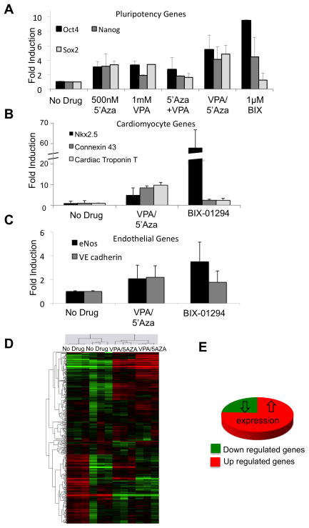
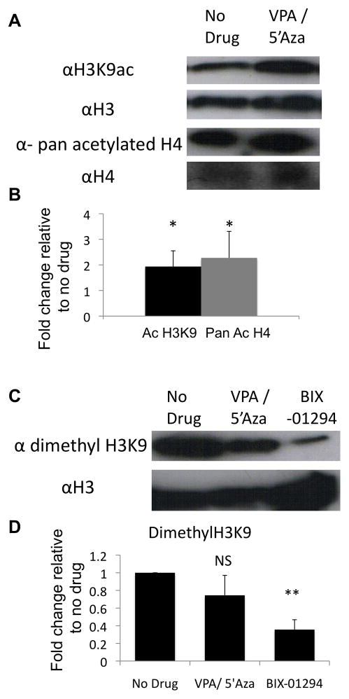
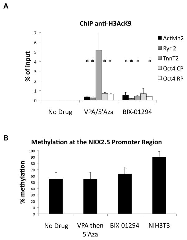
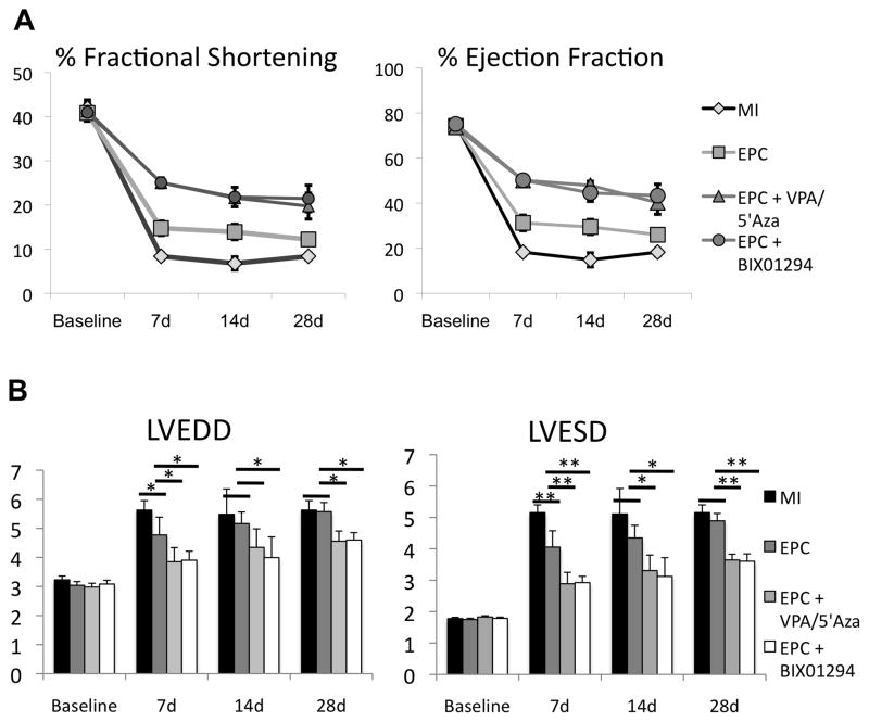
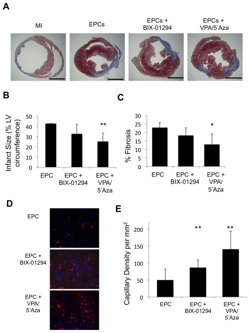
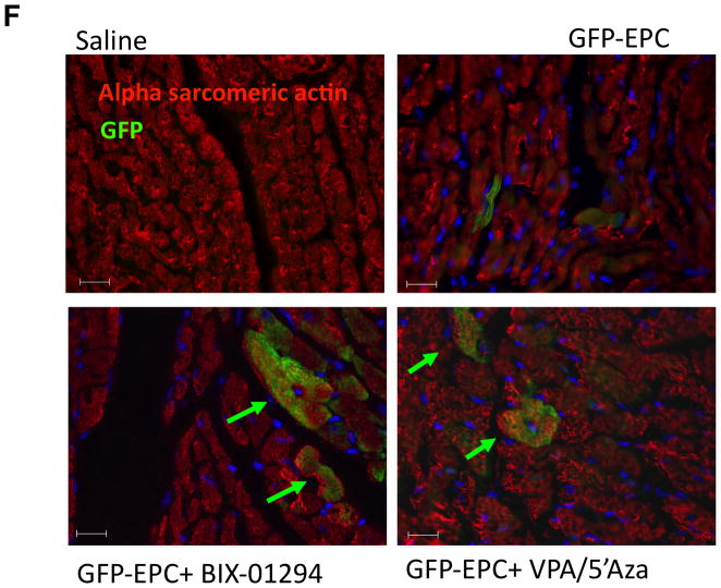
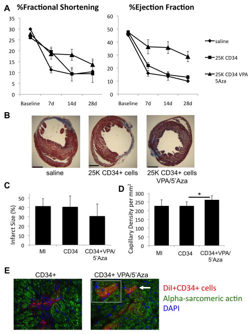
Method and results: Mouse Lin-Sca1(+)CD31(+) EPCs and human CD34(+) cells were treated with inhibitors of DNA methyltransferases (5-Azacytidine), histone deacetylases (valproic acid), and G9a histone dimethyltransferase. A 48-hour treatment led to global increase in active transcriptome, including the reactivation of pluripotency-associated and cardiomyocyte-specific mRNA expression, whereas endothelial cell-specific genes were significantly upregulated. When cultured under appropriate differentiation conditions, reprogrammed EPCs showed efficient differentiation into cardiomyocytes. Treatment with epigenetic-modifying agents show marked increase in histone acetylation on cardiomyocyte and pluripotent cell-specific gene promoters. Intramyocardial transplantation of reprogrammed mouse and human EPCs in an acute myocardial infarction mouse model showed significant improvement in ventricular functions, which was histologically supported by their de novo cardiomyocyte differentiation and increased capillary density and reduced fibrosis. Importantly, cell transplantation was safe and did not form teratomas.

Conclusions: Taken together, our results suggest that epigenetically reprogrammed EPCs display a safe, more plastic phenotype and improve postinfarct cardiac repair by both neocardiomyogenesis and neovascularization.


Figures

Figure 1
Figure 1
Drug treatment of sorted EPCs induces gene expression compared to the untreated cells based on Real-time PCR analysis. (a) pluripotency genes; Oct4, Nanog, and Sox2. (b) cardiomyocyte genes; Nkx2.5, Cx43 and Tnnt2. (c) endothelial genes; eNOS and VE cadherin. Values are all fold change compared to the untreated cells. (n=3) (d) Clustering analysis of microarray data comparing human CD34+ cells to those treated with VPA/5′Aza. Up-and down-regulated genes are represented in red and green colors, respectively. CD34+ cells from 2 healthy patient donors were run in triplicate for both conditions. (e) Pie graph depicting pattern of expression changes of statistically significantly affected genes upon VPA/5′Aza treatment of CD34+ cells.
Figure 2
Figure 2
VPA/5′Aza or BIX-01294 treatment effects histone modifications in ECs. (a) Western blot analysis of acetylated H3K9, or pan acetylated H4, or (c) di-methyl H3K9 levels from 10 million SVEC cells after 24 hours treatment with 1mM VPA followed by an additional 24 hours with 500nM 5′Azacytidine or 1μM BIX-01294. (b, d) Quantitative assessment of modified histone levels relative to total histone levels. Values are fold change compared to untreated SVEC cells. (n=3)
Figure 3
Figure 3
Epigenetically reprogrammed EPCs have increased acH3K9 associated with cardiac-specific promoters. (a) ChIP assay at CMC-associated gene promoters and 2 regulatory regions of Oct4 CP=core promoter, RP=regulatory promoter. Values represent percent of total input. * p<0.05 (b) Pyro-sequencing of the Nkx2.5 promoter from isolated and bisulfite converted genomic DNA from SVECs with either no drug, VPA/5′Aza or BIX-01294. Converted NIH-3T3 gDNA was used as a positive control.
Figure 4
Figure 4
Drug treated mouse EPCs improve left ventricular function post myocardial infarction (AMI). (a) Echocardiographic analysis of left ventricular heart function assessed by percent fractional shortening and ejection fraction prior to surgery and at 7, 14 and 28 days post-surgery. MI=saline negative control, EPC=Lin-Sca-1+CD31+ mouse bone marrow cells. (n≥6 at each time point). (b) Left ventricle end-systolic and end-diastolic diameter measured from short-axis m-mode echocardiography. *p≤0.05, **p≤0.01 (n≥6)
Figure 5
Figure 5
Histological evaluation of infarcted hearts indicates drug treated EPCs confer less severe disease and allow for CMC trans-differentiation in vivo. (a) Masson’s trichrome-stained heart sections (28 d post-AMI). Scale bar is 5mm. (b) Quantitative analysis of infarct size and (c) fibrotic area (%LV area) analysis at 28 d post-AMI. *p≤0.05, **p≤0.01 (n≥3) (d, e) Capillary density per mm2 calculated from 3 high power fields per heart of the border zone of myocardial infarcted mice, minimally 3 mice per condition. (f) IF of heart sections at day 14 post-AMI for GFP and alpha-sarcomeric actin. DAPI was used to stain nuclei. Arrows point to double stained cells. Representative images shown of n=4 animals.
Figure 5
Figure 5
Histological evaluation of infarcted hearts indicates drug treated EPCs confer less severe disease and allow for CMC trans-differentiation in vivo. (a) Masson’s trichrome-stained heart sections (28 d post-AMI). Scale bar is 5mm. (b) Quantitative analysis of infarct size and (c) fibrotic area (%LV area) analysis at 28 d post-AMI. *p≤0.05, **p≤0.01 (n≥3) (d, e) Capillary density per mm2 calculated from 3 high power fields per heart of the border zone of myocardial infarcted mice, minimally 3 mice per condition. (f) IF of heart sections at day 14 post-AMI for GFP and alpha-sarcomeric actin. DAPI was used to stain nuclei. Arrows point to double stained cells. Representative images shown of n=4 animals.
Figure 6
Figure 6
Drug treated human CD34+ cells improve LV function based on %FS and %EF. (a) Echocardiographic analysis at baseline and at days 7, 14 and 28 post-AMI for all groups. (b) Masson’s trichrome stained 5μm sections 1mm below suture of infracted hearts 28 days post-AMI. Scale bar is 5mm. (c) Measurements from Masson’s trichrome stained sections for infarct size as a percent of the circumference. (d) Capillary density calculated per mm2 from 3 high-powered fields. (n≥5) for each condition. (e) In vivo cardiomyocyte differentiation identified by immunofluorescence in d7 hearts of mice receiving AMI and DiI labeled CD34+ or VPA/5′Aza treated CD34+ cells in the border zone. DiI labeled donor cells (red), alpha-sarcomeric actin and nuclei (DAPI). White arrow indicates potential donor-derived cardiomyocyte and shown bigger in inset. Scale bar represents 20μm. Representative images from each group are shown.

Comment in

References

    1. Britten MB, Abolmaali ND, Assmus B, Lehmann R, Honold J, Schmitt J, Vogl TJ, Martin H, Schachinger V, Dimmeler S, Zeiher AM. Infarct remodeling after intracoronary progenitor cell treatment in patients with acute myocardial infarction (topcare-ami): Mechanistic insights from serial contrast-enhanced magnetic resonance imaging. Circulation. 2003;108:2212–2218. - PubMed
    1. Losordo DW, Dimmeler S. Therapeutic angiogenesis and vasculogenesis for ischemic disease: Part ii: Cell-based therapies. Circulation. 2004;109:2692–2697. - PubMed
    1. Losordo DW, Dimmeler S. Therapeutic angiogenesis and vasculogenesis for ischemic disease. Part i: Angiogenic cytokines. Circulation. 2004;109:2487–2491. - PubMed
    1. Schachinger V, Erbs S, Elsasser A, Haberbosch W, Hambrecht R, Holschermann H, Yu J, Corti R, Mathey DG, Hamm CW, Suselbeck T, Werner N, Haase J, Neuzner J, Germing A, Mark B, Assmus B, Tonn T, Dimmeler S, Zeiher AM. Improved clinical outcome after intracoronary administration of bone-marrow-derived progenitor cells in acute myocardial infarction: Final 1-year results of the repair-ami trial. Eur Heart J. 2006;27:2775–2783. - PubMed
    1. Strauer BE, Brehm M, Zeus T, Kostering M, Hernandez A, Sorg RV, Kogler G, Wernet P. Repair of infarcted myocardium by autologous intracoronary mononuclear bone marrow cell transplantation in humans. Circulation. 2002;106:1913–1918. - PubMed

Publication types

MeSH terms