. 2012 Jun 20;53(7):3792-800.
doi: 10.1167/iovs.11-9235.

ER stress is involved in T17M rhodopsin-induced retinal degeneration

Affiliations

ER stress is involved in T17M rhodopsin-induced retinal degeneration

Mansi M Kunte et al. Invest Ophthalmol Vis Sci. .

Abstract

Purpose: The human rhodopsin (Rho) mutation T17M leads to autosomal dominant retinitis pigmentosa (adRP). The goal of our study was to elucidate the role of endoplasmic reticulum (ER) stress in retinal degeneration in hT17M Rho mice and identify potential candidates for adRP gene therapy.

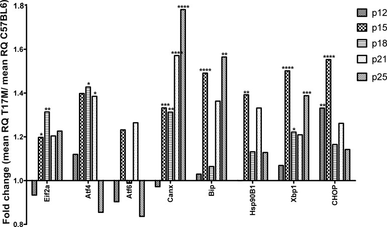
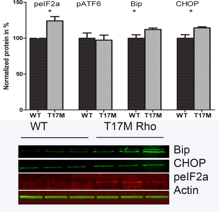
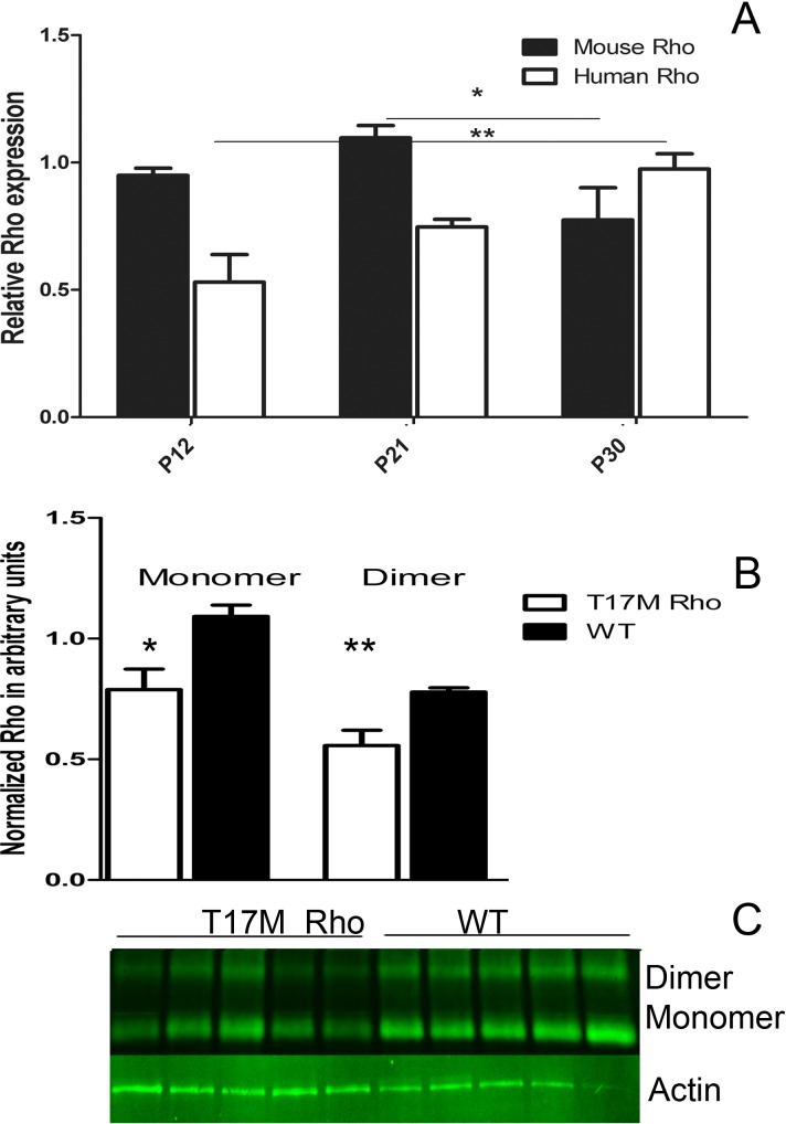
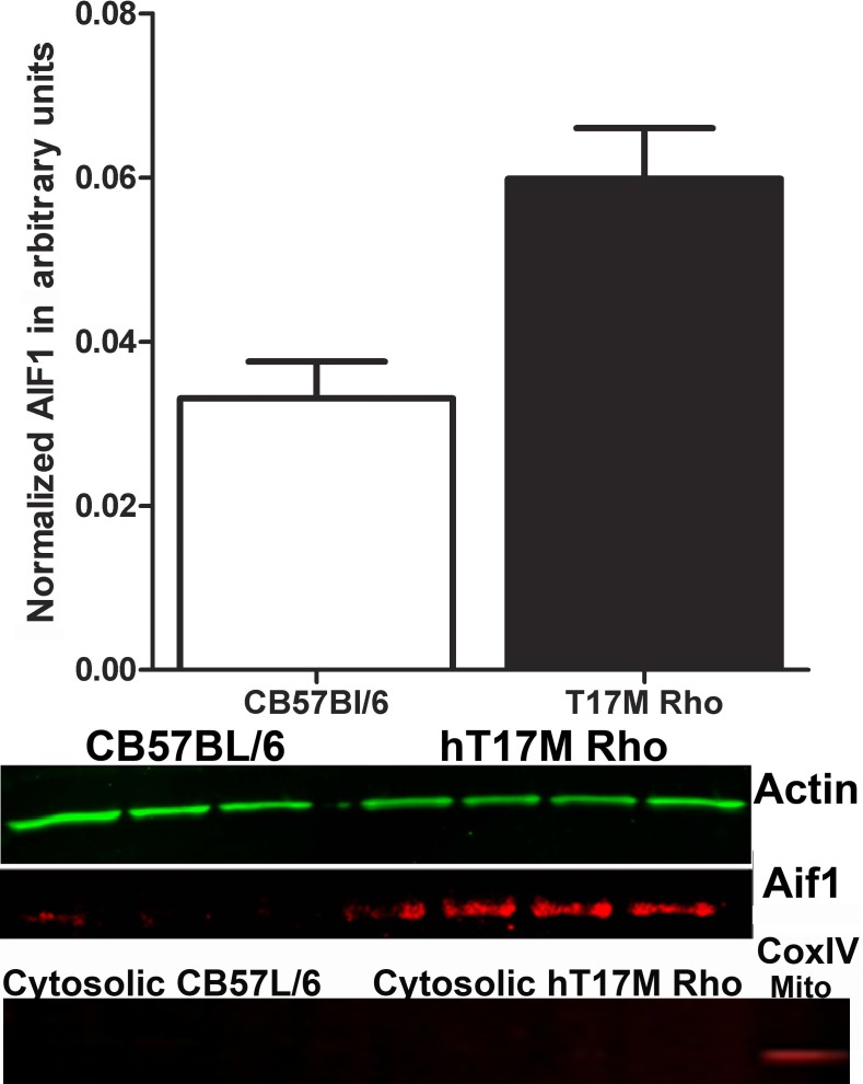
Methods: We used transgenic mice expressing the ER stress-activated indicator (ERAI) and hT17M Rho to evaluate the activation of ER stress responses. Quantitative reverse transcription PCR (qRT-PCR) was used to analyze changes in the expression of 30 unfolded protein response (UPR)-associated genes at P12, 15, 18, 21, and 25. The cytosolic fraction of hT17M Rho retinal cells was used to measure the release of cytochrome C and apoptotic inducing factor-1 (AIF1) by Western blotting. Optical coherence tomography (OCT) analysis was performed for 1-month-old hT17M Rho mice.

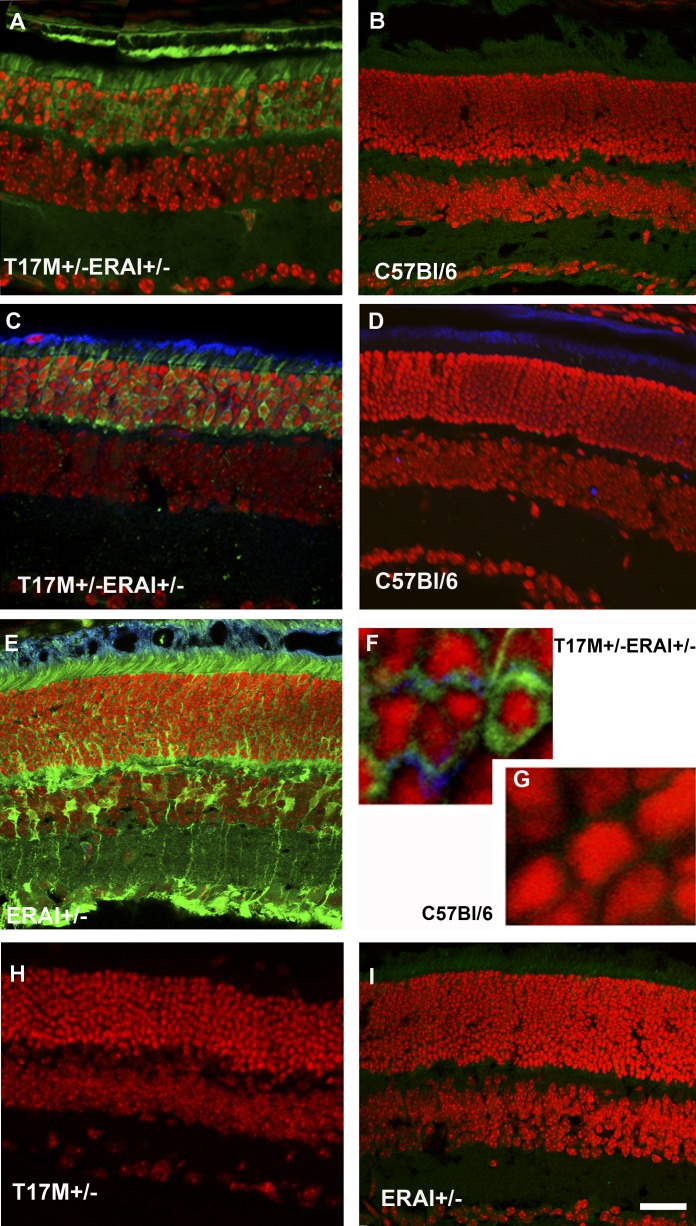
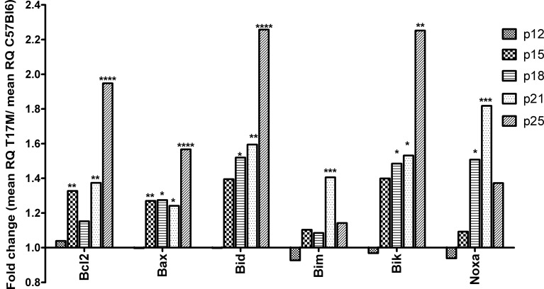
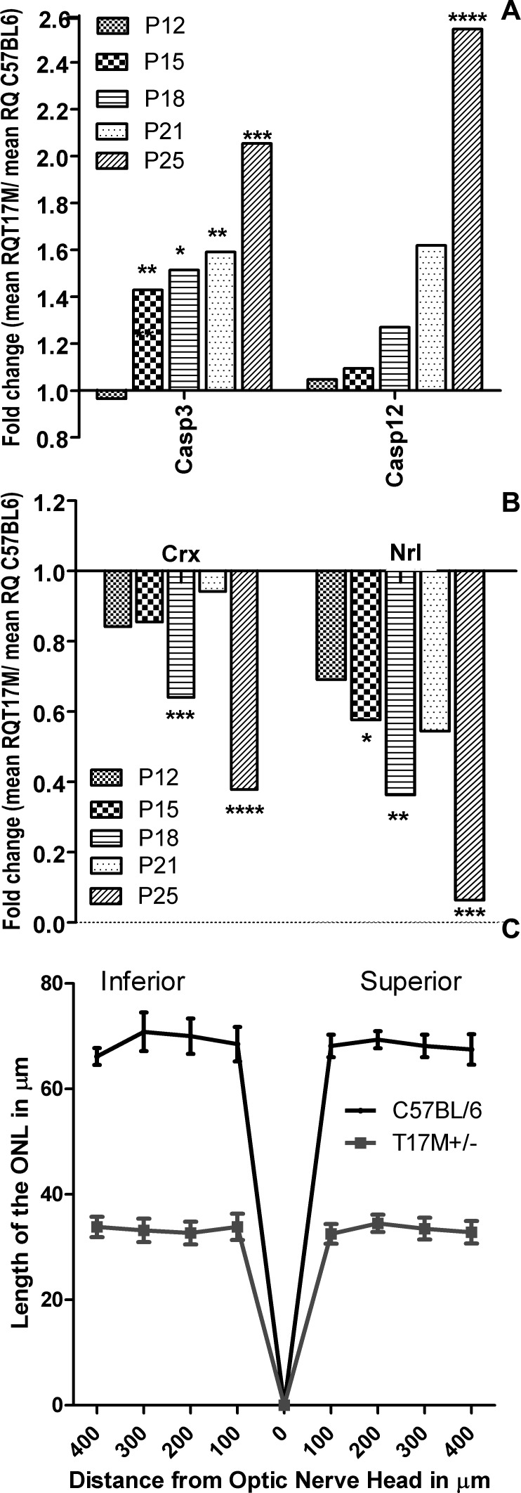
Results: hT17M Rho was localized in the outer nuclear layer (ONL) of T17M(+/-)ERAI(+/-) photoreceptors as well as C57BL/6 retinas injected with AAV-hT17M Rho-GFP. In P15 hT17M Rho retinas, we observed an up-regulation of UPR genes (Atf4, Eif2α, Xbp1, Bip, Canx, and Hsp90), autophagy genes and proapoptotic Bcl2 genes. OCT, and the downregulation of Nrl and Crx gene expression confirmed that cell death occurs in 55% of photoreceptors via the up-regulation of caspase-3 and caspase-12, and the release of AIF1 from the mitochondria.

Conclusions: The ER stress response is involved in retinal degeneration in hT17M Rho mice. The final demise of photoreceptors occurs via apoptosis involving ER stress-associated and mitochondria-induced caspase activation. We identified Atg5, Atg7, Bax, Bid, Bik, and Noxa as potential therapeutic targets for adRP treatment.


Conflict of interest statement

Disclosure: M.M. Kunte, None; S. Choudhury, None; J.F. Manheim, None; V.M. Shinde, None; M. Miura, None; V.A. Chiodo, None; W.W. Hauswirth, None; O.S. Gorbatyuk, None; M.S. Gorbatyuk, None

Figures

Figure 1.
Figure 1.
hT17M rhodopsin leads to activation of the ER stress response at P15 in T17M+/− ERAI+/− mice. Confocal microscopy is used to obtain images of mouse retinas. Propidium iodide is used to stain the nuclei (red). GFP protein expression (green) results from the splicing of the Xbp1-GFP transcription factor (detected by direct fluorescence or immunohistochemical analysis). For immunohistochemistry, an anti-GFP antibody is used to stain the sXbp1-GFP. The primary anti-rhodopsin antibody 1D4 and secondary anti-mouse Cy5-conjugated antibodies are applied to detect the localization of rhodopsin (blue). T17M+/− ERAI+/− (A) and C57BL/6 (B) images of retinas are obtained via direct observation under a fluorescence microscope. T17M+/− ERAI+/− (C), C57BL/6 (D), ERAI+/− positive control with tunicamycin injection (E), T17M+/− ERAI+/− (F), and C57BL/6 (G) images of retinas are obtained after immunostaining with anti-GFP and anti-rhodopsin antibodies. GFP fluorescence is observed in images T17M+/− ERAI+/− (A), T17M+/− ERAI+/− (C), ERAI+/− positive control with tunicamycin injection (E), and T17M+/− ERAI+/−(F), and not in the negative controls C57BL/6 (B), C57BL/6 (D), C57BL/6 (G), T17M+/− (H), and ERAI+/− (I). Accumulation of rhodopsin is observed in T17M+/− ERAI+/− (F) but is not detected in C57BL/6 (G). Scale bar indicates 30 μm (AE, H, I) and 300 μm (F, G).
Figure 2.
Figure 2.
UPR genes are up-regulated at P15. Wild-type gene expression is normalized to 1, and the ratio of hT17M Rho to the wild-type is shown. Induction of the expression of the molecular chaperones BiP, Cnx, and Hsp90 in hT17M Rho retinas was observed at P15; the observed increases were 1.4-, 1.3-, and 1.4-fold compared to the control. Markers of the PERK (eiF2a and ATF4) and IRE (Xbp1) pathways are up-regulated strongly (1.2-, 1.4-, 1.5-fold increases) at P15, whereas the expression of the CHOP gene is up-regulated earlier at P12 (1.3-fold; *P < 0.05, **P < 0.01, ***P < 0.001, and ****P < 0.0001).
Figure 3.
Figure 3.
Activation of UPR in hT17M Rho retina. The UPR (peIF2α, Bip and Chop) markers were elevated in P15 hT17M Rho retina by 1.24-fold (100% ± 4.84% in wild-type vs. 124% ± 5.98% in hT17M Rho, P = 0.048), by 1.13-fold (100% ± 4.66% in wild-type vs. 113% ± 2.11% in hT17M Rho, P = 0.032), and by 1.14-fold (100% ± 4.89% in wild-type vs. 114% ± 1.48% in hT17M Rho, P = 0.042), correspondingly. The pATF6 (50kD) protein is not elevated in hT17M Rho retina, suggesting that the ATF6 signaling does not have a prominent role in the pathogenesis of hT17M Rho photoreceptors.
Figure 4.
Figure 4.
Rho gene expression in hT17M Rho retina. (A) Mouse and human Rho gene expression in P12, P21, and P30 hT17M Rho retina. Mouse gene expression declines in time from P21 to P30 by 30% (from 1.12 to 0.87, Supplementary Table S2, http://www.iovs.org/content/53/7/3792/suppl/DC1), while human Rho gene expression is elevated steadily from P12 to P30 by 83% (from 1.21 to 2.22, Supplementary Table S2, http://www.iovs.org/content/53/7/3792/suppl/DC1). (B) Modulation in mouse and human Rho expressions leads to reduction in Rho protein level by 28%. (C) Image of Western blot treated with anti-Rho antibody.
Figure 5.
Figure 5.
Anti-apoptotic and pro-apoptotic members of the Bcl2 gene family are up-regulated at P15. Wild-type gene expression is normalized to 1, and the ratio of hT17M Rho to the wild-type is shown. The BH3-only proteins Bax, Bid, Bim, Bik, and Noxa are up-regulated in hT17M Rho retinas starting from P15. Bax, Bid, and Bik gene expression is up-regulated continuously over time. At P18, the expression of these genes is increased 1.3-, 1.5-, and 1.5-fold; at P21, the expression of these genes is increased 1.2-, 1.6-, and 1.5-fold; and at P25, the expression of these genes is increased 1.5-, 2.2-, and 2.2-fold, Bim and Noxa expression, however, declined over time. Surprisingly, expression of the anti-apoptotic Bcl2 gene also is increased at P15 and remained elevated steadily at subsequent time points. At P15, P18, P21, and P25 its expression is increased 1.3-, 1.1-, 1.3-, and 1.9-fold, respectively. (*P < 0.05, **P < 0.01, ***P < 0.001, and ****P < 0.0001).
Figure 6.
Figure 6.
Release of AIF1 from the mitochondria of hT17M Rho retinas. We isolated the cytosolic fraction of the hT17M Rho retinas and performed Western blot analysis to detect AIF1; β-actin is detected as an internal control. A 55% increase in the level of AIF1 was observed in hT17M Rho mice. The upper panel shows the results of the quantitative analysis of the intensities of the bands corresponding to AIF1 and β-actin, using the AIF1:β-actin ratio to measure the level of the AIF1 in the cytoplasm. The bottom panel shows images of blots stained for AIF1, β-actin, and the COXIV protein, which was used as a mitochondrial positive control.
Figure 7
Figure 7
hT17M Rho photoreceptor cell death occurs via caspase-dependent apoptosis. (A) Caspase-12 (ER-associated) and caspase-3 (executioner) gene expression is up-regulated in hT17M Rho retinas. Caspase-3 expression already was increased at P15 (1.4-fold increase) and remained up-regulated until P25 (2-fold increase), while caspase-12 is up-regulated significantly only at P25 (2.5-fold increase), suggesting that ER-stress-induced apoptotic signaling is not the only cause of retinal degeneration in hT17M Rho retinas. (B) Reductions in the rod-specific genes Nrl and Crx is observed at P15 and P18, respectively. Wild-type gene expression is normalized to 1, and the ratio of hT17M Rho to the wild-type is shown. Nrl gene expression is downregulated by 42%, 67%, and 93% at P15, P18, and P25, respectively. Crx gene expression is decreased by 38% and 62% at P18 and P25, respectively. The reduction in the expression of photoreceptor-specific genes suggests that degeneration of the hT17M Rho photoreceptors starts as early as P15. (C) Early apoptosis in T17M Rho retinas results in photoreceptor damage in the inferior and superior hemispheres. A 50% (n = 6, P < 0.0001 for all time points) reduction in the thickness of the ONL is observed in the inferior and superior hemispheres. The graph shows the average ± SEM. (*P < 0.05, **P < 0.01, ***P < 0.001, and ****P < 0.0001).

References

    1. Dryja TP, McGee TL, Hahn LB, et al. Mutations within the rhodopsin gene in patients with autosomal dominant retinitis pigmentosa. N Engl J Med. 1990;323:1302–1307 - PubMed
    1. Krebs MP, Holden DC, Joshi P, Clark CL, III, Lee AH, Kaushal S. Molecular mechanisms of rhodopsin retinitis pigmentosa and the efficacy of pharmacological rescue. J Mol Biol. 2010;395:1063–1078 - PubMed
    1. Tam BM, Moritz OL. The role of rhodopsin glycosylation in protein folding, trafficking, and light-sensitive retinal degeneration. J Neurosci. 2009;29:15145–15154 - PMC - PubMed
    1. Kaushal S, Khorana HG. Structure and function in rhodopsin. 7. Point mutations associated with autosomal dominant retinitis pigmentosa. Biochemistry. 1994;33:6121–6128 - PubMed
    1. Li T, Sandberg MA, Pawlyk BS, et al. Effect of vitamin A supplementation on rhodopsin mutants threonine-17 ≥ methionine and proline-347 ≥ serine in transgenic mice and in cell cultures. Proc Natl Acad Sci U S A. 1998;95:11933–11938 - PMC - PubMed

Publication types