. 2012 Jun 29;287(27):22749-58.
doi: 10.1074/jbc.M111.291872. Epub 2012 May 15.

Molecular determinants of Gem protein inhibition of P/Q-type Ca2+ channels

Affiliations

Molecular determinants of Gem protein inhibition of P/Q-type Ca2+ channels

Mingming Fan et al. J Biol Chem. .

Abstract

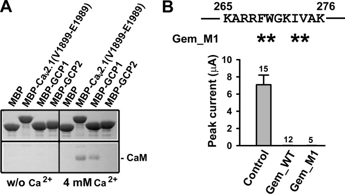
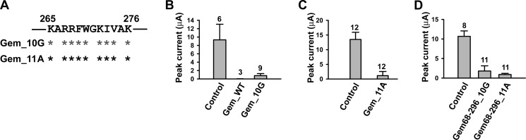
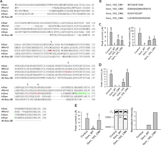
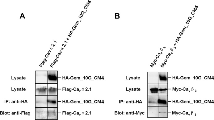
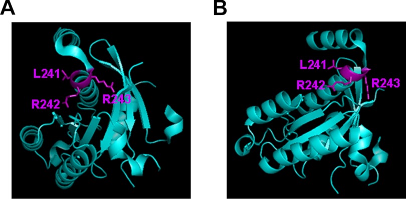
The RGK family of monomeric GTP-binding proteins potently inhibits high voltage-activated Ca(2+) channels. The molecular mechanisms of this inhibition are largely unclear. In Xenopus oocytes, Gem suppresses the activity of P/Q-type Ca(2+) channels on the plasma membrane. This is presumed to occur through direct interactions of one or more Gem inhibitory sites and the pore-forming Ca(v)2.1 subunit in a manner dependent on the Ca(2+) channel subunit β (Ca(v)β). In this study we investigated the molecular determinants in Gem that are critical for this inhibition. Like other RGK proteins, Gem contains a conserved Ras-like core and extended N and C termini. A 12-amino acid fragment in the C terminus was found to be crucial for and sufficient to produce Ca(v)β-dependent inhibition, suggesting that this region forms an inhibitory site. A three-amino acid motif in the core was also found to be critical, possibly forming another inhibitory site. Mutating either site individually did not hamper Gem inhibition, but mutating both sites together completely abolished Gem inhibition without affecting Gem protein expression level or disrupting Gem interaction with Ca(v)2.1 or Ca(v)β. Mutating Gem residues that are crucial for interactions with previously demonstrated RGK modulators such as calmodulin, 14-3-3, and phosphatidylinositol lipids did not significantly affect Gem inhibition. These results suggest that Gem contains two candidate inhibitory sites, each capable of producing full inhibition of P/Q-type Ca(2+) channels.


Figures

FIGURE 1.
FIGURE 1.
Mapping regions of Gem critical for the inhibition of P/Q-type Ca2+ channels. A, shown is a schematic diagram of WT Gem and various truncation mutants. B–D, left panels, shown are whole oocyte currents recorded at +10 mV by TEVC from oocytes expressing Cav2.1, α2δ, and β3 without Gem (control), with WT Gem (Gem_WT), or with the indicated truncated Gem. In these and the following panels showing TEVC currents the number of recordings is indicated above the bar, and all the results shown in each panel were obtained from the same batch of oocytes. B and D, middle panels, a Western blot shows protein expression of WT Gem and the indicated mutant Gem in the lysates of the oocytes recorded in the corresponding left panels. β3 expression is shown as the loading control. B and D, right panels, bar graphs show the normalized intensity of the corresponding Gem bands in the middle panels.
FIGURE 2.
FIGURE 2.
A 12-amino acid region in the C-terminus is crucial for Gem inhibition of P/Q-type Ca2+ channels on plasma membrane. A and B, left panels, shown is time course of inhibition of currents (recorded at +20 mV) by 5 μm purified Gem-(Ser-68–Lys-276) protein (A) or Gem-(Ser-68–Arg-264) protein (B) in inside-out membrane patches from oocytes expressing Cav2.1, α2δ and β3_GK. n = 5. Right panels, current traces selected from a representative patch for each condition are shown. Currents were evoked by a depolarization to +20 mV from a holding potential of −80 mV and were obtained immediately before (red), during (blue), or after (green) perfusion of the indicated purified mutant Gem proteins. The dashed line indicates zero current level.
FIGURE 3.
FIGURE 3.
A 12-amino acid Gem peptide is sufficient to inhibit P/Q-type Ca2+ channels on plasma membrane. A, shown is an amino acid sequence of three Gem-derived peptides. GCP1 and GCP2 correspond to Lys-265–Lys-276 and Asn-277–Leu-296 of Gem, respectively; GCP1_S is a sequence-scrambled version of GCP1. B–D and F, left panels, shown is time course of inhibition of currents (recorded at +20 mV) by 100 μm GCP1 (B), 200 μm GCP2 (C), 5 μm GCP1 (D), or 100 μm GCP1_S (F) in inside-out membrane patches from oocytes expressing Cav2.1, α2δ, and β3. n = 5. E, left panel, shown is time course of inhibition of β-less channels by 100 μm GCP1. Currents (recorded at +20 mV) were obtained from inside-out membrane patches excised from oocytes expressing Cav2.1, α2δ, and β3_Mut2. Before time 0, the patch had been perfused for 5 min such that the channels had lost β3_Mut2 and become β-less. n = 4. B–F, right panels, shown are current traces selected from a representative patch for each condition. Currents were evoked by a depolarization to +20 mV from a holding potential of −80 mV and were obtained immediately before (red), during (blue), or after (green) perfusion of the indicated Gem peptides. The dashed line indicates zero current level. G, left panel, whole oocyte currents were recorded at +10 mV by TEVC from oocytes expressing Cav2.1, α2δ, and β3, without Gem (control), with WT Gem (Gem_WT), or with the indicated mutant Gem. *** p < 0.01 (compared with control). Middle panel, a Western blot shows protein expression of WT Gem and the indicated mutant Gem in the lysates of the oocytes recorded in the left panel. β3 expression was shown as the loading control. Right panel, the bar graph shows the normalized intensity of the corresponding Gem bands in the middle panel.
FIGURE 4.
FIGURE 4.
CaM binding is not required for Gem inhibition of P/Q-type Ca2+ channels. A, MBP-fused GCP1 peptide pulls down purified CaM in a Ca2+-dependent manner. In the presence of 4 mm Ca2+, CaM interacted with MBP-tagged GCP1 and MBP-tagged Cav2.1-(Val-1899–Glu-1989), which served as a positive control, but not with MBP itself and MBP-tagged GCP2. B, shown are whole-oocyte currents recorded at +10 mV in oocytes expressing Cav2.1, α2δ, and β3 without Gem (control), with WT Gem, or with Gem_M1, in which four hydrophobic residues (asterisks) in the indicated region were mutated to alanine.
FIGURE 5.
FIGURE 5.
The Gem Lys-265–Lys-276 region is not the sole inhibitory site in Gem. A, a schematic shows mutations in the Lys-265–Lys-276 region of full-length Gem. Dark and gray asterisks mark residues mutated to alanine and glycine, respectively. B–D, whole-oocyte currents are recorded at +10 mV in oocytes expressing Cav2.1, α2δ, and β3 without Gem (control) or with the indicated Gem mutants.
FIGURE 6.
FIGURE 6.
The C terminus of Gem does not contain a second inhibitory site. A, shown is the amino acid sequence of the last 41 residues (Gln-256–Leu-296) of Gem and Gem_10G. The indicated full-length Gem mutants were all made from Gem_10G, with the asterisks indicating residues that were mutated to alanine. B and C, shown are whole-oocyte currents recorded at +10 mV in oocytes expressing Cav2.1, α2δ, and β3 without Gem (control) or with the indicated Gem mutants. *, p < 0.05; ***, p < 0.01 (compared with Gem_10G).
FIGURE 7.
FIGURE 7.
Identification of a triamino acid motif in the core important for Gem inhibition. A, shown is amino acid sequence alignment of human Rad (BC057815), rat Rem2 (GenBankTM accession number AF084464), human Gem/Kir (BC022010), human Rem (AF084465), and rat K-Ras (BC126086). Red residues are conserved among RGK proteins but not in Ras and were mutated in the Gem_10G background to residues indicated in B. Green residues mark the Lys-265–Lys-276 region. The numbers above the hRad sequence correspond to the names of the mutants in B. B, shown are the name and the mutations of the mutants. CM, core mutation. C and D, and left panel of E, whole-oocyte currents were recorded at +10 mV in oocytes expressing Cav2.1, α2δ, and β3 without Gem (control), with WT Gem, or with the indicated Gem mutants. E, middle panel, a Western blot shows protein expression of WT Gem and Gem_10G_CM4 in the lysates of the oocytes recorded in the corresponding left panel. β3 expression was shown as the loading control. Right panel, a bar graph shows the normalized intensity of the corresponding Gem bands in the middle panel.
FIGURE 8.
FIGURE 8.
Gem_10G_CM4 can still associate with Cav2.1 and β3. A and B, a Western blot shows co-immunoprecipitation of HA-Gem_10G_CM4 and FLAG-Cav2.1 (A) or HA-Gem_10G_CM4 and Myc-β3 (B). Immunoprecipitation (IP) of Gem was carried out using an anti-HA antibody from the lysates of HEK 293T cells expressing FLAG-Cav2.1 alone (A, left lane) or Myc-β3 alone (B, left lane), co-expressing HA-Gem_10G_CM4 and FLAG-Cav2.1 (A, right lane), or co-expressing HA-Gem_10G_CM4 and Myc-β3 (B, right lane). Cav2.1 was detected with an anti-FLAG antibody and β3 with an anti-Myc antibody. Each experiment was repeated twice.
FIGURE 9.
FIGURE 9.
Location and side-chain projection of Leu-241/Arg-242/Arg-243. Two different orientations of a crystal structure (Protein Data Bank ID 2CJW) of a GDP-bound Gem protein fragment (Met-73–Arg-264) are shown. The figures were generated using the program PyMOL.
FIGURE 10.
FIGURE 10.
The distal C terminus is necessary for the function of the core inhibitory site. Left panel, whole-oocyte currents were recorded at +10 mV in oocytes expressing Cav2.1, α2δ, and β3 without Gem (control), with WT Gem, or with Gem_10GΔC. Middle panel, a Western blot shows protein expression of WT Gem and Gem_10GΔC in the lysates of the oocytes recorded in the corresponding left panel. β3 expression was shown as loading controls. Right panel, a bar graph shows normalized intensity of the corresponding Gem bands in the middle panel.

References

    1. Felix R. (2005) Molecular regulation of voltage-gated Ca2+ channels. J. Recept. Signal Transduct. Res. 25, 57–71 - PubMed
    1. Catterall W. A. (2000) Structure and regulation of voltage-gated Ca2+ channels. Annu. Rev. Cell Dev. Biol. 16, 521–555 - PubMed
    1. Kelly K. (2005) The RGK family. A regulatory tail of small GTP-binding proteins. Trends Cell Biol. 15, 640–643 - PubMed
    1. Correll R. N., Pang C., Niedowicz D. M., Finlin B. S., Andres D. A. (2008) The RGK family of GTP-binding proteins. Regulators of voltage-dependent calcium channels and cytoskeleton remodeling. Cell. Signal. 20, 292–300 - PMC - PubMed
    1. Buraei Z., Yang J. (2010) The β subunit of voltage-gated Ca2+ channels. Physiol. Rev. 90, 1461–1506 - PMC - PubMed

Publication types

MeSH terms

Substances