. 2012;8(5):e1002675.
doi: 10.1371/journal.ppat.1002675. Epub 2012 May 10.

The lipopolysaccharide core of Brucella abortus acts as a shield against innate immunity recognition

Affiliations

The lipopolysaccharide core of Brucella abortus acts as a shield against innate immunity recognition

Raquel Conde-Álvarez et al. PLoS Pathog. 2012.

Abstract

Innate immunity recognizes bacterial molecules bearing pathogen-associated molecular patterns to launch inflammatory responses leading to the activation of adaptive immunity. However, the lipopolysaccharide (LPS) of the gram-negative bacterium Brucella lacks a marked pathogen-associated molecular pattern, and it has been postulated that this delays the development of immunity, creating a gap that is critical for the bacterium to reach the intracellular replicative niche. We found that a B. abortus mutant in the wadC gene displayed a disrupted LPS core while keeping both the LPS O-polysaccharide and lipid A. In mice, the wadC mutant induced proinflammatory responses and was attenuated. In addition, it was sensitive to killing by non-immune serum and bactericidal peptides and did not multiply in dendritic cells being targeted to lysosomal compartments. In contrast to wild type B. abortus, the wadC mutant induced dendritic cell maturation and secretion of pro-inflammatory cytokines. All these properties were reproduced by the wadC mutant purified LPS in a TLR4-dependent manner. Moreover, the core-mutated LPS displayed an increased binding to MD-2, the TLR4 co-receptor leading to subsequent increase in intracellular signaling. Here we show that Brucella escapes recognition in early stages of infection by expressing a shield against recognition by innate immunity in its LPS core and identify a novel virulence mechanism in intracellular pathogenic gram-negative bacteria. These results also encourage for an improvement in the generation of novel bacterial vaccines.


Conflict of interest statement

The authors have declared that no competing interests exist.

Figures

Figure 1
Figure 1. BaΔwadC carries a partially defective LPS core oligosaccharide.
Western blot analyses were performed with monoclonal antibodies Cby-33H8 (O-polysaccharide C/Y epitope) and A68/24D08/G09 (core oligosaccharide) and SDS-proteinase K LPS extracts of Ba-parental, BaΔwadC and BaΔwadC-compl (complemented mutant), and the SDS-PAGE electropherogram of LPS phenol-water extracts was silver stained.
Figure 2
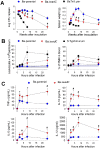
Figure 2. BaΔwadC is attenuated in mice and induces transitory splenomegaly, local leukocyte recruitment and cytokine secretion.
(A), Left panel, infection kinetics in the spleens of mice intraperitoneally inoculated with 5×10⁴ Ba-parental, BaΔwadC or 1×10⁸ BaTn5::per; right panel, spleen weights. (B), Leukocyte and PMN levels in the peritoneum (left panel) and blood (right panel) of mice after intraperitoneal infection with 1×10⁶ Ba-parental, BaΔwadC or 1×10⁵ S. Typhimurium SL1344. (C), TNF-α, IL-6, IL-10 and IL-12 p40/p70 levels in the sera of mice infected with Ba-parental or BaΔwadC. Each point is the mean ± standard deviation (n = 5). Differences between Ba-parental and BaΔwadC are indicated with symbols (*, p<0.05; #, p<0.001).
Figure 3
Figure 3. BaΔwadC is attenuated in BMDC.
Upper panel, intracellular replication of Ba-parental, BaΔwadC, BaΔwadC-compl and virB mutant in BMDC and BMDM. Each point is the mean ± standard error of an experiment performed in triplicate, and the results representative of three independent experiments. Differences between Ba-parental and BaΔwadC are indicated with symbols (#, p<0.001). Lower panel, confocal imaging of Ba-parental and BaΔwadC carrying a GFP plasmid at 24 h post-infection (calnexin and LAMP-1 are in red).
Figure 4
Figure 4. BaΔwadC induces a cytokine release that is reproduced by purified LPS in a TLR-4 dependent fashion.
(A), IL-12 p40/p70 and TNF-α released by BMDC 24 h after infection (left panel) or stimulation with 10 µg/mL of Ba-parental or BaΔwadC LPS, or with 100 ng/mL of E. coli LPS (right panel) measured by ELISA; (B), IL-12 p40/p70 (left panel) and TNF-α (right panel) released by BMDC from wild-type (WT), TLR9−/− TLR4−/− or TLR2−/− mice 24 h after stimulation with 10 µg/mL of Ba-parental or BaΔwadC LPS, or with 100 ng/mL of E. coli LPS measured by ELISA; (C), Surface expression of CD40, CD86 and MHCII in LPS-stimulated BMDC measured by flow cytometry. Each point is the mean ± standard error (n = 3). But for TLR4−/− mice differences between Ba-parental and BaΔwadC or their corresponding LPS were statistically significant (p<0.001). Results are representative of two independent experiments.
Figure 5
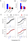
Figure 5. BaΔwadC shows increased sensitivity to normal serum and bactericidal peptides that relates to the LPS core defect.
(A), Survival of Ba-parental, BaΔwadC and BaTn5::per after incubation in non-immune calf serum for 90 min. Each point is the mean ± standard error of an experiment performed in triplicate, and the results representative of five independent experiments (differences between Ba-parental and BaΔwadC are indicated as #, p<0.001). (B), minimal inhibitory concentrations (MIC) of polymyxin B (determined by the serial dilution method) and colistin (determined by the E-test) (results representative of three independent experiments). (C), Crystalline to fluid (β↔α) phase transition of the hydrocarbon chains of Ba-parental LPS and BaΔwadC LPS measured in the presence (filled circles) or absence (empty circles) of normal human serum (red circles) or polymyxin B (black circles; LPS∶PMB 1∶0.1 molar ratio). The plots represent the position (wavenumber) of the peak of the symmetric stretching vibration of the methylene groups νs(CH2) versus temperature. Tc, transition temperature (results are representative of three independent experiments).
Figure 6
Figure 6. BaΔwadC LPS binds to MD-2 and increases NF-κB translocation and S6 phosphorylation.
(A) LPS binding to hMD-2. After incubation of 0.75 µM hMD-2 with increasing LPS concentrations, the fraction of hMD-2 not bound to LPS was detected with free-hMD-2 specific antibody 9B4 by ELISA (results are representative of three independent experiments run in triplicate; differences between Ba-parental and BaΔwadC are indicated as *, p<0.05 or #, p<0.001) (B) NF-κB translocation to the nucleus. BMDC were stimulated for 1 h with media, E. coli LPS (100 ng/mL), Ba-parental LPS (10 µg/mL) or BaΔwadC LPS (10 µg/mL), cells fixed in 3% paraformaldehyde at 37°C for 15 min, immunostained for CD11c and MHCII, and the % of cells showing nuclear translocation of the NF-κB subunit p65/RelA recorded (results are representative of three independent experiments run in triplicate; differences between Ba-parental and BaΔwadC are indicated as #, p<0.001). (C) S6 phosphorylation. BMDC were stimulated for 30 min, 1, 4, or 6 h with (left to right) media, E. coli LPS (100 ng/mL), Ba-parental LPS (100 ng/mL), Ba-parental LPS (10 µg/mL) or BaΔwadC LPS (10 µg/mL). Cell lysates (30 µg protein) were analyzed by Western blot with a polyclonal antibody against S6-P or an anti-actin antibody as control.

References

    1. Gorvel JP, Moreno E. Brucella intracellular life: from invasion to intracellular replication. Vet Microbiol. 2002;90:281–297. - PubMed
    1. Barquero-Calvo E, Chaves-Olarte E, Weiss DS, Guzman-Verri C, Chacón-Díaz C, et al. Brucella abortus uses a stealthy strategy to avoid activation of the innate immune system during the onset of infection. PLoS One. 2007;2:e631. - PMC - PubMed
    1. Martirosyan A, Moreno E, Gorvel JP. An evolutionary strategy for a stealthy intracellular Brucella pathogen. Immunol Rev. 2011;240:211–234. - PubMed
    1. Lapaque N, Moriyón I, Moreno E, Gorvel JP. Brucella lipopolysaccharide acts as a virulence factor. Curr Opin Microbiol. 2005;8:60–66. - PubMed
    1. Palacios-Chaves L, Conde-Álvarez R, Gil-Ramírez Y, Zúñiga-Ripa A, Barquero-Calvo E, et al. Brucella abortus ornithine lipids are dispensable outer membrane components devoid of a marked pathogen-associated molecular pattern. PLoS One. 2011;6:e16030. - PMC - PubMed

Publication types

MeSH terms

LinkOut - more resources