. 2012;8(5):e1002689.
doi: 10.1371/journal.ppat.1002689. Epub 2012 May 10.

Vitamin D inhibits human immunodeficiency virus type 1 and Mycobacterium tuberculosis infection in macrophages through the induction of autophagy

Affiliations

Vitamin D inhibits human immunodeficiency virus type 1 and Mycobacterium tuberculosis infection in macrophages through the induction of autophagy

Grant R Campbell et al. PLoS Pathog. 2012.

Abstract

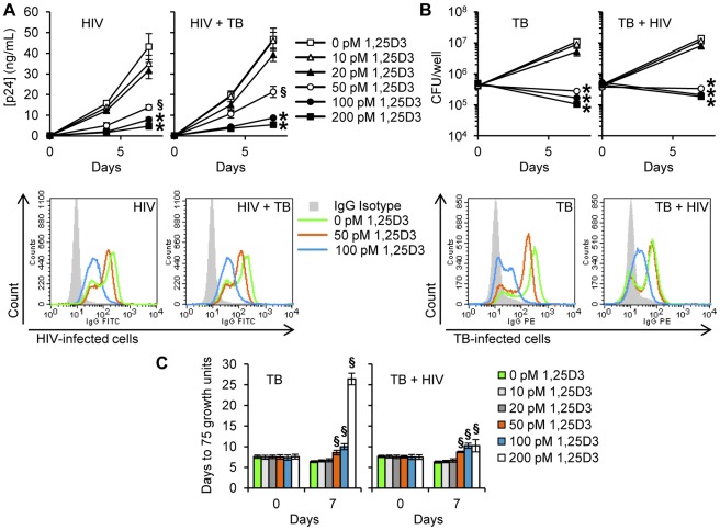
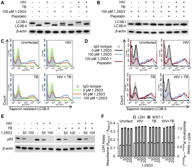
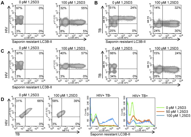
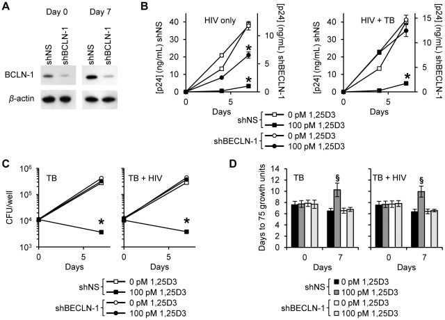
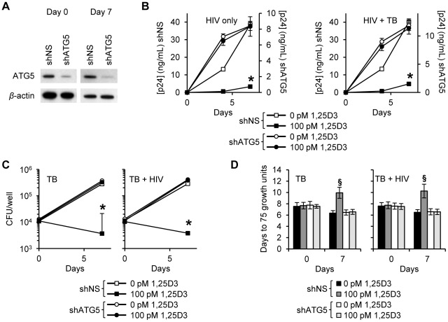
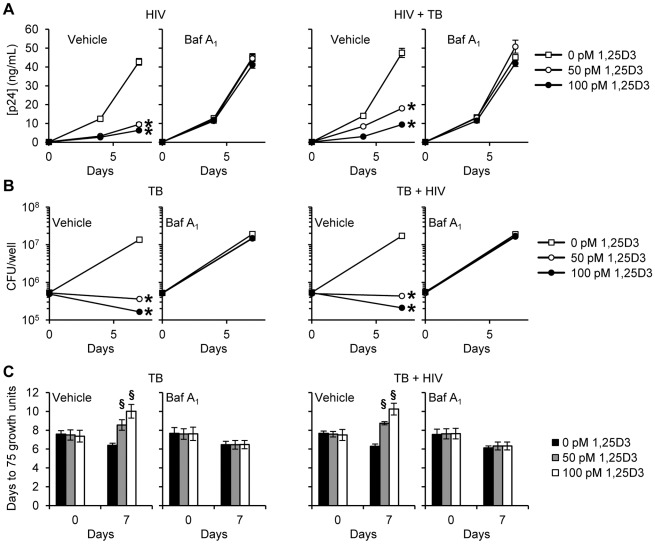
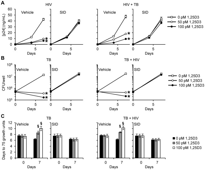
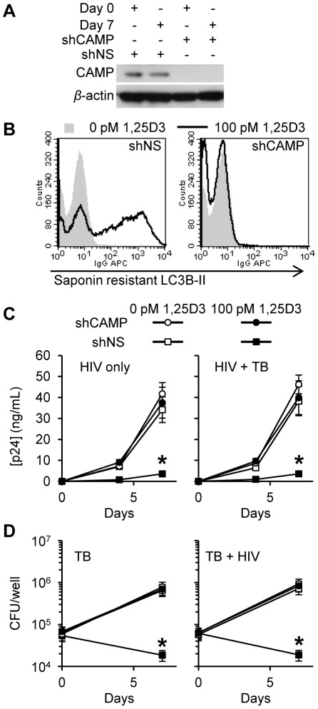
Low vitamin D levels in human immunodeficiency virus type-1 (HIV) infected persons are associated with more rapid disease progression and increased risk for Mycobacterium tuberculosis infection. We have previously shown that 1α,25-dihydroxycholecalciferol (1,25D3), the active form of vitamin D, inhibits HIV replication in human macrophages through the induction of autophagy. In this study, we report that physiological concentrations of 1,25D3 induce the production of the human cathelicidin microbial peptide (CAMP) and autophagic flux in HIV and M. tuberculosis co-infected human macrophages which inhibits mycobacterial growth and the replication of HIV. Using RNA interference for Beclin-1 and the autophagy-related 5 homologue, combined with the chemical inhibitors of autophagic flux, bafilomycin A₁, an inhibitor of autophagosome-lysosome fusion and subsequent acidification, and SID 26681509 an inhibitor of the lysosome hydrolase cathepsin L, we show that the 1,25D3-mediated inhibition of HIV replication and mycobacterial growth during single infection or dual infection is dependent not only upon the induction of autophagy, but also through phagosomal maturation. Moreover, through the use of RNA interference for CAMP, we demonstrate that cathelicidin is essential for the 1,25D3 induced autophagic flux and inhibition of HIV replication and mycobacterial growth. The present findings provide a biological explanation for the benefits and importance of vitamin D sufficiency in HIV and M. tuberculosis-infected persons, and provide new insights into novel approaches to prevent and treat HIV infection and related opportunistic infections.


Conflict of interest statement

The authors have declared that no competing interests exist.

Figures

Figure 1
Figure 1. 1,25D3 inhibits HIV and M. tuberculosis replication.
MDM were incubated with 1,25D3 for 4 h before infection with HIV and/or M. tuberculosis (TB) for 3 h, washed then incubated with or without 1,25D3 for 7 days. (A) Top, extracellular release of HIV p24 antigen into the cell supernatant at days 0, 4 and 7 was detected by ELISA. Bottom, MDM were harvested and stained for HIV p17. Histograms are shown for a representative donor. (B) Top, cells were lysed after 3 h exposure to infectious agents or at the completion of the infection phase. Intracellular mycobacteria were harvested and assayed for mycobacterial growth at day 0 and day 7. Bottom, MDM were harvested and stained for mycobacterium. Histograms are shown for a representative donor. (C) Cell lysates from panel B were diluted and run in a MGIT 960. Bar and line graphs are reported as mean ± s.e.m. of three independent experiments performed in triplicate. § P<0.05; * P<0.001.
Figure 2
Figure 2. 1,25D3 induces autophagy in human macrophages co-infected with HIV and M. tuberculosis.
HIV and/or M. tuberculosis (TB) infected MDM were treated for 7 days with 100 pmol/L 1,25D3. (A) Cells were lysed and immunoblots of LC3B isoforms using antibody to LC3B or β-actin performed. (B) Cells were incubated with 10 µg/mL pepstatin A for 4 h on day 7 prior to lysis. Immunoblots of LC3B isoforms using antibody to LC3B or β-actin. (C) Flow cytometry analysis of saponin-resistant LC3B-II in macrophages after 1,25D3 treatment for 7 d. Representative histograms of cells displaying saponin-resistant LC3B-II from three donors are shown. (D) Flow cytometry analysis of saponin-resistant LC3B-II in macrophages after 1,25D3 treatment for 7 d followed by 10 µg/mL pepstatin A for a further 4 h. Representative histograms of cells displaying saponin-resistant LC3B-II from three donors are shown. (E) Immunoblots of p62 using antibody to p62 or β-actin 7 d after macrophages treated with 1,25D3. (F) At 7 d post-infection, aliquots of supernatant taken before the addition of WST-1 were tested for lactate dehydrogenase (LDH) spectrophotometrically using the LDHPLUS assay. For the last hour cells were incubated with WST-1, and the reduction of the WST-1 reagent to its formazan product was monitored spectrophotometrically.
Figure 3
Figure 3. 1,25D3 induces autophagy in human macrophages co-infected with HIV and M. tuberculosis.
HIV (A), M. tuberculosis (TB) (B), and HIV/TB dual infected MDM (C) were treated for 7 days with 100 pmol/L 1,25D3, harvested and stained for HIV p17, mycobacteria, and saponin-resistant LC3B-II. Representative density plots from three donors are shown. (D) HIV/M. tuberculosis infected MDM (C) were treated for 7 days with 100 pmol/L 1,25D3, harvested and stained for HIV p17, mycobacteria, and saponin-resistant LC3B-II. Left, representative density plots from three donors from HIV/M. tuberculosis infected MDM are shown for HIV/M. tuberculosis co-infection. Right, histograms of saponin-resistant LC3B-II in macrophages that were HIV+ M. tuberculosis or HIV+ M. tuberculosis + at day 7 post-infection.
Figure 4
Figure 4. 1,25D3 inhibition of HIV and M. tuberculosis is Beclin-1 dependent.
MDM were transduced with non-specific scrambled shRNA (shNS) or Beclin-1 shRNA (shBCLN1) and selected using puromycin resistance. Five days later, cells were incubated with 100 pmol/L 1,25D3 or vehicle control for 4 h before infection with HIV and/or M. tuberculosis (TB) for 3 h. Cells were then washed and incubated with 100 pmol/L 1,25D3 or vehicle control for 7 days. (A) Immunoblot analysis performed using antibodies raised to Beclin-1 or β-actin after initial pathogen exposure (Day 0) or after 7 days. (B) ELISA performed for HIV p24 antigen release over 7 d. (C) Intracellular mycobacteria were harvested and assayed for mycobacterial growth by cfu enumeration at day 0 and day 7. (D) Intracellular mycobacteria harvested and assayed for viability based on time to positivity (75 growth units) at day 0 and day 7 using the MGIT 960. All bar and line graphs are reported as mean ± s.e.m. of three independent experiments performed in triplicate. § P<0.05; * P<0.001.
Figure 5
Figure 5. 1,25D3 inhibition of HIV and M. tuberculosis is ATG5 dependent.
MDM were transduced with non-specific scrambled shRNA (shNS) or ATG5 shRNA (shATG5) and selected using puromycin resistance. Five days later, cells were incubated with 100 pmol/L 1,25D3 or vehicle control for 4 h before infection with HIV and/or M. tuberculosis (TB) for 3 h. Cells were then washed and incubated with 100 pmol/L 1,25D3 or vehicle control for 7 days. (A) Immunoblot analysis performed using antibodies raised to ATG5 or β-actin after initial pathogen exposure (Day 0) or after 7 days. (B) ELISA performed for HIV p24 antigen release over 7 d. (C) Intracellular mycobacteria were harvested and assayed for mycobacterial growth by cfu enumeration at day 0 and day 7. (D) Intracellular mycobacteria harvested and assayed for viability based on time to positivity (75 growth units) at day 0 and day 7 using the MGIT 960. All bar and line graphs are reported as mean ± s.e.m. of three independent experiments performed in triplicate. § P<0.05; * P<0.001.
Figure 6
Figure 6. Bafilomycin A1 inhibits the 1,25D3 mediated inhibition of HIV and M. tuberculosis replication.
MDM were pretreated with bafilomycin A1 (Baf A1) before treatment with 1,25D3 and subsequent infection with HIV and/or M. tuberculosis (TB). (A) ELISA performed for HIV p24 antigen release at days 0, 4 and 7. (B) Intracellular mycobacteria were harvested and assayed for mycobacterial growth by cfu enumeration at day 0 and 7. (C) Intracellular mycobacteria harvested and assayed for viability using the MGIT 960 based on time to positivity at day 0 and 7. All bar and line graphs are reported as mean ± s.e.m. of three independent experiments performed in triplicate. § P<0.05; * P<0.01.
Figure 7
Figure 7. SID 26681509 inhibits the 1,25D3 mediated inhibition of HIV and M. tuberculosis replication.
MDM were pretreated with SID 26681509 (SID) before treatment with 1,25D3 and subsequent infection with HIV and/or M. tuberculosis (TB). (A) ELISA performed for HIV p24 antigen release at days 0, 4 and 7. (B) Intracellular mycobacteria were harvested and assayed for mycobacterial growth by cfu enumeration at day 0 and 7. (C) Intracellular mycobacteria harvested and assayed for viability using the MGIT 960 based on time to positivity at day 0 and 7. All bar and line graphs are reported as mean ± s.e.m. of three independent experiments performed in triplicate. § P<0.05; * P<0.01.
Figure 8
Figure 8. Inhibition of HIV and M. tuberculosis by 1,25D3 is CAMP and autophagy dependent.
MDM were transduced with non-specific scrambled shRNA (shNS) or CAMP shRNA (shCAMP) and selected using puromycin resistance. Five days later, cells were incubated with 100 pmol/L 1,25D3 or vehicle control for 4 h before infection with HIV and/or M. tuberculosis (TB) for 3 h. Cells were then washed and incubated with 100 pmol/L 1,25D3 or vehicle control for 7 days. (A) Immunoblot analysis performed using antibodies raised to CAMP or β-actin after initial pathogen exposure (Day 0) or after 7 days. (B) Day 7 HIV-1Ba-L and TB co-infected cells were harvested and stained for saponin-resistant LC3B-II at day 7. A representative histogram from three donors are shown. (C) ELISA performed for HIV p24 antigen release over 7 d. (D) Intracellular mycobacteria were harvested and assayed for mycobacterial growth by cfu enumeration at day 0 and day 7. All bar and line graphs are reported as mean ± s.e.m. of three independent experiments performed in triplicate. * P<0.001.

References

    1. Bezuidenhout J, Roberts T, Muller L, van Helden P, Walzl G. Pleural tuberculosis in patients with early HIV infection is associated with increased TNF-alpha expression and necrosis in granulomas. PLoS One. 2009;4:e4228. - PMC - PubMed
    1. de Noronha AL, Bafica A, Nogueira L, Barral A, Barral-Netto M. Lung granulomas from Mycobacterium tuberculosis/HIV-1 co-infected patients display decreased in situ TNF production. Pathol Res Pract. 2008;204:155–161. - PubMed
    1. Lawn SD, Butera ST, Shinnick TM. Tuberculosis unleashed: the impact of human immunodeficiency virus infection on the host granulomatous response to Mycobacterium tuberculosis. Microbes Infect. 2002;4:635–646. - PubMed
    1. Safi H, Gormus BJ, Didier PJ, Blanchard JL, Lakey DL, et al. Spectrum of manifestations of Mycobacterium tuberculosis infection in primates infected with SIV. AIDS Res Hum Retroviruses. 2003;19:585–595. - PubMed
    1. Shen JY, Barnes PF, Rea TH, Meyer PR. Immunohistology of tuberculous adenitis in symptomatic HIV infection. Clin Exp Immunol. 1988;72:186–189. - PMC - PubMed

Publication types

MeSH terms