

Glycoproteins as targets of autoantibodies in CNS inflammation: MOG and more
- PMID: 22590479
- PMCID: PMC3349079
- DOI: 10.1177/1756285611433772
Glycoproteins as targets of autoantibodies in CNS inflammation: MOG and more
Abstract
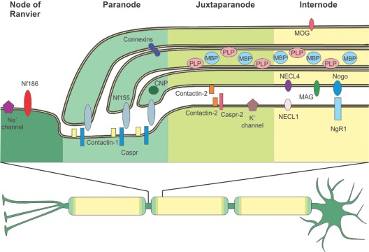
B cells and antibodies constitute an important element in different inflammatory diseases of the central nervous system (CNS). Autoantibodies can serve as a biomarker to identify disease subgroups and may in addition contribute to the pathogenic process. One candidate autoantigen for multiple sclerosis (MS) is myelin oligodendrocyte glycoprotein (MOG). MOG is localized at the outermost surface of myelin in the CNS and has been the focus of extensive research for more than 30 years. Its role as an important autoantigen for T cells and as a target of demyelinating autoantibodies has been established in several variants of experimental autoimmune encephalomyelitis (EAE), an animal model of MS. The literature regarding antibodies to MOG in MS patients is confusing and contradictory. Recent studies, however, have described high levels of antibodies to conformationally correct MOG in pediatric acquired demyelination, both acute disseminated encephalomyelitis (ADEM) and MS. In adult MS, such antibodies are rarely found and then only at low levels. In this review, we summarize key findings from animal models and patient studies, discuss challenges in detecting anti-MOG antibodies in patients and present recent approaches to identifying new autoantigens in MS.
Keywords: ADEM; Myelin oligodendrocyte glycoprotein; detection methods of autoantibodies; multiple sclerosis.
Conflict of interest statement
The authors declare no conflicts of interest in preparing this article.
Figures
References
-
- Adelmann M., Wood J., Benzel I., Fiori P., Lassmann H., Matthieu J.M., et al. (1995) The N-terminal domain of the myelin oligodendrocyte glycoprotein (MOG) induces acute demyelinating experimental autoimmune encephalomyelitis in the Lewis rat. J Neuroimmunol 63: 17–27 - PubMed
-
- Berger T., Rubner P., Schautzer F., Egg R., Ulmer H., Mayringer I., et al. (2003) Antimyelin antibodies as a predictor of clinically definite multiple sclerosis after a first demyelinating event. N Engl J Med 349: 139–145 - PubMed
-
- Bourquin C., Schubart A., Tobollik S., Mather I., Ogg S., Liblau R., et al. (2003) Selective unresponsiveness to conformational B cell epitopes of the myelin oligodendrocyte glycoprotein in H-2b mice. J Immunol 171: 455–461 - PubMed
-
- Bradl M., Misu T., Takahashi T., Watanabe M., Mader S., Reindl M., et al. (2009) Neuromyelitis optica: pathogenicity of patient immunoglobulin in vivo. Ann Neurol 66: 630–643 - PubMed
LinkOut - more resources
Full Text Sources
Other Literature Sources
