. 2012;7(5):e36136.
doi: 10.1371/journal.pone.0036136. Epub 2012 May 9.

Maternal Wnt/β-catenin signaling coactivates transcription through NF-κB binding sites during Xenopus axis formation

Affiliations

Maternal Wnt/β-catenin signaling coactivates transcription through NF-κB binding sites during Xenopus axis formation

Neil J Armstrong et al. PLoS One. 2012.

Abstract

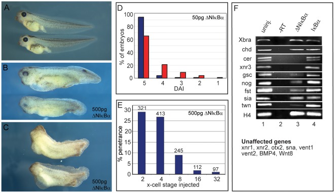
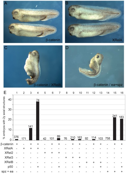
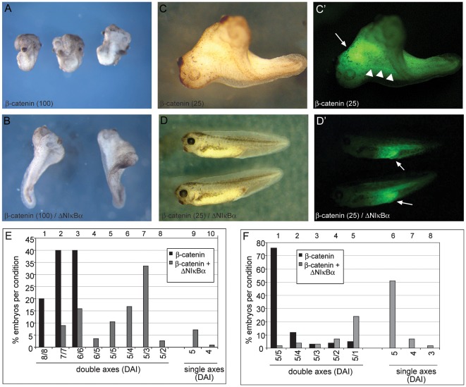
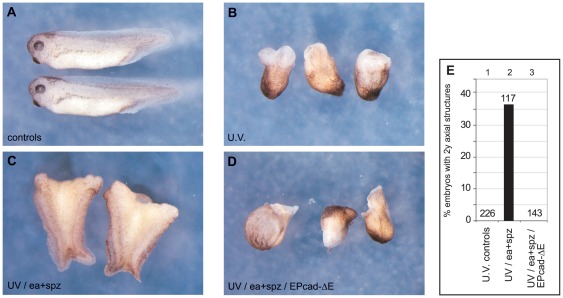
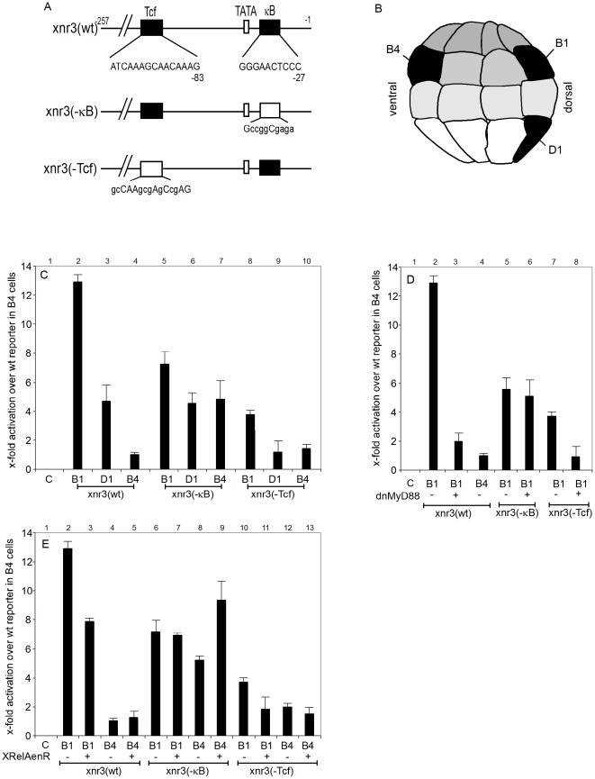
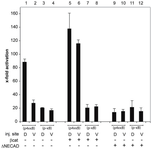
Maternal Wnt/β-Catenin signaling establishes a program of dorsal-specific gene expression required for axial patterning in Xenopus. We previously reported that a subset of dorsally expressed genes depends not only on Wnt/β-Catenin stimulation, but also on a MyD88-dependent Toll-like receptor/IL1-receptor (TLR/IL1-R) signaling pathway. Here we show that these two signal transduction cascades converge in the nucleus to coactivate gene transcription in blastulae through a direct interaction between β-Catenin and NF-κB proteins. A transdominant inhibitor of NF-κB, ΔNIκBα, phenocopies loss of MyD88 protein function, implicating Rel/NF-κB proteins as selective activators of dorsal-specific gene expression. Sensitive axis formation assays in the embryo demonstrate that dorsalization by Wnt/β-Catenin requires NF-κB protein activity, and vice versa. Xenopus nodal-related 3 (Xnr3) is one of the genes with dual β-Catenin/NF-κB input, and a proximal NF-κB consensus site contributes to the regional activity of its promoter. We demonstrate in vitro binding of Xenopus β-Catenin to several XRel proteins. This interaction is observed in vivo upon Wnt-stimulation. Finally, we show that a synthetic luciferase reporter gene responds to both endogenous and exogenous β-Catenin levels in an NF-κB motif dependent manner. These results suggest that β-Catenin acts as a transcriptional co-activator of NF-κB-dependent transcription in frog primary embryonic cells.


Conflict of interest statement

Competing Interests: The current affiliations of NJA (DNA Logic Ltd) and CP (Nycomed US) are not connected to the work presented in this study and do not alter the authors' adherence to all the PLoS ONE policies on sharing data and materials. The authors have declared that no other competing interests exist.

Figures

Figure 1
Figure 1. NF-κB activity is required for head formation.
A) Wild type embryos at stage 40/41. B, C) Sibling embryos injected sub-equatorially on the dorsal side with 500 pg of ΔNIκBα RNA at the 4-cell stage show pleiomorphic phenotypes: B) Mild phenotypes (DAI 4) are characterized by reduction in forebrain and eyes. C) Stronger phenotypes (DAI 3) show reduced head and dorsal structures. (D) Graphical representation of dorsoanterior index (DAI) of control embryos (blue columns) versus embryos injected dorsally with 50 pg of ΔNIκBα (red columns). (E) Time course analysis of embryos injected dorsally with a total of 500 pg ΔNIκBα RNA at the two, four, eight, 16- and 32-cell stage; the accompanying decrease in cell size was compensated by injecting multiple dorsal cells from the 8-cell stage on (see materials and methods). Y-axis represents the percentage of embryos with anterior truncations. (F) Semi-quantitative RT-PCR analysis for marker genes in stage 10 marginal zone explants. Lane 1: uninjected embryos; lane 2: minus reverse transcriptase control; lane 3: 500 pg ΔNIκBα RNA injected dorsally; lane 4: 500 pg wildtype IκBα RNA injected dorsally.
Figure 2
Figure 2. Synergism of XRel and β-Catenin proteins in ectopic axis formation.
Suboptimal levels of β-Catenin synergize with XRel proteins or Drosophila Easter/Spätzle to induce axial structures on the ventral side. Embryos injected ventrally with low levels (4 pg) of β-Catenin (A) or 500 pg of XRelA (B), XRel2, XRel3, XrelB RNAs were indistinguishable from uninjected sibling embryos. Co-injection of 500 pg XRelA with 3 pg of β-Catenin could induce rudimentary axial structures (C), as could co-injection of 100 pg each of easter and spätzle with 3 pg of β-Catenin (D). Graphical representation (E) of all injected combinations, indicating a selective synergism between ectopic β-Catenin and exogenous XrelA/p50 heterodimers, as well as with endogenous NF-κB proteins released by ectopic Ea/Spz.
Figure 3
Figure 3. Dorsalisation by ectopic β-Catenin depends on endogenous NF-κB activity.
Embryos were injected ventrally with high doses (100 pg, panels A,B) or low doses (25 pg C,D) of β-Catenin RNA, alone (A,C), or with 500 pg ΔNIκBα RNA (B,D). All injections included GFP RNA for lineage tracing (C′,D′). A) Embryos injected with high β-Catenin RNA were highly dorsalized (average DAI 6.8 for each axis), almost always lacking posterior structures (n = 160). B) Co-injection of 500 pg of ΔNIκBα reduced the secondary axis to an average DAI = 3.98, with a small number (8%) being single axis embryos (n = 210). C) The majority of embryos injected with low β-Catenin RNA produced pairs of complete axes, both of which scored as “5” on the DAI scale (n = 155). D) Coinjecting 500 pg of ΔNIκBα produced mostly single axis embryos (n = 162). (C′,D′) Lineage tracing showed that the β-Catenin RNA-injected cell progeny accumulated in the anterior region of one of the two axis (white arrow) with some posterior trailing in axial tissue (white arrowheads) (C′), while it was localized in posterior ventral tissues when ΔNIκBα was co-injected (D′). Panels E and F summarize the phenotypic penetrance for respectively high (100 pg) and low (25 pg) β-Catenin RNA (3 independent experiments in each case). Numbers on the X-axis indicate DAI-values of each embryonic Anlage (e.g. 5/5 defining a twinned embryo with two complete heads).
Figure 4
Figure 4. ea/spz-dependent dorsalization requires β-Catenin activity.
(A) Untreated controls at tadpole stage (NF 35/36). (B) UV-ventralized siblings (average DAI = 0.30). (C) UV-ventralized embryos injected sub-equatorially with 100 pg each of Drosophila easter and spätzle RNA produced embryos with 2 rudimentary trunk axes (37%). (D) Embryos injected with 50 pg EPcadΔE, in addition to easter and spätzle, did not show any axial rescue (0%). (E) Graphical representation of axial rescue, giving total numbers for each experimental cohort.
Figure 5
Figure 5. The Xnr3 promoter is regulated through a consensus NF-κB DNA binding site.
Panel (A) - Pictorial representation of the Xnr3 promoter and its κB and Tcf binding sites, as well as representations of mutated promoters, which were used in the luciferase reporter gene assays. Bases at mutated nucleotide positions are shown in lower-case letters. (B) Lateral schematic view of a 32-cell stage embryo locating the B1, B4 and D1 blastomeres in black, whose progeny will later contribute to the BCNE, the ventral gastrula center (VGC) and the Nieuwkoop center (NC), respectively. (C) Results of injection of a group of wildtype and mutant Xnr3 promoter driven luciferase constructs, each injected into the B1, B4 and D1 blastomeres of 32-cell stage embryos; y-axis describes x-fold production of luciferase, normalized to an internal renilla control, relative to the luciferase activity of uninjected embryos (lane 1). (D) and (E) Luciferase activities of wildtype and mutant Xnr3 reporter constructs, injected into B1 or B4 blastomeres as indicated, under conditions, in which endogenous NF-κB activity is either antagonized through coninjected XrelAenR (D) or reduced through dominant-negative XMyD88 (E). Luciferase activity was measured at midgastrula (NF11).
Figure 6
Figure 6. Interaction of β-Catenin and Rel/NF-κB proteins.
A–C: in vitro pull down using his-tagged bacterial recombinant β-Catenin and 35S-labelled in vitro translated Rel proteins. (A) XrelA, 2, 3 and B, but not p50, co-precipitated with full length β-Catenin fused to 6His-Trx-S-tag, but not with a control 6His-Trx-S-tag fragment (Tag abbreviated as “6His” in panels). Ax(194–762), an Axin fragment containing the β-Catenin binding domain and Myc-YFP were used as positive and negative controls for the in vitro translated proteins, respectively (n≥3 independent experiments). (B) XRelA interacts with β-Catenin via its RHD domain. (C) RelA interaction is mediated by the first eight arm-repeats of β-Catenin: XRelA was found to bind full length β-Catenin and the fragments arm1–12 and arm1–8, but neither the N-terminal nor the C-terminal fragments (B and C: n = 2 independent experiments each). (D) Co-Immunoprecipitation of endogenous p65/NF-κB with β-Catenin in Wnt-stimulated REH cells. Extracts from control cells and cells stimulated with Wnt3a-containing medium were immunoprecipitated using an anti-β-Catenin antibody. Immunoprecipitates were analyzed for β-Catenin and p65/NF-κB by Western blots (n = 3 independent experiments).
Figure 7
Figure 7. A synthetic NF-κB reporter gene is coactivated by β-Catenin.
Luciferase activity at midgastrula (NF11) produced by injection of the reporter constructs pNF-κB+ (containing 4 κB binding sites) and pNF-κB− (containing none) into both blastomeres of either the dorsal or ventral side of 4-cell stage embryos. β-Catenin (25 pg; lanes 5–8) and E-cadΔE (50 pg; lanes 9–12) RNAs were co-injected with the reporter. Y-axis shows x-fold stimulation of luciferase activity over uninjected controls after normalization to the internal Renilla control.

References

    1. Schneider S, Steinbeisser H, Warga RM, Hausen P. ß-Catenin translocation into nuclei demarcates the dorsalizing centers in frog and fish embryos. Mechanisms of Development. 1996;57:191–198. - PubMed
    1. Tao Q, Yokota C, Puck H, Kofron M, Birsoy B, et al. Maternal wnt11 activates the canonical wnt signaling pathway required for axis formation in Xenopus embryos. Cell. 2005;120:857–871. - PubMed
    1. Houston DW, Kofron M, Resnik E, Langland R, Destree O, et al. Repression of organizer genes in dorsal and ventral Xenopus cells mediated by maternal XTcf3. Development. 2002;129:4015–4025. - PubMed
    1. Standley HJ, Destree O, Kofron M, Wylie C, Heasman J. Maternal XTcf1 and XTcf4 have distinct roles in regulating Wnt target genes. Dev Biol. 2006;289:318–328. - PubMed
    1. Wardle FC, Smith JC. Transcriptional regulation of mesendoderm formation in Xenopus. Semin Cell Dev Biol. 2006;17:99–109. - PubMed

Publication types

MeSH terms