. 2012;7(5):e36555.
doi: 10.1371/journal.pone.0036555. Epub 2012 May 9.

An allograft glioma model reveals the dependence of aquaporin-4 expression on the brain microenvironment

Affiliations

An allograft glioma model reveals the dependence of aquaporin-4 expression on the brain microenvironment

Susan Noell et al. PLoS One. 2012.

Abstract

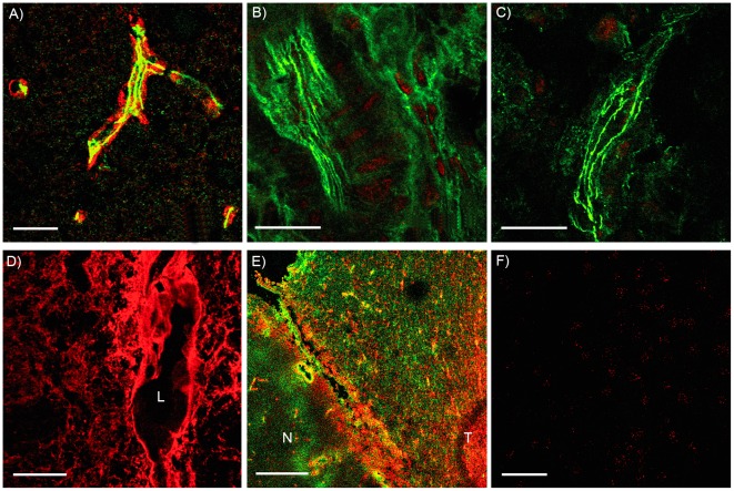
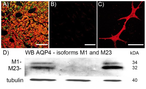
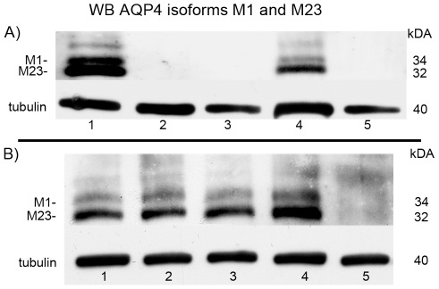
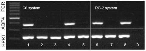
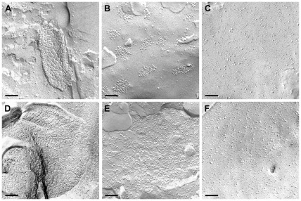
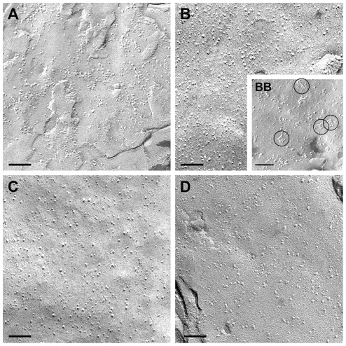
Aquaporin-4 (AQP4), the main water channel of the brain, is highly expressed in animal glioma and human glioblastoma in situ. In contrast, most cultivated glioma cell lines don't express AQP4, and primary cell cultures of human glioblastoma lose it during the first passages. Accordingly, in C6 cells and RG2 cells, two glioma cell lines of the rat, and in SMA mouse glioma cell lines, we found no AQP4 expression. We confirmed an AQP4 loss in primary human glioblastoma cell cultures after a few passages. RG-2 glioma cells if grafted into the brain developed AQP4 expression. This led us consider the possibility of AQP4 expression depends on brain microenvironment. In previous studies, we observed that the typical morphological conformation of AQP4 as orthogonal arrays of particles (OAP) depended on the extracellular matrix component agrin. In this study, we showed for the first time implanted AQP4 negative glioma cells in animal brain or flank to express AQP4 specifically in the intracerebral gliomas but neither in the extracranial nor in the flank gliomas. AQP4 expression in intracerebral gliomas went along with an OAP loss, compared to normal brain tissue. AQP4 staining in vivo normally is polarized in the astrocytic endfoot membranes at the glia limitans superficialis and perivascularis, but in C6 and RG2 tumors the AQP4 staining is redistributed over the whole glioma cell as in human glioblastoma. In contrast, primary rat or mouse astrocytes in culture did not lose their ability to express AQP4, and they were able to form few OAPs.


Conflict of interest statement

Competing Interests: The authors have declared that no competing interests exist.

Figures

Figure 1
Figure 1. Immunoreactivity against AQP4.
(A) Rat control brain, AQP4 (red) is restricted to astrocytic endfeet contacting the blood vessel (green ZO-1). (B) Extracranial implanted C6-tumor, no AQP4 could be detected, green ZO-1. (C) C6-tumor implanted into the flank, no AQP4 could be detected, green ZO-1. (D) Intracerebral implanted C6-tumor, immunofluorescence (red) shows an intensive staining for AQP4 (L: blood vessel lumen). (E) Right side: Intracerebral implanted RG-2-tumor (T); AQP4 (red) is present in astrocytes all over the tumor tissue and stronger fluorescence in the reactive astrocytes, compared to the healthy part of the brain (N), GFAP (green). Left side: healthy part of the brain (N), AQP4 (red) is restricted to astrocytic membranes contacting the blood vessels. (F) No AQP4 could be detected in C6 cell cultures. Scale bars each 20 µm.
Figure 2
Figure 2. Immunoreactivity against AQP4 (red) and GFAP (green).
(A) In human glioblastoma tissue AQP4 (red) shows an intensive staining whereas in primary cell culture of this glioblastoma AQP4 could not be detected (B). (C) Primary cell culture of astrocytes stained for AQP4. (D) Immunoblot against AQP4; the lower band (32 kDA) represents the AQP4 isoform M23 and the upper band (34 kDA) the isoform M1. Tubulin was used as loading control for these samples (40 kDA). The western blot is positive for AQP4 in the glioblastoma tissue (left lane), whereas the primary glioma cell cultures were negative (middle). Primary mouse astrocytes are positive for AQP4 (right lane). The M23-AQP4 isoform always shows a stronger band than the M1 isoform.
Figure 3
Figure 3. Western blot against AQP4.
(A) C6-system: the lower band (32 kDA) represents the AQP4 isoform M23 and the upper band (34 kDA) the isoform M1. Rat control brain (lane 1), C6 cell culture (lane 2), extracranial implanted C6 cells (lane 3), intracerebral implanted C6 cells (lane 4) and C6 cells implanted into the rat flank (lane 5). Tubulin was used as loading control for these samples (40 kDA). (B) RG-2- system: Control rat brains (lane 1 and 3) and Intracerebral RG-2 cell tumors (lane 2 and 4) are positive for AQP4. The RG-2 cell culture (lane 5) is negative. The M23 AQP4 isoform always shows a stronger band than the M1 isoform. Tubulin was used as loading control for these samples (40 kDA).
Figure 4
Figure 4. Representative RT-PCR on an agarose gel showing bands corresponding to AQP4 mRNA and the internal standard HPRT.
AQP4 mRNA is only expressed in normal rat brain (lane 1 and 6) and intracerebral implanted C6 and RG-2 cell tumor (lane 4 and 8) compared to the C6 and RG-2 cell culture (lane 2 and 7) as well as C6 implanted extracranial and flank tumor (lane 3 and 5). Lane 9 shows the negative H2O control.
Figure 5
Figure 5. Freeze fracture analysis.
(A) Normal human brain tissue and rat brain tissue (D) showing astrocytic endfoot membranes studded with OAPs containing AQP4. (B) Altered morphology of clustered OAPs in human glioblastoma tissue. (C) Primary cell culture membranes of human glioblastoma are devoid of OAPs as well as intracerebral RG-2 tumor (E), and the RG-2 cell line (F). Scale bars 100 nm.
Figure 6
Figure 6. Freeze fracture analysis of the C6 system.
(A) C6 implanted intracerebral tumor, (B), C6 cell culture. C6 cells implanted extracranial (C) and flank tumor (D). There are no OAPs. In contrast (BB) shows a freeze fracture replica of primary astrocytes in culture revealing a few OAPs (encircled). Scale bars 100 nm.

References

    1. Moe SE, Sorbo JG, Sogaard R, Zeuthen T, Petter Ottersen, et al. New isoforms of rat Aquaporin-4. Genomics. 2008;91:367–377. - PubMed
    1. Silberstein C, Bouley R, Huang Y, Fang P, Pastor-Soler N, et al. Membrane organization and function of M1 and M23 isoforms of aquaporin-4 in epithelial cells. Am J Physiol Renal Physiol. 2004;287:F501–511. - PubMed
    1. Wolburg H, Wolburg-Buchholz K, Fallier-Becker P, Noell S, Mack AF. Structure and functions of aquaporin-4-based orthogonal arrays of particles. Int Rev Cell Mol Biol. 2011;287:1–41. - PubMed
    1. Dermietzel R. Visualization by freeze-fracturing of regular structures in glial cell membranes. Naturwissenschaften. 1973;60:208. - PubMed
    1. Furman CS, Gorelick-Feldman DA, Davidson KG, Yasumura T, Neely JD, et al. Aquaporin-4 square array assembly: opposing actions of M1 and M23 isoforms. Proc Natl Acad Sci U S A. 2003;100:13609–13614. - PMC - PubMed

Publication types

MeSH terms