

Loss of the urothelial differentiation marker FOXA1 is associated with high grade, late stage bladder cancer and increased tumor proliferation
- PMID: 22590586
- PMCID: PMC3349679
- DOI: 10.1371/journal.pone.0036669
Loss of the urothelial differentiation marker FOXA1 is associated with high grade, late stage bladder cancer and increased tumor proliferation
Abstract
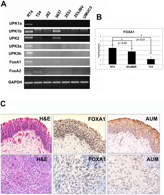
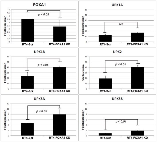
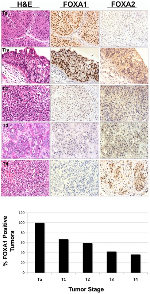
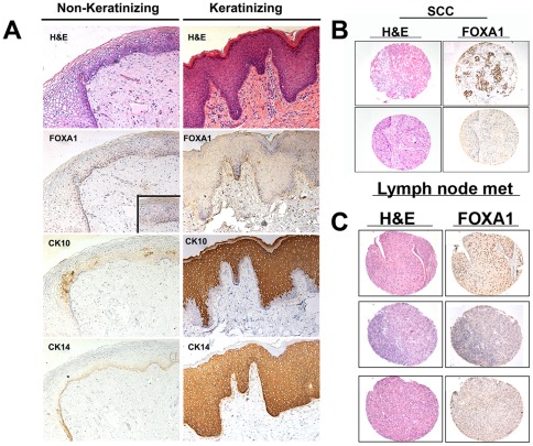
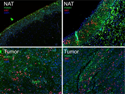
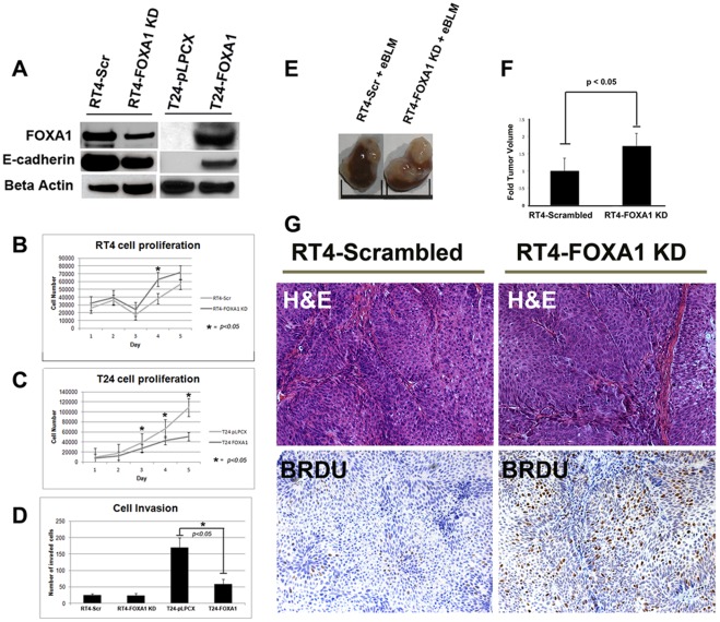
Approximately 50% of patients with muscle-invasive bladder cancer (MIBC) develop metastatic disease, which is almost invariably lethal. However, our understanding of pathways that drive aggressive behavior of MIBC is incomplete. Members of the FOXA subfamily of transcription factors are implicated in normal urogenital development and urologic malignancies. FOXA proteins are implicated in normal urothelial differentiation, but their role in bladder cancer is unknown. We examined FOXA expression in commonly used in vitro models of bladder cancer and in human bladder cancer specimens, and used a novel in vivo tissue recombination system to determine the functional significance of FOXA1 expression in bladder cancer. Logistic regression analysis showed decreased FOXA1 expression is associated with increasing tumor stage (p<0.001), and loss of FOXA1 is associated with high histologic grade (p<0.001). Also, we found that bladder urothelium that has undergone keratinizing squamous metaplasia, a precursor to the development of squamous cell carcinoma (SCC) exhibited loss of FOXA1 expression. Furthermore, 81% of cases of SCC of the bladder were negative for FOXA1 staining compared to only 40% of urothelial cell carcinomas. In addition, we showed that a subpopulation of FOXA1 negative urothelial tumor cells are highly proliferative. Knockdown of FOXA1 in RT4 bladder cancer cells resulted in increased expression of UPK1B, UPK2, UPK3A, and UPK3B, decreased E-cadherin expression and significantly increased cell proliferation, while overexpression of FOXA1 in T24 cells increased E-cadherin expression and significantly decreased cell growth and invasion. In vivo recombination of bladder cancer cells engineered to exhibit reduced FOXA1 expression with embryonic rat bladder mesenchyme and subsequent renal capsule engraftment resulted in enhanced tumor proliferation. These findings provide the first evidence linking loss of FOXA1 expression with histological subtypes of MIBC and urothelial cell proliferation, and suggest an important role for FOXA1 in the malignant phenotype of MIBC.
Conflict of interest statement
Figures
Comment in
-
Re: loss of the urothelial differentiation marker FOXA1 is associated with high grade, late stage bladder cancer and increased tumor proliferation.J Urol. 2013 Apr;189(4):1597. doi: 10.1016/j.juro.2012.12.067. Epub 2012 Dec 28. J Urol. 2013. PMID: 23561391 No abstract available.
References
-
- Siegel R, Ward E, Brawley O, Jemal A. Cancer statistics, 2011: the impact of eliminating socioeconomic and racial disparities on premature cancer deaths. CA: a cancer journal for clinicians. 2011;61:212–236. - PubMed
-
- Thomas JC, Oottamasathien S, Makari JH, Honea L, Sharif-Afshar AR, et al. Temporal-spatial protein expression in bladder tissue derived from embryonic stem cells. J Urol. 2008;180:1784–1789. - PubMed
-
- Varley CL, Bacon EJ, Holder JC, Southgate J. FOXA1 and IRF-1 intermediary transcriptional regulators of PPARgamma-induced urothelial cytodifferentiation. Cell Death Differ. 2009;16:103–114. - PubMed
Publication types
MeSH terms
Substances
Grants and funding
- K08 CA113452/CA/NCI NIH HHS/United States
- TL1 RR024978/RR/NCRR NIH HHS/United States
- KL2 RR024977/RR/NCRR NIH HHS/United States
- CA143971/CA/NCI NIH HHS/United States
- K08-CA113452/CA/NCI NIH HHS/United States
- UL1RR024975-01/RR/NCRR NIH HHS/United States
- R01 CA143971/CA/NCI NIH HHS/United States
- UL1 RR024975/RR/NCRR NIH HHS/United States
- T32 DK007563/DK/NIDDK NIH HHS/United States
- U54 CA163072/CA/NCI NIH HHS/United States
- T32 CA119925/CA/NCI NIH HHS/United States
- R01-DK55748/DK/NIDDK NIH HHS/United States
- R01 DK055748/DK/NIDDK NIH HHS/United States
LinkOut - more resources
Full Text Sources
Other Literature Sources
Medical
Research Materials
