. 2012;7(5):e36703.
doi: 10.1371/journal.pone.0036703. Epub 2012 May 8.

Strain-dependent differences in bone development, myeloid hyperplasia, morbidity and mortality in ptpn2-deficient mice

Affiliations

Strain-dependent differences in bone development, myeloid hyperplasia, morbidity and mortality in ptpn2-deficient mice

Florian Wiede et al. PLoS One. 2012.

Abstract

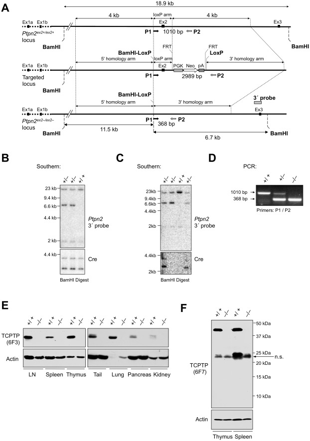
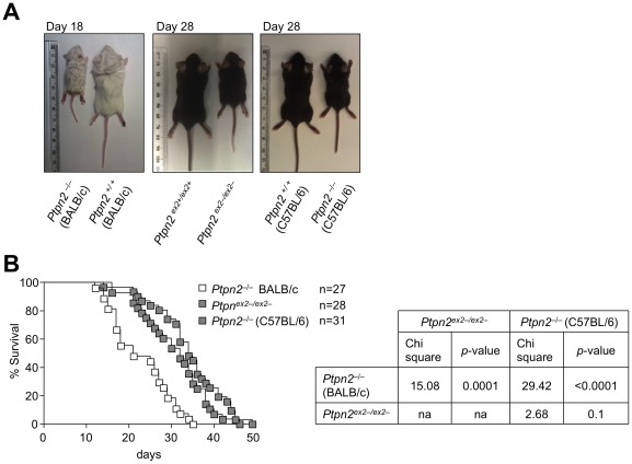
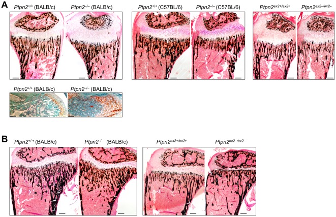
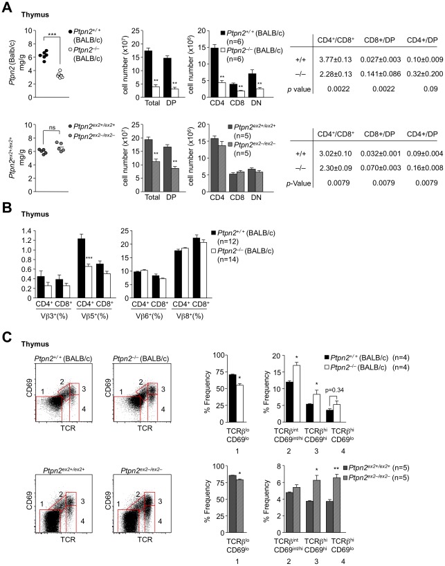
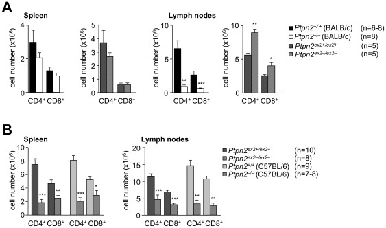
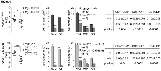
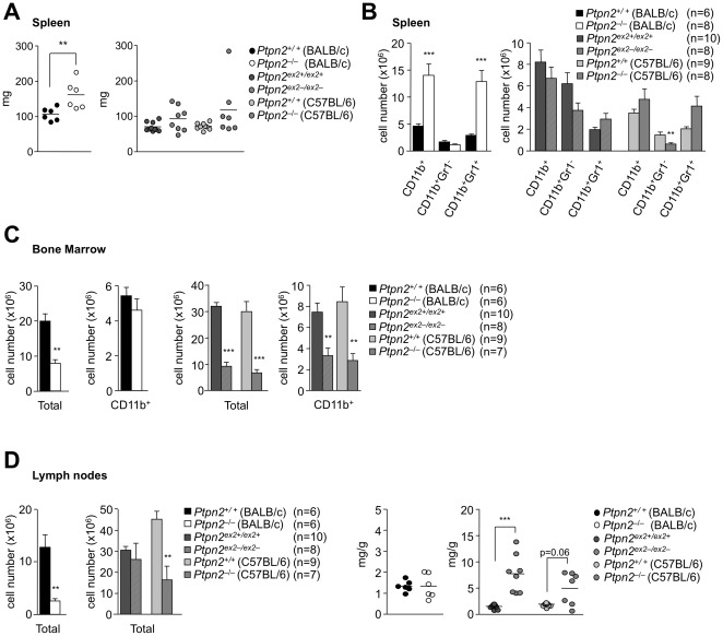
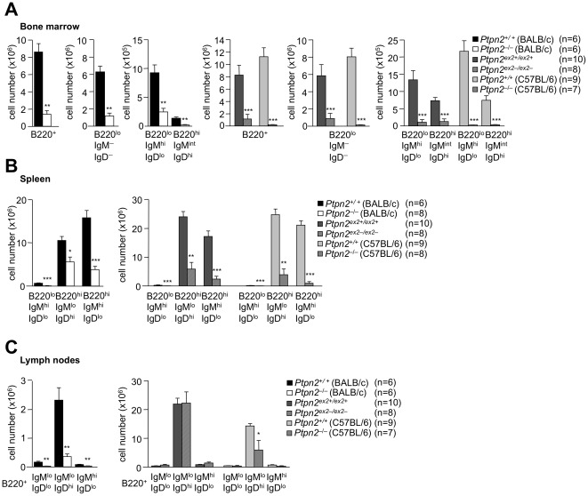
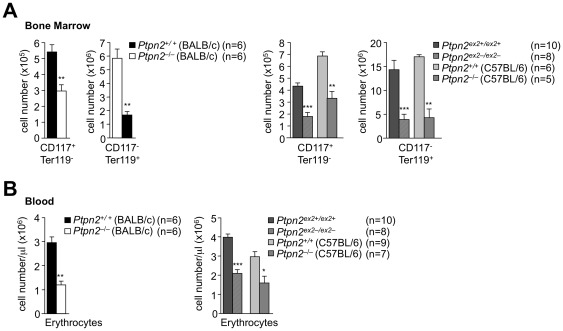
Single nucleotide polymorphisms in the gene encoding the protein tyrosine phosphatase TCPTP (encoded by PTPN2) have been linked with the development of autoimmunity. Here we have used Cre/LoxP recombination to generate Ptpn2(ex2-/ex2-) mice with a global deficiency in TCPTP on a C57BL/6 background and compared the phenotype of these mice to Ptpn2(-/-) mice (BALB/c-129SJ) generated previously by homologous recombination and backcrossed onto the BALB/c background. Ptpn2(ex2-/ex2-) mice exhibited growth retardation and a median survival of 32 days, as compared to 21 days for Ptpn2(-/-) (BALB/c) mice, but the overt signs of morbidity (hunched posture, piloerection, decreased mobility and diarrhoea) evident in Ptpn2(-/-) (BALB/c) mice were not detected in Ptpn2(ex2-/ex2-) mice. At 14 days of age, bone development was delayed in Ptpn2(-/-) (BALB/c) mice. This was associated with increased trabecular bone mass and decreased bone remodeling, a phenotype that was not evident in Ptpn2(ex2-/ex2-) mice. Ptpn2(ex2-/ex2-) mice had defects in erythropoiesis and B cell development as evident in Ptpn2(-/-) (BALB/c) mice, but not splenomegaly and did not exhibit an accumulation of myeloid cells in the spleen as seen in Ptpn2(-/-) (BALB/c) mice. Moreover, thymic atrophy, another feature of Ptpn2(-/-) (BALB/c) mice, was delayed in Ptpn2(ex2-/ex2-) mice and preceded by an increase in thymocyte positive selection and a concomitant increase in lymph node T cells. Backcrossing Ptpn2(-/-) (BALB/c) mice onto the C57BL/6 background largely recapitulated the phenotype of Ptpn2(ex2-/ex2-) mice. Taken together these results reaffirm TCPTP's important role in lymphocyte development and indicate that the effects on morbidity, mortality, bone development and the myeloid compartment are strain-dependent.


Conflict of interest statement

Competing Interests: The authors have declared that no competing interests exist.

Figures

Figure 1
Figure 1. Generation of Ptpn2ex2+/ex2− mice.
(a) Ptpn2 genomic locus and targeting design. (b–c) Southern blot analysis of Ptpn2ex2+/ex2+ (+/+) and Ptpn2ex2+/ex2 (+/−) mice. (b) Exon (Ex) 2 floxed mice mated with Oz-Cre mice for the germline deletion of exon 2 and removal of the Neomycin (Neo) resistance cassette. (c) Ex2-deleted mice were subsequently mated with C57BL/6 mice for the elimination of the Cre transgene. (d) PCR analysis of Ptpn2ex2+/ex2+ (+/+), Ptpn2ex2+/ex2 (+/−) and Ptpn2ex2 /ex2 (−/−) mice. (e–f) Immunoblot analysis of TCPTP expression in lymphoid and non-lymphoid tissues detected with antibodies to the TCPTP non-catalytic C-terminus (6F3) and TCPTP N-terminus (6F7; raised against the first 38 residues of TCPTP); a non-specific (n.s.) protein detected by 6F7 is indicated. Results are representative of three independent experiments.
Figure 2
Figure 2. Runtiness and mortality in Ptpn2−/− (BALB/c), Ptpn2ex2−/ex2− and Ptpn2−/− (C57BL/6) mice.
(a) Images of representative 18 day-old Ptpn2 / (BALB/c), 28 day-old Ptpn2ex2−/ex2− and 28 day-old Ptpn2 / (C57BL/6) mice along with corresponding littermate wild type control mice. (b) Kaplan-Meier survival curves for Ptpn2 / (BALB/c), Ptpn2ex2−/ex2− and Ptpn2 / (C57BL/6) mice. Statistical analyses were performed using a Log Rank (Mantel-Cox) test with one degree of freedom.
Figure 3
Figure 3. Bone development in Ptpn2−/− (BALB/c) mice.
Representative longitudinal sections of tibiae from (a) 14 day-old Ptpn2 / (BALB/c), Ptpn2 / (C57BL/6) and Ptpn2ex2−/ex2− mice, or (b) 21 day-old Ptpn2 / (BALB/c) and Ptpn2ex2−/ex2− mice and their corresponding littermate wild type controls stained with the von Kossa technique (mineralized bone stained black). In a) safranin O staining [only shown for 14 day-old Ptpn2+ /+ (BALB/c) and Ptpn2 / (BALB/c)] highlights the delayed cartilage (in orange) destruction; scale bar = 100 micron.
Figure 4
Figure 4. Thymocyte development in 18 day-old Ptpn2−/− (BALB/c) and Ptpn2ex2−/ex2− mice.
(a) Thymi from the indicated 18 day-old littermates were weighed using an analytical balance and normalised to body weight. Thymocytes were stained with fluorochrome-conjugated antibodies against CD4 and CD8 and analyzed by flow cytometry. Cells were gated for the DP, SP and DN stages and absolute numbers and the indicated ratios determined. (b) Thymocytes from Ptpn2+/+ and Ptpn2 / (BALB/c) littermates were stained with fluorochrome-conjugated antibodies against CD4, CD8, TCR-Vβ3, -5, -6 and -8 and analyzed by flow cytometry. Cells were gated for the CD4 and CD8 SP stages and the percentages of TCR-Vβ3, -5, -6 and -8 T cells determined. (c) Thymocytes from the indicated 18 day-old mice were stained with fluorochrome-conjugated antibodies against CD4, CD8, TCRβ and CD69 and analyzed by flow cytometry. Cells were gated for the different developmental stages according to the expression of the positive selection markers TCRβ and CD69. Results in (a–c) are means ± SEM for the indicated number of mice and are representative of at least two independent experiments; significance determined using a two-tailed Mann-Whitney U Test; *p<0.05 ** p<0.01 *** p<0.001.
Figure 5
Figure 5. Peripheral T cells in Ptpn2-deficient mice.
(a) Splenocytes or peripheral lymph node cells from 18 day-old Ptpn2 / (BALB/c) mice, 18 day-old Ptpn2ex2−/ex2− mice and their corresponding littermates were stained with fluorochrome-conjugated antibodies against CD4, CD8 and TCRβ and analysed by flow cytometry and absolute numbers determined. (b) Splenocytes or peripheral lymph node cells from 28 day-old Ptpn2ex2−/ex2− mice, 29 day-old Ptpn2−/− (C57BL/6) mice and their corresponding littermates were stained with fluorochrome-conjugated antibodies against CD4, CD8 and TCRβ and analysed by flow cytometry and absolute numbers determined. Results shown in (a–b) are means ± SEM for the indicated number of mice and are representative of at least two independent experiments; significance determined using a two-tailed Mann-Whitney U Test; *p<0.05 ** p<0.01 *** p<0.001.
Figure 6
Figure 6. Thymoycte development in 28 day-old Ptpn2ex2−/ex2− and Ptpn2−/− (C57BL/6) mice.
Thymi from the indicated littermate mice were weighed using an analytical balance and normalised to body weight. Thymocytes were stained with fluorochrome-conjugated antibodies against CD4 and CD8 and analyzed by flow cytometry. Cells were gated for the DP, SP and DN stages and absolute numbers and the indicated ratios determined. Results shown are means ± SEM for the indicated number of mice and are representative of at least two independent experiments; significance determined using a two-tailed Mann-Whitney U Test; *p<0.05 *** p<0.001.
Figure 7
Figure 7. Splenomegaly, myeloid development and lymphadenopathy in Ptpn2-deficient mice.
(a) Spleen weights from 18 day-old Ptpn2 / (BALB/c) and 28 day-old Ptpn2ex2−/ex2− and Ptpn2 / (C57BL/6) mice and corresponding wild type littermate mice. (b) Splenocytes or (c) bone marrow (tibia and femur) cells were stained with fluorochrome-conjugated antibodies to CD11b and Gr1 and analysed by flow cytometry; absolute numbers of CD11b+, CD11b+Gr1+ and CD11b+Gr1 were determined (d) Weights of pooled peripheral lymph nodes from 18 day-old Ptpn2 / (BALB/c) and 28 day-old Ptpn2ex2−/ex2− and Ptpn2 / (C57BL/6) mice and corresponding wild type littermate mice. Results shown in (a–d) are means ± SEM for the indicated number of mice and are representative of at least two independent experiments; significance determined using a two-tailed Mann-Whitney U Test; *p<0.05 ** p<0.01 *** p<0.001.
Figure 8
Figure 8. B cell development in Ptpn2-deficient mice.
Bone marrow cells (pooled from one tibia and one femur), splenocytes or pooled peripheral lymph node cells from 18 day-old Ptpn2 / (BALB/c), 28 day-old Ptpn2ex2−/ex2− and 29 day-old Ptpn2 / (C57BL/6) mice and their corresponding littermates (a–c) were stained with fluorochrome-conjugated antibodies to CD45R(B220), IgM and IgD and analysed by flow cytometry. Absolute numbers of progenitor (B220loIgMloIgDlo), immature (B220loIgMhi IgDlo), mature (B220hiIgMintIgDhi) B cells and B1 (B220lo IgMhiIgDlo), follicular (B220hiIgMloIgDhi) and marginal zone (B220hiIgMhiIgDlo) B cells were determined. Results shown are means ± SEM for the indicated number; significance determined using a two-tailed Mann-Whitney U Test; *p<0.05 ** p<0.01 *** p<0.001.
Figure 9
Figure 9. Erythrocyte development in Ptpn2-deficient mice.
(a) Bone marrow cells (pooled from one tibia and one femur) from 18 day-old Ptpn2 / (BALB/c) and 28 day-old Ptpn2ex2−/ex2− and Ptpn2 / (C57BL/6) mice and corresponding littermate wild type control mice were stained with fluorochrome-conjugated antibodies to CD117 and Ter119 and analysed by flow cytometry. Absolute numbers of progenitor (CD117+ Ter119) and erythroid (CD117Ter119+) cells were determined. (b) Red blood cells were quantified (identified in the forward and side scatter according to size and granularity) by flow cytometry. Results shown are means ± SEM for the indicated number of mice and are representative of two independent experiments; significance determined using a two-tailed Mann-Whitney U Test; *p<0.05 ** p<0.01 *** p<0.001.

References

    1. Tiganis T, Bennett AM. Protein tyrosine phosphatase function: the substrate perspective. Biochem J. 2007;402:1–15. - PMC - PubMed
    1. Todd JA, Walker NM, Cooper JD, Smyth DJ, Downes K, et al. Robust associations of four new chromosome regions from genome-wide analyses of type 1 diabetes. Nat Genet. 2007;39:857–864. - PMC - PubMed
    1. Smyth DJ, Plagnol V, Walker NM, Cooper JD, Downes K, et al. Shared and distinct genetic variants in type 1 diabetes and celiac disease. N Engl J Med. 2008;359:2767–2777. - PMC - PubMed
    1. The Wellcome Trust Case Control Consortium. Genome-wide association study of 14,000 cases of seven common diseases and 3,000 shared controls. Nature. 2007;447:661–678. - PMC - PubMed
    1. Long SA, Cerosaletti K, Wan JY, Ho JC, Tatum M, et al. An autoimmune-associated variant in PTPN2 reveals an impairment of IL-2R signaling in CD4(+) T cells. Genes Immun. 2011;12:116–125. - PMC - PubMed

Publication types

Substances