. 2012;7(5):e36714.
doi: 10.1371/journal.pone.0036714. Epub 2012 May 9.

Chitin binding proteins act synergistically with chitinases in Serratia proteamaculans 568

Affiliations

Chitin binding proteins act synergistically with chitinases in Serratia proteamaculans 568

Pallinti Purushotham et al. PLoS One. 2012.

Abstract

Genome sequence of Serratia proteamaculans 568 revealed the presence of three family 33 chitin binding proteins (CBPs). The three Sp CBPs (Sp CBP21, Sp CBP28 and Sp CBP50) were heterologously expressed and purified. Sp CBP21 and Sp CBP50 showed binding preference to β-chitin, while Sp CBP28 did not bind to chitin and cellulose substrates. Both Sp CBP21 and Sp CBP50 were synergistic with four chitinases from S. proteamaculans 568 (Sp ChiA, Sp ChiB, Sp ChiC and Sp ChiD) in degradation of α- and β-chitin, especially in the presence of external electron donor (reduced glutathione). Sp ChiD benefited most from Sp CBP21 or Sp CBP50 on α-chitin, while Sp ChiB and Sp ChiD had major advantage with these Sp CBPs on β-chitin. Dose responsive studies indicated that both the Sp CBPs exhibit synergism ≥ 0.2 µM. The addition of both Sp CBP21 and Sp CBP50 in different ratios to a synergistic mixture did not significantly increase the activity. Highly conserved polar residues, important in binding and activity of CBP21 from S. marcescens (Sm CBP21), were present in Sp CBP21 and Sp CBP50, while Sp CBP28 had only one such polar residue. The inability of Sp CBP28 to bind to the test substrates could be attributed to the absence of important polar residues.


Conflict of interest statement

Competing Interests: The authors have declared that no competing interests exist.

Figures

Figure 1
Figure 1. Binding of Sp CBPs to insoluble polymeric substrates.
The reaction mixture (1 mL) containing 100 µg of Sp CBP21/Sp CBP50 and 1 mg of one of the insoluble substrates (α-chitin, β-chitin, colloidal chitin and Avicel) was incubated in 50 mM sodium phosphate buffer pH 7.0 under constant shaking at 1300 rpm at 37°C for 24 h. (A and B) The amount of bound protein was calculated as the difference in protein concentration before and after incubation with the insoluble substrates, (C and D) Decrease in free protein concentration after binding to α- and β-chitin was determined at different time points till 24 h. (A and C) Sp CBP21, (B and D) Sp CBP50. Vertical bars represent standard deviation of triplicate experiments.
Figure 2
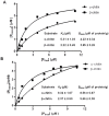
Figure 2. Equilibrium adsorption isotherms of Sp CBP21 and Sp CBP50 to α- and β-chitin.
The reaction assay (1 mL) containing 1.0 mg substrates (α- and β- chitin) and varied concentrations of Sp CBP21 and Sp CBP50 starting from 0 to 10.0 µM was incubated (Sp CBP21 with α- and β-chitin, 12 h and 6 h, respectively; Sp CBP50 with α- and β-chitin, 12 h) at 37°C. The reaction mixtures were centrifuged and concentration of bound protein (Pbound) and un-bound free protein (Pfree) was determined and plotted to fit into GraphPad Prism software version 5.0. All data sets were fitted to the equation for one-site binding by non-linear regression function, and to calculate B max and K d using GraphPad Prism software version 5.0. (A and B) The K d and B max values of Sp CBP21 and Sp CBP50 were shown in the inset table.
Figure 3
Figure 3. Sequence alignment and domain organisation for Sp CBPs.
(A) Full-length sequences of Sp CBP21, Sp CBP28, Sp CBP50 and Sm CBP21 (CBP21 from S. marcescens) were aligned using clustalw2. Residues that are thought to be located in the binding surface for chitin present in Sm CBP21, Sp CBP21, Sp CBP50 and not present in Sp CBP28 are shaded in yellow (as derived from the crystal structure of Sm CBP21, as well as mutagenesis studies [7], [8]). Residue involved in the chitin-binding and functional properties of Sm CBP21 but also conserved in Sp CBP28 are shaded grey. The arrow indicates the terminal amino acid of the N-terminal signal sequence for respective CBPs. (B) The sequences of Sp CBPs were submitted to SMART domain data base (http://smart.embl-heidelberg.de/). The part indicated in red colour shows the signal peptide and the region Chitin_bind_3 indicates the chitin binding domain.
Figure 4
Figure 4. The 3D models of Sp CBP21 and Sp CBP50.
(A and B) The models Sp CBP21 and ChBD region of Sp CBP50 were generated by Modeller9v8 (http://www.salilab.org/modeller/) using Sm CBP21 (PDB ID: 2BEM) as structure template. Residues important for chitin binding were shown in sticks representation with carbon, oxygen and nitrogen atoms colored light green, red and dark blue, respectively. The figures were prepared using PyMOL (http://www.pymol.org/), (C and D) Stereo view of the superimposed structure of Sp CBP21 and Sp CBP50 (green) with Sm CBP21 (red), respectively.
Figure 5
Figure 5. Degradation of α- and β-chitin by Sp chitinases in the absence or presence of Sp CBP21, Sp CBP50 and reduced glutathione.
Reaction mixture (1 mL) containing 0.25 mg/mL of chitin substrates (α- and β-chitin), 1 µM of Sp chitinase (Sp ChiA/Sp ChiB/Sp ChiC/Sp ChiD) were incubated with 0.3 µM Sp CBP21 or Sp CBP50 and 1.0 mM reduced glutathione in 50 mM sodium phosphate buffer pH 7.0. After incubation at 37°C for 7 days at 1000 rpm, after every 24 h, 100 µL of reaction mixture was transferred. To this 100 µL of 0.02N NaOH was added to stop the reaction and stored at −20°C until products quantification by standard reducing end assay. Vertical bars represent standard deviation of triplicate experiments. (A and B) Degradation of α- and β-chitin by Sp chitinases in the presence/absence of Sp CBP21 and reduced glutathione (RG), (C and D) degradation of α- and β-chitin by Sp chitinases in the presence/absence of Sp CBP50 and reduced glutathione (RG). Sp CBP21+RG or Sp CBP50+RG: Sp CBP21/Sp CBP50 and reduced glutathione, Sp CBP21+ or Sp CBP50+: only Sp CBP21/Sp CBP50 without reduced gluthathione, Sp CBP21- or Sp CBP50 -: without Sp CBP21/Sp CBP50 and reduced glutathione.
Figure 6
Figure 6. β-chitin hydrolysis enhancing effects of Sp CBP21 and Sp CBP50 with Sp ChiD.
Reaction mixture (1 mL) containing 0.25 mg/mL of β-chitin, 0.25 µM/0.50 µM/0.75 µM/1.0 µM Sp ChiD incubated individually with 0.3 µM of Sp CBP21/Sp CBP50 or combining both Sp CBP21 and Sp CBP50 (0.15 µM +0.15 µM/0.30 µM +0.30 µM), in 50 mM sodium phosphate buffer pH 7.0. After incubation at 37°C for 24 h at 1000 rpm, 100 µL of reaction mixture was transferred. To this 100 µL of 0.02N NaOH was added to stop the reaction and stored at −20°C until products quantification by standard reducing end assay. Vertical bars represent standard deviation of triplicate experiments.

References

    1. Rinaudo M. Chitin and chitosan: Properties and applications. Prog Polym Sci. 2006;31:603–632.
    1. Neeraja Ch, Anil K, Purushotham P, Suma K, Sarma PVSRN, et al. Biotechnological approaches to develop bacterial chitinases as a bioshield against fungal diseases of plants. Crit Rev Biotechnol. 2010c;30:231–241. - PubMed
    1. Zamil SS, Ahmad S, Choi MH, Yoon SC. Production of poly-N-acetylglucosamine by Staphylococcus saprophyticus BMSZ711: Characterization and production optimization. Bioresour Technol. 2010;101:7177–7180. - PubMed
    1. Vaaje-Kolstad G, Bunæs AC, Mathiesen G, Eijsink VGH. The chitinolytic system of Lactococcus lactis ssp. Lactis comprises a nonprocessive chitinase and a chitin-binding protein that promotes the degradation of α- and β-chitin. FEBS J. 2009;276:2402–2415. - PubMed
    1. Orikoshi H, Nakayama S, Miyamoto K, Hanato C, Yasuda M, et al. Roles of four chitinases (ChiA, ChiB, ChiC, and ChiD) in the chitin degradation system of marine bacterium Alteromonas sp. Strain O-7. Appl Environ Microbiol. 2005;71:1811–1815. - PMC - PubMed

Publication types

MeSH terms

LinkOut - more resources