. 2012;7(5):e37075.
doi: 10.1371/journal.pone.0037075. Epub 2012 May 10.

Soluble epoxide hydrolase activity determines the severity of ischemia-reperfusion injury in kidney

Affiliations

Soluble epoxide hydrolase activity determines the severity of ischemia-reperfusion injury in kidney

Jung Pyo Lee et al. PLoS One. 2012.

Abstract

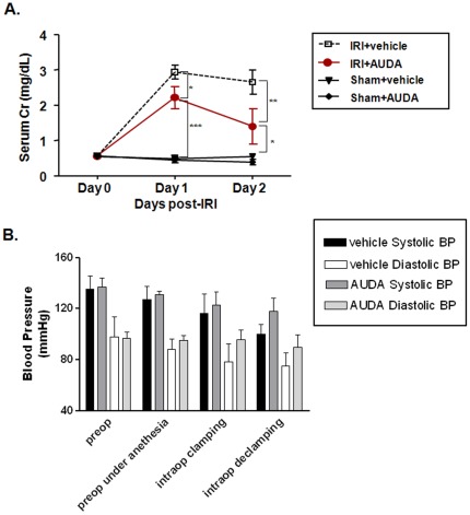
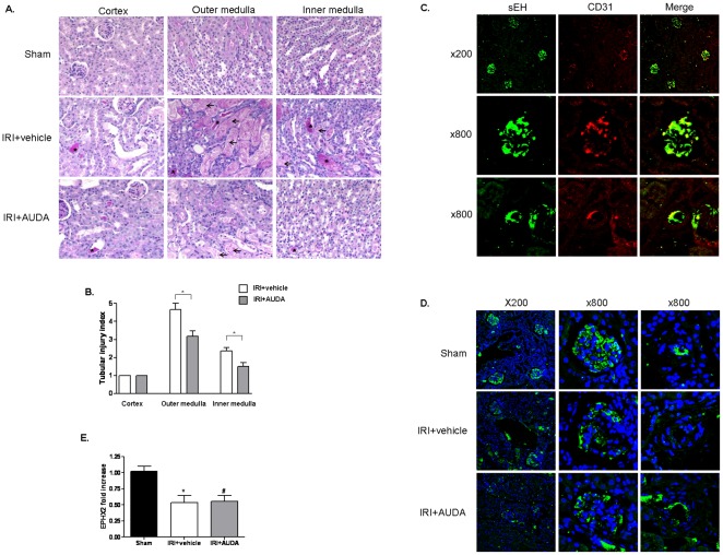
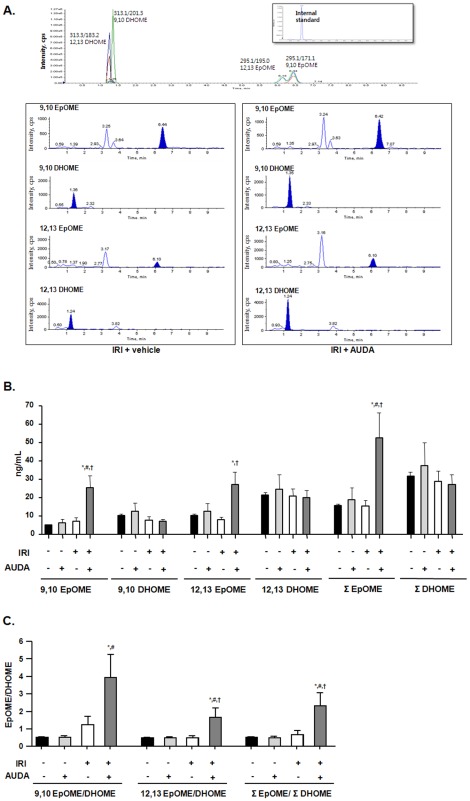
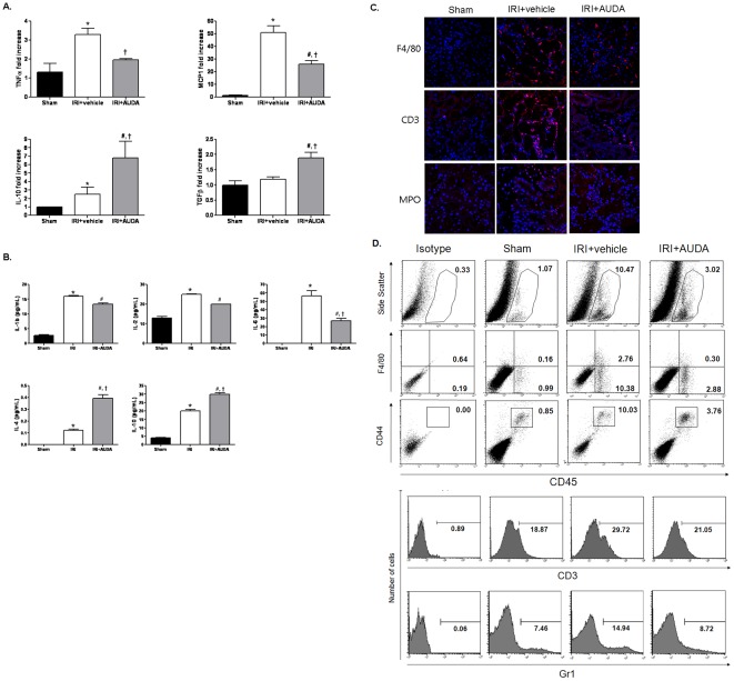
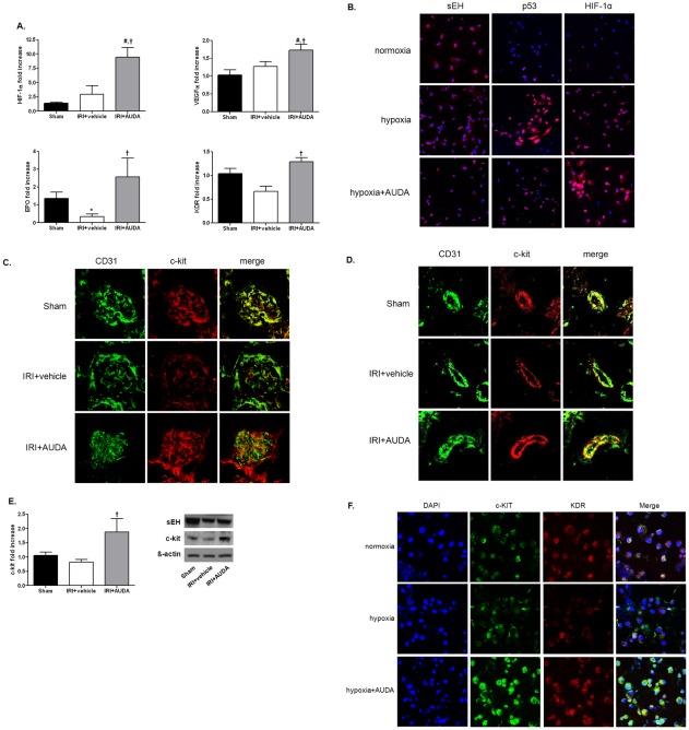
Soluble epoxide hydrolase (sEH) in endothelial cells determines the plasma concentrations of epoxyeicosatrienoic acids (EETs), which may act as vasoactive agents to control vascular tone. We hypothesized that the regulation of sEH activity may have a therapeutic value in preventing acute kidney injury by controlling the concentration of EETs. In this study, we therefore induced ischemia-reperfusion injury (IRI) in C57BL/6 mice and controlled sEH activity by intraperitoneal administration of the sEH inhibitor 12-(3-adamantan-1-ylureido)-dodecanoic acid (AUDA). The deterioration of kidney function induced by IRI was partially moderated and prevented by AUDA treatment. In addition, AUDA treatment significantly attenuated tubular necrosis induced by IRI. Ischemic injury induced the down-regulation of sEH, and AUDA administration had no effect on the expression pattern of sEH induced by IRI. In vivo sEH activity was assessed by measuring the substrate epoxyoctadecenoic acid (EpOME) and its metabolite dihydroxyoctadec-12-enoic acid (DHOME). Ischemic injury had no effects on the plasma concentrations of EpOME and DHOME, but inhibition of sEH by AUDA significantly increased plasma EpOME and the EpOME/DHOME ratio. The protective effect of the sEH inhibitor was achieved by suppression of proinflammatory cytokines and up-regulation of regulatory cytokines. AUDA treatment prevented the intrarenal infiltration of inflammatory cells, but promoted endothelial cell migration and neovascularization. The results of this study suggest that treatment with sEH inhibitors can reduce acute kidney injury.


Conflict of interest statement

Competing Interests: The authors have declared that no competing interests exist.

Figures

Figure 1
Figure 1. Role of soluble epoxide hydrolase (sEH) activity in ischemia-reperfusion injury (IRI) in kidneys.
A: The adamantyl alkyl urea-based sEH inhibitor, 12-(3-adamantan-1-ylureido)-dodecanoic acid (AUDA) reduced IRI in the kidney. All values are given as means±S.E. (n = 6 per group for each experiment). Data represent one of three independent experiments. Day 0, before bilateral IRI; day 1, 24 h after bilateral IRI; day 2, 48 h after bilateral IRI (two-way ANOVA with Bonferroni post-testing; *P<0.05; **P<0.01; ***P<0.001). B: Administration of AUDA had no effect on blood pressure during the procedure.
Figure 2
Figure 2. Effects of 12-(3-adamantan-1-ylureido)-dodecanoic acid (AUDA) on renal expression of soluble epoxide hydrolase (sEH) in ischemia-reperfusion injury (IRI) in kidneys.
A: Histological changes were consistent with the functional changes (×200). IRI induced tubular necrosis, consisting of disruption and sloughing of tubular epithelial cells. Arrows indicate necrotic tubules, and asterisks indicate tubular casts. Tubular injury was increased in disease-control mice compared to AUDA-treated mice. B: Expression was quantified by a renal pathologist in a blinded fashion (*P<0.05). Scores ranged from 1–5, based on the percentage of tubules affected (1: <10%; 2: 10–25%; 3: 25–50%; 4: 50–75%; 5: >75%). C: sEH was expressed in the endothelium of intraglomerular capillary loops and peritubular capillaries. D and E: Ischemic injury induced the down-regulation of sEH, but AUDA administration had no effect on sEH expression. DAPI was used as counterstaining. EPHX2, gene encoding sEH.
Figure 3
Figure 3. Regulation of soluble epoxide hydrolase (sEH) activity by adamantyl alkyl urea-based sEH inhibitor (AUDA) in renal ischemia-reperfusion injury (IRI).
A: Plasma epoxyoctadecenoic acid (EpOME) and dihydroxyoctadec-12-enoic acid (DHOME) levels were quantified to investigate the enzyme activity of sEH. B: 9,10-, 12,13-, and total EpOME plasma concentrations were significantly increased in response to AUDA in renal IRI. C: The EpOME/DHOME ratio was significantly increased (*P<0.05 compared to sham+vehicle; #P<0.05 compared to sham+AUDA; †P<0.05 compared to IRI+vehicle).
Figure 4
Figure 4. Effects of soluble epoxide hydrolase (sEH) inhibitor on the pro-/anti-inflammatory microenvironment in injured kidneys.
A: Proinflammatory cytokines TNF-α and MCP-1 were significantly suppressed, while IL-10 and TGF-β were enhanced by treatment with 12-(3-adamantan-1-ylureido)-dodecanoic acid (AUDA), as shown by real-time PCR. B: The proinflammatory cytokine IL-6 was decreased and the regulatory cytokines IL-4 and IL-10 were augmented by AUDA, as shown by multiplex cytokine assay. (*P<0.05 compared to sham; #P<0.05 compared to sham; †P<0.05 compared to IRI+vehicle). C: AUDA decreased the infiltration of inflammatory cells (macrophages (F4/80), lymphocytes (CD3), and neutrophils (MPO)) mainly trafficked in the interstitial area. D: AUDA attenuated the infiltration of macrophages/monocytes and T cells expressing CD3, as shown by flow cytometry. F4/80, marker for pan-macrophage; CD44, indicative marker for effector-memory T-cells; CD45, leukocyte common antigen; Gr1, myeloid differentiation antigen.
Figure 5
Figure 5. Protective effects of soluble epoxide hydrolase (sEH) inhibitor on hypoxic damage via neovascularization.
A: Hypoxia inducible factor (HIF)-1α, vascular endothelial growth factor (VEGF), VEGF receptor-2 (KDR), and erythropoietin (EPO) were enhanced by 12-(3-adamantan-1-ylureido)-dodecanoic acid (AUDA) administration. (*P<0.05 compared to sham; #P<0.05 compared to sham; †P<0.05 compared to ischemia-reperfusion injury (IRI) +vehicle). B: Hypoxia induced the down-regulation of sEH in human umbilical vein endothelial cells (HUVECs). Cells were incubated with or without AUDA (10 µM) under hypoxic (1% O2) or normoxic conditions (20% O2) for 24 h. Apoptosis of HUVECs was assessed by p53 expression. Hypoxia induced apoptosis in HUVECs, but AUDA treatment reduced apoptosis associated with enhancement of HIF-1α. DAPI was used for counterstaining (magnification, ×400). C and D: c-kit (CD117) expression decreased after IRI, but was amplified by AUDA administration (magnification, ×800). E: AUDA treatment significantly enhanced c-kit expression. Data represent the results of one of three independent experiments (n = 6 per group; †P<0.05 compared to IRI+vehicle). F: AUDA treatment increased c-kit and KDR expression levels in HUVECs exposed to hypoxia.

References

    1. Santos WJ, Zanetta DM, Pires AC, Lobo SM, Lima EQ, et al. Patients with ischaemic, mixed and nephrotoxic acute tubular necrosis in the intensive care unit–a homogeneous population? Crit Care. 2006;10:R68. - PMC - PubMed
    1. Levy EM, Viscoli CM, Horwitz RI. The effect of acute renal failure on mortality. A cohort analysis. JAMA. 1996;275:1489–1494. - PubMed
    1. Yang SH, Lee JP, Jang HR, Cha RH, Han SS, et al. Sulfatide-Reactive Natural Killer T Cells Abrogate Ischemia-Reperfusion Injury. J Am Soc Nephrol. 2011;22:1305–1314. - PMC - PubMed
    1. Jang HR, Ko GJ, Wasowska BA, Rabb H. The interaction between ischemia-reperfusion and immune responses in the kidney. J Mol Med. 2009;87:859–864. - PubMed
    1. Campbell WB, Gebremedhin D, Pratt PF, Harder DR. Identification of epoxyeicosatrienoic acids as endothelium-derived hyperpolarizing factors. Circ Res. 1996;78:415–423. - PubMed

Publication types