. 2012 Apr 28;4(4):128-34.
doi: 10.4329/wjr.v4.i4.128.

The 7th lung cancer TNM classification and staging system: Review of the changes and implications

Affiliations

The 7th lung cancer TNM classification and staging system: Review of the changes and implications

Saeed Mirsadraee et al. World J Radiol. .

Abstract

Lung cancer is the most common cause of death from cancer in males, accounting for more than 1.4 million deaths in 2008. It is a growing concern in China, Asia and Africa as well. Accurate staging of the disease is an important part of the management as it provides estimation of patient's prognosis and identifies treatment sterategies. It also helps to build a database for future staging projects. A major revision of lung cancer staging has been announced with effect from January 2010. The new classification is based on a larger surgical and non-surgical cohort of patients, and thus more accurate in terms of outcome prediction compared to the previous classification. There are several original papers regarding this new classification which give comprehensive description of the methodology, the changes in the staging and the statistical analysis. This overview is a simplified description of the changes in the new classification and their potential impact on patients' treatment and prognosis.

Keywords: Lung cancer staging; Lung neoplasms; Neoplasm staging.
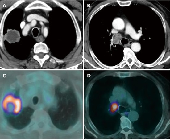
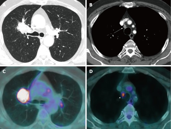
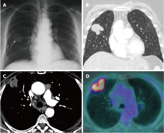
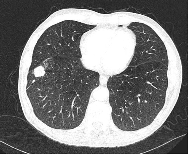

Figures

Figure 1
Figure 1
Contrast enhanced computed tomography of the chest viewed with mediastinal window settings demonstrated a large necrotic mass in the right upper lobe (A) with enlarged necrotic paratracheal lymph nodes in station 4R (arrowhead) (B). C, D: Positron emission tomography-computed tomography demonstrated high [18F]-2-fluoro-2-deoxy-d-glucose-uptake in the mass and lymph nodes confirming a T2aN2 lung cancer.
Figure 2
Figure 2
Contrast enhanced thoracic computed tomography viewed with lung window settings shows a 5.2 cm ill-defined mass suspected of lung cancer abutting the right lung hilum, causing narrowing of the upper lobe bronchus (A). B: Review of images on mediastinal window settings showed a paratracheal lymph node with short axis smaller than 1 cm (arrow) in station 2R; C and D: Positron emission tomography-computed tomography demonstrated a high [18F]-2-fluoro-2-deoxy-d-glucose-uptake of both mass and 2R node (arrowhead), resulting in an up-staging of the disease from T2bN1 (CT classification) to T2bN2. This finding was further confirmed by mediastinoscopy sampling.
Figure 3
Figure 3
Example of cross sectional imaging nodal staging pitfalls. A: A mass of the right lung (arrow) was identified on chest radiograph; B: A 4 cm spiculated mass suspected of lung cancer in the right upper lobe was confirmed by computed tomography (CT); C, D: Contrast enhanced CT demonstrated enlarged lymph nodes (> 1 cm in short axis; arrowheads) in ipsi- and contra-lateral mediastinal nodal stations (C) (T2aN3), positron emission tomography-computed tomography (PET-CT) (D) showed high metabolic activity of the parenchymal lesion but no nodal [18F]-2-fluoro-2-deoxy-d-glucose uptake (PET-CT staging: T2aN0M0). No metastatic nodes were demonstrated by endoscopic ultrasound-needle aspiration and mediastinoscopy, and surgical staging was in full agreement with PET-CT (pT2N0 adenocarcinoma).
Figure 4
Figure 4
A 2.5 cm mass (arrow) in the right lower lobe was classified as T1 in 6th edition and is described as T1b in the 7th classification. This did not change the staging (see Table 3).
Figure 5
Figure 5
A 7.5 cm necrotic mass in the right lower lobe (arrow). As far as size is concerned, the T classification is upstaged from T2 to T3 and therefore a worse prognosis is expected.
Figure 6
Figure 6
Example of change in classification status. A: A mass in the right upper lobe and a satellite nodule (arrow) in the right middle lobe. This tumour is reclassified from M1 to T4. Patient may be considered for treatment (e.g., pneumonectomy) and a better prognosis is to be expected; B: A tumour with a satellite nodule (arrowhead) both in right lower lobe; this was therefore downstaged from T4 to T3 in the new classification and surgical treatment was considered.
Figure 7
Figure 7
A patient with a necrotic mass in the right lower lobe (short arrow). As seen in this axial contrast enhanced computed tomography, there is pleural (p) and pericardial (pc) effusions which were confirmed to be malignant. This will be re-classified from T4 to M1a indicating worse estimated prognosis.
Figure 8
Figure 8
Lymphangitis caused by a large right upper lobe mass, note thickening of interlobular septa (arrowhead) and perilobular nodules (arrow). There is no provision for lymphangitis in the tumour, node, metastasis classification.

References

    1. Available from: http://www.ons.gov.uk/ons/rel/vsob1/mortality-statistics--deaths-registe....
    1. Jemal A, Bray F, Center MM, Ferlay J, Ward E, Forman D. Global cancer statistics. CA Cancer J Clin. 2011;61:69–90. - PubMed
    1. Ferlay J, Bray F, Pisani P, Parkin DM. GLOBOCAN 2000: cancer incidence, mortality and prevalence worldwide. IARC CancerBase No. 5 [CD-ROM]. Version 1.1. Lyon: IARC Press; 2001.
    1. Rami-Porta R, Crowley JJ, Goldstraw P. The revised TNM staging system for lung cancer. Ann Thorac Cardiovasc Surg. 2009;15:4–9. - PubMed
    1. Sobin LH, Wittekind CH. TNM classification of malignant tumours. 6th ed. Hoboken, NJ: John Wiley&Sons; 2002.