

Review
doi: 10.3390/v4030363.
Epub 2012 Mar 8.
The coronavirus E protein: assembly and beyond
Affiliations
- PMID: 22590676
- PMCID: PMC3347032
- DOI: 10.3390/v4030363
Item in Clipboard
Review
The coronavirus E protein: assembly and beyond
Viruses.
2012 Mar.
Abstract
The coronavirus E protein is a small membrane protein that has an important role in the assembly of virions. Recent studies have indicated that the E protein has functions during infection beyond assembly, including in virus egress and in the host stress response. Additionally, the E protein has ion channel activity, interacts with host proteins, and may have multiple membrane topologies. The goal of this review is to highlight the properties and functions of the E protein, and speculate on how they may be related.
Keywords: Golgi complex; coronavirus assembly; envelope protein; ion channel; membrane protein topology.
Figures
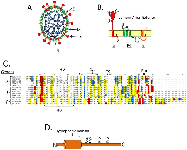
Primary structure of the Coronavirus (CoV) E protein. (A) A cartoon depicting a CoV virion. The structural proteins are labeled. (B) The three major CoV structural proteins in the virion envelope. Oligosaccharides are shown on S and M. A single topology is shown for E, see below for discussion on E protein topology. (C) A multiple sequence alignment of several different CoV E proteins. The hydrophobic domain (HD) is bracketed. The CoV genera (alpha, beta, and gamma) are denoted on the left of the multiple sequence alignment. Positively charged residues are shown in blue, negatively charged residues are shown in red, and polar uncharged residues are shown in yellow. The conserved Cys and Pro residues are labeled with a bracket or an asterisk, respectively. The multiple sequence alignment of CoV E proteins was carried out with ClusalW2 at the European Bioinformatics Institutes server, and Jalview software version 2 was used to generate the figure [16,17]. (D) Cartoon depiction of the E protein with the hydrophobic domain shown as a cylinder and the conserved Cys and Pro residues labeled.
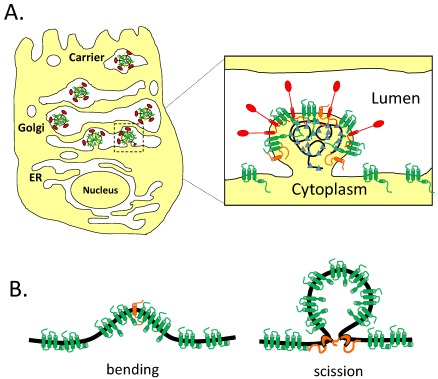
CoVs assemble and bud intracellularly at the ERGIC. (A) Newly formed virions bud into the lumen of the ERGIC and traverse the secretory pathway for egress. (B) Potential roles for E in assembly. The E protein is shown in orange and the M protein is shown in green. CoV E could help to bend membranes or play a role in membrane scission.
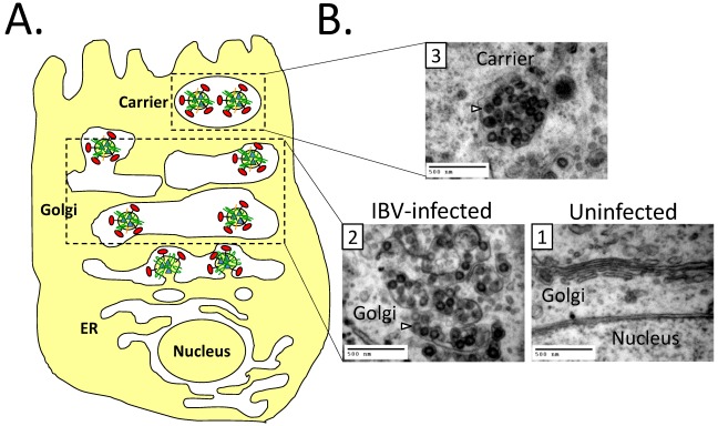
CoVs use the secretory pathway for egress. (A) A cartoon depicting virions within the Golgi complex. The Golgi cisternae are enlarged and fragmented during infection, possibly to aid in the trafficking of virions. (B) Transmission electron micrographs of uninfected or IBV infected Vero cells. (1) The Golgi complex of an uninfected cell; (2) The Golgi complex of an IBV infected cell with enlarged Golgi cisternae; (3) A putative virion carrier. Arrows denote virions. Scale bar is 500 nm. For images (2) and (3) Vero cells were infected at an moi of 0.1, and samples were fixed and processed 14 hrs post infection as described in [31].
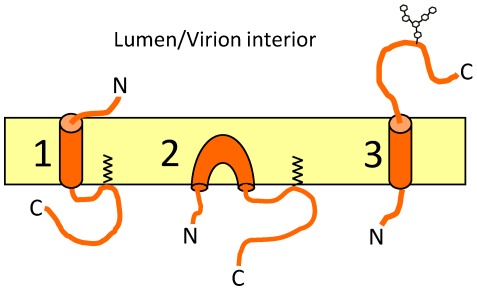
Topologies of the CoV E protein. The three proposed topologies of CoV E. (1) shows a type III membrane protein; (2) shows a membrane hairpin; and (3) shows a type II membrane protein with a putative N-linked oligosaccharide.
References
-
- Hogue B.G., Machamer C.E. Coronavirus structural proteins and virus assembly. In: Perlman S., Gallagher T., Snijder E.J., editors. Nidoviruses. ASM Press; Washington, DC, USA: 2008. pp. 179–200.
-
- Boursnell M.E., Binns M.M., Brown T.D. Sequencing of coronavirus IBV genomic RNA: Three open reading frames in the 5' 'unique' region of mRNA D. J. Gen. Virol. 1985;66:2253–2258. - PubMed
Publication types
MeSH terms
Substances
Grants and funding
LinkOut - more resources
Full Text Sources
Molecular Biology Databases
