Review
. 2012 Apr;4(4):613-36.
doi: 10.3390/v4040613. Epub 2012 Apr 19.

Paramyxovirus fusion and entry: multiple paths to a common end

Affiliations
Review

Paramyxovirus fusion and entry: multiple paths to a common end

Andres Chang et al. Viruses. 2012 Apr.

Abstract

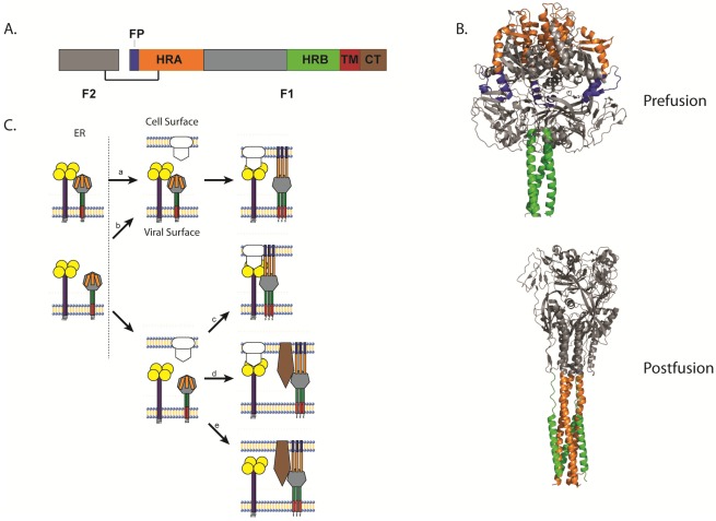
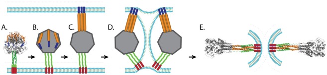
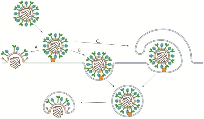
The paramyxovirus family contains many common human pathogenic viruses, including measles, mumps, the parainfluenza viruses, respiratory syncytial virus, human metapneumovirus, and the zoonotic henipaviruses, Hendra and Nipah. While the expression of a type 1 fusion protein and a type 2 attachment protein is common to all paramyxoviruses, there is considerable variation in viral attachment, the activation and triggering of the fusion protein, and the process of viral entry. In this review, we discuss recent advances in the understanding of paramyxovirus F protein-mediated membrane fusion, an essential process in viral infectivity. We also review the role of the other surface glycoproteins in receptor binding and viral entry, and the implications for viral infection. Throughout, we concentrate on the commonalities and differences in fusion triggering and viral entry among the members of the family. Finally, we highlight key unanswered questions and how further studies can identify novel targets for the development of therapeutic treatments against these human pathogens.

Keywords: membrane fusion; paramyxovirus; viral entry.


Figures

Figure 1
Figure 1
(A) Classification of representative members of the paramyxovirus family. (B) Schematic of a paramyxovirus. Genomic RNA is wrapped by nucleocapsid core proteins (brown), which are connected to the viral envelope (red) by the matrix protein (blue). The attachment (green), small hydrophobic (present only in certain paramyxoviruses, orange), and fusion proteins (cyan) are depicted at the virus surface.
Figure 2
Figure 2
(A) Schematic of the cleaved, disulfide-linked paramyxovirus fusion protein. (B) Structure of the uncleaved form of PIV5 F in its prefusion conformation [62] and structure of the hPIV3 F in its postfusion conformation [69]. (C) Schematic of the different models of paramyxovirus triggering. (a) The attachment and F proteins could interact while trafficking through the different biosynthetic compartments and dissociate upon receptor interaction with the attachment protein, allowing the F protein to trigger. (b) Alternatively, the attachment and F proteins could travel separately through the biosynthetic pathway, associate at the cell surface, and dissociate after attachment protein interaction with receptor, triggering the F protein. (c) The attachment and F proteins could also travel separately and not associate until the attachment protein interacts with its receptor. The association between the attachment protein and the F protein allows the latter to trigger. (d) Direct interaction between the attachment and F proteins may not be required for some viruses as receptor binding by the attachment protein could facilitate binding of another receptor by the F protein, allowing the F protein to trigger. (e) Finally, some paramyxoviruses do not require the expression of an attachment protein. For these viruses, the F protein binds to its receptor and then promotes membrane fusion, which in some cases can be triggered by low pH. For all images, the fusion peptide is represented in blue, heptad repeat A (HRA) in orange, heptad repeat B (HRB) in green, the transmembrane (TM) domain in red, and brown represents the cytoplasmic tail.
Figure 3
Figure 3
Schematic of membrane fusion mediated by paramyxovirus fusion proteins. After the prefusion form of the fusion protein (A) is triggered, the coiled-coil HRB domain melts leading to the formation of the open stalk form (B). The FP is then projected towards the target membrane and HRA refolds to a trimeric coiled coil (pre-hairpin intermediate, C). HRB then translocates toward HRA, forcing the viral membrane and the target membrane into a hemifusion state (D). The complete refolding of HRB around HRA forms the stable 6HB and allows for the formation of the fusion pore (E). Blue: FP, Orange: HRA, Green: HRB, Red: TM.
Figure 4
Figure 4
Schematic of different pathways of viral entry. (A) Viral-cell membrane fusion of pH-independent viruses could occur at the plasma membrane under neutral pH after binding to receptor. (B) A viral particle could enter the cell through an endocytic pathway after receptor binding. Viral-cell membrane fusion could then occur in an intracellular compartment. Low pH may or may not be required for membrane fusion to occur. (C) viruses could enter cells through macropinocytosis, where the viral particle is engulfed by the cell after receptor binding. Fusion between the viral and cellular membranes would then take place at an intracellular compartment.

References

    1. Mebatsion T., Koolen M.J.M., de Vaan L.T.C., de Haas N., Braber M., Romer-Oberdorfer A., van den Elzen P., van der Marel P. Newcastle Disease Virus (NDV) marker vaccine: An immunodominant epitope on the nucleoprotein gene of NDV can be deleted or replaced by a foreign epitope. J. Virol. 2002;76:10138–10146. - PMC - PubMed
    1. Govindarajan D., Buchholz U.J., Samal S.K. Recovery of avian metapneumovirus subgroup C from cDNA: Cross-recognition of avian and human metapneumovirus support proteins. J. Virol. 2006;80:5790–5797. - PMC - PubMed
    1. Larsen L., Tegtmeier C., Pedersen E. Bovine Respiratory Syncytial Virus (BRSV) pneumonia in beef calf herds despite vaccination. Acta Vet. Scand. 2001;42:113–121. - PMC - PubMed
    1. Lamb R.A., Parks G.D. Paramyxoviridae: The viruses and their replication. In: Knipe D.M., Howley P.M., editors. Fields Virology. 5th. Vol. 1. Lippincott Williams & Wilkins; Philadelphia, PA: 2007. pp. 1449–1646.
    1. Chua K.B., Bellini W.J., Rota P.A., Harcourt B.H., Tamin A., Lam S.K., Ksiazek T.G., Rollin P.E., Zaki S.R., Shieh W., et al. Nipah virus: A recently emergent deadly paramyxovirus. Science. 2000;288:1432–1435. - PubMed

Publication types

Substances

LinkOut - more resources