

Case Reports
doi: 10.1186/1477-7819-10-85.
A late recurring and easily forgotten tumor: ovarian granulosa cell tumor
Affiliations
- PMID: 22591557
- PMCID: PMC3536587
- DOI: 10.1186/1477-7819-10-85
Item in Clipboard
Case Reports
A late recurring and easily forgotten tumor: ovarian granulosa cell tumor
World J Surg Oncol.
.
Abstract
Ovarian granulosa cell tumor (GCT) is a malignant tumor with slow progression. The recurrence of granulosa cell tumor often happens after 5 years, leading to a 'forgotten tumor' by the patient. We present the case of a 64-year-old woman with a presentation of left flank pain. An initial computed tomography scan revealed a single tumor with multiple adjacent organ invasions. Surgical intervention was prescribed and the pathological results revealed a metastatic granulosa cell tumor. We also review the literature for the follow-up and further management of this tumor.
Figures
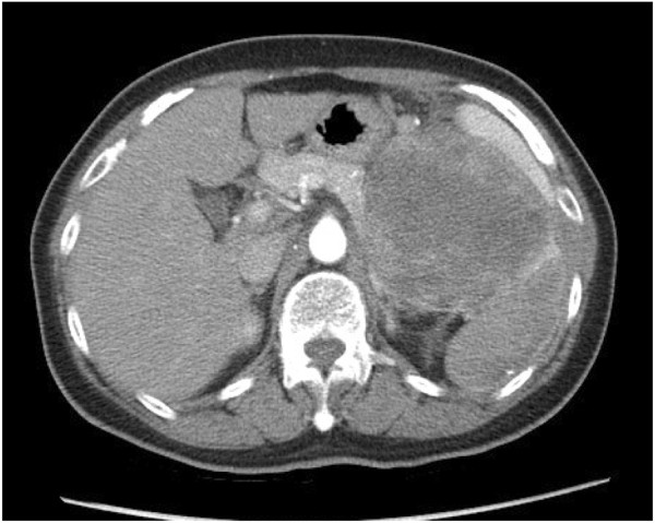
Abdominal computer tomography (CT) showing a single tumor measuring 20 × 15 cm in the perirenal space with spleen, pancreatic tail, and high gastric body invasion.
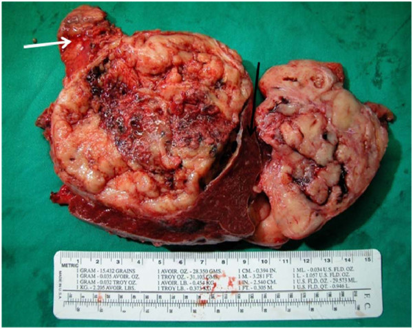
The gross pathology of the tumor was well encapsulated, yellowish in color, and friable with multiple areas of hemorrhage and necrosis. The black arrow shows the spleen; the white arrow is the pancreas tail.
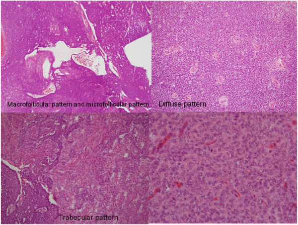
Under hematoxylin and eosin stain, the tumor showed small round to oval cells with multiple distributive patterns, including macrofollicular, microfollicular, diffuse and trabecullar patterns. The tumor cells also showed scanty cytoplasm with a coffee-bean-like nucleus.
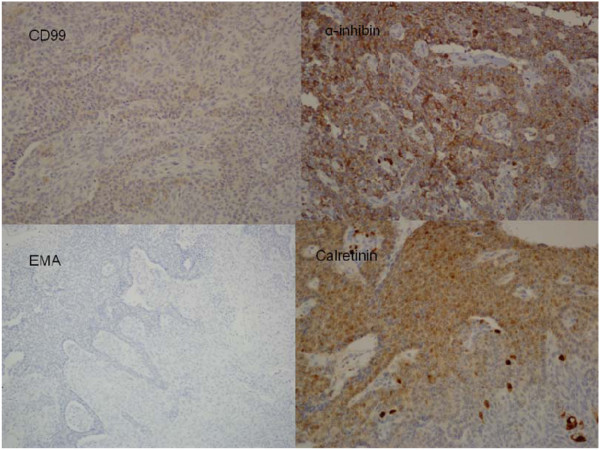
An immunohistological stain was positive for CD99, α-inhibin, calretinin (sex cord stromal tumor markers) and negative for epithelial membrane antigen (EMA) (sarcoma and mesenchymal tissue marker).
References
Publication types
MeSH terms
LinkOut - more resources
Full Text Sources
Medical
