. 2012 May 16:10:95.
doi: 10.1186/1479-5876-10-95.

Small molecule inhibition of 6-phosphofructo-2-kinase suppresses t cell activation

Affiliations

Small molecule inhibition of 6-phosphofructo-2-kinase suppresses t cell activation

Sucheta Telang et al. J Transl Med. .

Abstract

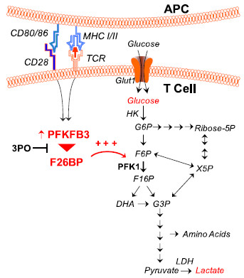
Background: T cell activation is associated with a rapid increase in intracellular fructose-2,6-bisphosphate (F2,6BP), an allosteric activator of the glycolytic enzyme, 6-phosphofructo-1-kinase. The steady state concentration of F2,6BP in T cells is dependent on the expression of the bifunctional 6-phosphofructo-2-kinase/fructose-2,6-bisphosphatases (PFKFB1-4) and the fructose-2,6-bisphosphatase, TIGAR. Of the PFKFB family of enzymes, PFKFB3 has the highest kinase:bisphosphatase ratio and has been demonstrated to be required for T cell proliferation. A small molecule antagonist of PFKFB3, 3-(3-pyridinyl)-1-(4-pyridinyl)-2-propen-1-one (3PO), recently has been shown to reduce F2,6BP synthesis, glucose uptake and proliferation in transformed cells. We hypothesized that the induction of PFKFB3 expression may be required for the stimulation of glycolysis in T cells and that exposure to the PFKFB3 antagonist, 3PO, would suppress T cell activation.

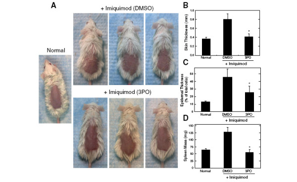
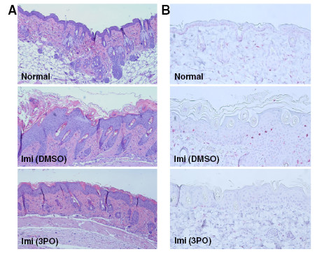
Methods: We examined PFKFB1-4 and TIGAR expression and F2,6BP concentration in purified CD3+ T cells stimulated with microbead-conjugated agonist antibodies specific for CD3 and the co-stimulatory receptor, CD28. We then determined the effect of 3PO on anti-CD3/anti-CD28-induced T cell activation, F2,6BP synthesis, 2-[1-14C]-deoxy-d-glucose uptake, lactate secretion, TNF-α secretion and proliferation. Finally, we examined the effect of 3PO administration on the development of delayed type hypersensitivity to methylated BSA and on imiquimod-induced psoriasis in mice.

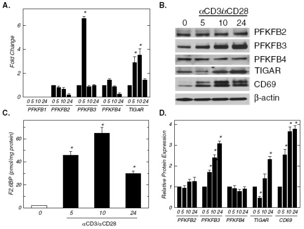
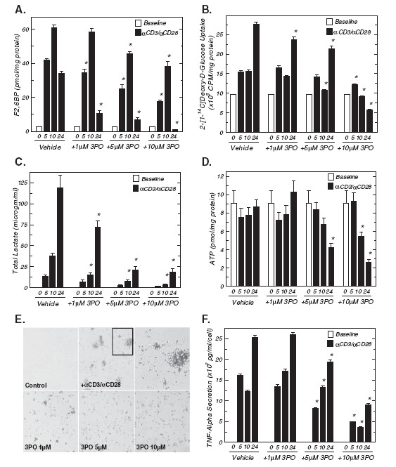
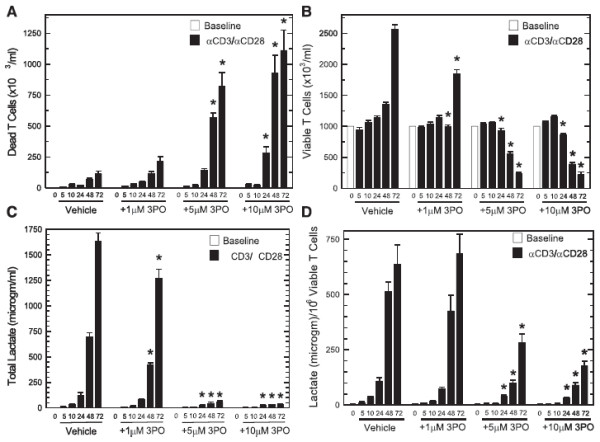
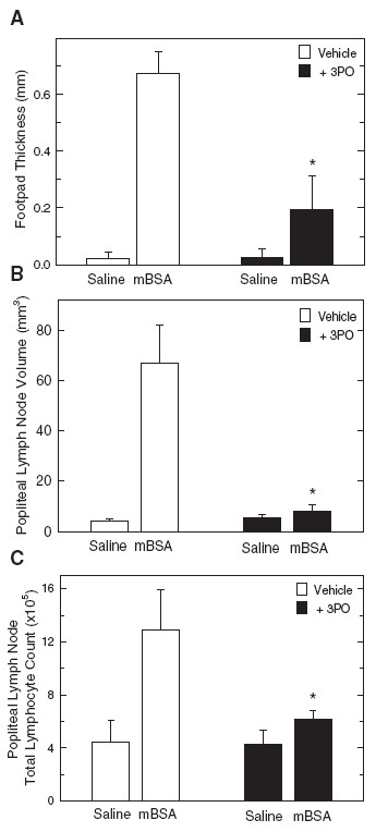
Results: We found that purified human CD3+ T cells express PFKFB2, PFKFB3, PFKFB4 and TIGAR, and that anti-CD3/anti-CD28 conjugated microbeads stimulated a >20-fold increase in F2,6BP with a coincident increase in protein expression of the PFKFB3 family member and a decrease in TIGAR protein expression. We then found that exposure to the PFKFB3 small molecule antagonist, 3PO (1-10 μM), markedly attenuated the stimulation of F2,6BP synthesis, 2-[1-14C]-deoxy-D-glucose uptake, lactate secretion, TNF-α secretion and T cell aggregation and proliferation. We examined the in vivo effect of 3PO on the development of delayed type hypersensitivity to methylated BSA and on imiquimod-induced psoriasis in mice and found that 3PO suppressed the development of both T cell-dependent models of immunity in vivo.

Conclusions: Our data demonstrate that inhibition of the PFKFB3 kinase activity attenuates the activation of T cells in vitro and suppresses T cell dependent immunity in vivo and indicate that small molecule antagonists of PFKFB3 may prove effective as T cell immunosuppressive agents.


Figures

Figure 1
Figure 1
Activation of human CD3+T cells increases PFKFB3 expression and intracellular F2,6BP. CD3+ T cells were isolated by negative selection and then plated at a final concentration of 1 x 10⁶ cells/ml in the absence or presence of anti-CD3/anti-CD28-conjugated microbeads for 5, 10 and 24 hours. (A), PFKFB1-4 and TIGAR mRNA expression was determined using real-time RT-PCR analyses. (B), PFKFB2-4, TIGAR, CD69 and β-actin protein expression was determined by Western blot analyses. (C), Intracellular F2,6BP concentration was determined using a coupled enzyme assay. (D), Densitometric analysis of PFKFB2-4, TIGAR, CD69 and β-actin protein expression. Data are representative of three independent experiments. *p < 0.05 (relative to expression or concentration at 0 hours).
Figure 2
Figure 2
A PFKFB3 small molecule antagonist inhibits stimulation of F2,6BP, glucose uptake, lactate secretion, ATP and TNF-α secretion caused by 10 hours of exposure to anti-CD3/anti-CD28-conjugated microbeads. (A), Either vehicle (DMSO) or 1–10 μM 3PO was added to human CD3+ T cells stimulated with anti-CD3/anti-CD28-conjugated microbeads for 5, 10 and 24 hours and F2,6BP was determined. (B), 2-[1-14 C]-deoxy-D-glucose uptake after addition of anti-CD3/anti-CD28-conjugated microbeads in the presence or absence of the indicated concentrations of 3PO for 5, 10 and 24 hours. (C), Lactate secretion after addition of anti-CD3/anti-CD28-conjugated microbeads in the presence or absence of the indicated concentrations of 3PO for 5, 10 and 24 hours. (D), Intracellular ATP after addition of anti-CD3/anti-CD28-conjugated microbeads in the presence or absence of the indicated concentrations of 3PO for 5, 10 and 24 hours. (E), Either vehicle (DMSO) or 1–10 μM 3PO was added to human CD3+ T cells stimulated with anti-CD3/anti-CD28-conjugated microbeads for 10 hours and cell aggregates were assessed using light microscopy. (F), Secreted TNF-α after addition of anti-CD3/anti-CD28-conjugated microbeads in the presence or absence of the indicated concentrations of 3PO for 5, 10 and 24 hours was determined by ELISA. Data are representative of three independent experiments. *p < 0.05 (relative to vehicle control).
Figure 3
Figure 3
A PFKFB3 small molecule antagonist causes cell death and a reduction of proliferation caused by anti-CD3/anti-CD28-conjugated microbeads after 24–72 hours. Either vehicle (DMSO) or 1–10 μM 3PO was added to human CD3+ T cells stimulated with anti-CD3/anti-CD28-conjugated microbeads and IL-2 for 5, 10, 24, 48 and 72 hours and dead (A) and viable (B) T cells were enumerated with a New Brunswick Nucleocounter based propidium iodide staining with and without lysis. (C), Lactate secretion after addition of anti-CD3/anti-CD28-conjugated microbeads in the presence or absence of the indicated concentrations of 3PO for 5–72 hours. (D), Lactate secretion after addition of anti-CD3/anti-CD28-conjugated microbeads in the presence or absence of the indicated concentrations of 3PO for 5–72 hours was normalized to viable T cell counts. Data are representative of three independent experiments. *p < 0.05 (relative to vehicle control).
Figure 4
Figure 4
Intraperitoneal administration of 3PO suppresses delayed type hypersensitivity to mBSA. (A), Six BALB/c mice were injected subcutaneously with 250 μg mBSA emulsified with CFA and, seven days following immunization, the mice were challenged with an injection of 50 μl of 0.5 mg/ml mBSA in saline in one rear foot pad and 50 μl saline in the other rear footpad and then administered either vehicle (DMSO) or 3PO (0.07 mg/gm) 30 minutes and 12 hrs after the challenge. Footpad thickness was measured 24 hours following mBSA challenge using microcalipers. After the mice were euthanized, the draining popliteal lymph nodes were removed and measured with microcalipers (B) and the total number of lymphocytes counted using light microscopy (C). Data are representative of three independent experiments. *p < 0.05 (relative to vehicle control).
Figure 5
Figure 5
Intraperitoneal administration of 3PO suppresses the development of imiquimod-induced psoriasis. (A) Three BALB/c mice were administered a daily topical dose of 62.5 mg of imiquimod cream (5%) over a shaved area on the back for 5 days. Thirty minutes following each daily administration of imiquimod, the mice were injected intraperitoneally with either DMSO or 3PO 0.07 mg/gm daily. After 5 days, photographs were taken that demonstrate gross skin folds (i.e. wrinkling), scaling and erythema. A photograph was captured of a shaved mouse that had not been treated with imiquimod (Normal). (B), After 5 daily topical applications of imiquimod, skin thickness was measured using microcalipers. (C) After 5 daily topical applications of imiquimod, the epidermal thickness relative to the total cutis was determined. (D) After 5 daily topical applications of imiquimod, spleens were resected and mass was determined. Data from nine mice were averaged from three independent experiments (n = 3 per experiment). *p < 0.05 (relative to vehicle control). Data are representative of three independent experiments.
Figure 6
Figure 6
3PO suppresses the increase in epidermal thickness and CD3+T cell infiltration caused by imiquimod. BALB/c mice were administered a daily topical dose of 62.5 mg of imiquimod cream (5%) over a shaved area on the back for 5 days and thirty minutes following each daily administration of imiquimod, the mice were injected intraperitoneally with either DMSO or 3PO 0.07 mg/gm daily. (A) After 5 daily topical applications of imiquimod with intraperitoneal administration of DMSO or 3PO, the exposed skin was resected, fixed in formalin, embedded in paraffin, sectioned and stained with hematoxylin and eosin. (B) After 5 daily topical applications of imiquimod with intraperitoneal administration of DMSO or 3PO, the exposed skin was resected, fixed in formalin, embedded in paraffin, sectioned and CD3+ T cells were detected by immunohistochemistry. Data are representative of three independent experiments.
Figure 7
Figure 7
3PO suppresses the CD3/CD28-induced activation of glycolysis in T cells. Activation of T cells by anti-CD3/anti-CD28-conjugated microbeads causes a marked increase in PFKFB3 expression, F2,6BP, glucose uptake and lactate secretion. The PFKFB3 antagonist, 3PO, blocks T cell activation-induced glycolysis which, in turn, results in suppression of T cell function.

References

    1. Cooper EH, Barkhan P, Hale AJ. Observations on the proliferation of human leucocytes cultured with phytohaemagglutinin. Br J Haematol. 1963;9:101–111. doi: 10.1111/j.1365-2141.1963.tb05446.x. - DOI - PubMed
    1. Culvenor JG, Weidemann MJ. Phytohaemagglutinin stimulation of rat thymus lymphocytes glycolysis. Biochim Biophys Acta. 1976;437:354–363. doi: 10.1016/0304-4165(76)90005-2. - DOI - PubMed
    1. Roos D, Loos JA. Changes in the carbohydrate metabolism of mitogenically stimulated human peripheral lymphocytes. II. Relative importance of glycolysis and oxidative phosphorylation on phytohaemagglutinin stimulation. Exp Cell Res. 1973;77:127–135. doi: 10.1016/0014-4827(73)90561-2. - DOI - PubMed
    1. Wang T, Marquardt C, Foker J. Aerobic glycolysis during lymphocyte proliferation. Nature. 1976;261:702–705. doi: 10.1038/261702a0. - DOI - PubMed
    1. Jones RG, Thompson CB. Revving the engine: signal transduction fuels T cell activation. Immunity. 2007;27:173–178. doi: 10.1016/j.immuni.2007.07.008. - DOI - PubMed

LinkOut - more resources