

Expression of heat shock protein 70 in nasopharyngeal carcinomas: different expression patterns correlate with distinct clinical prognosis
- PMID: 22591702
- PMCID: PMC3478221
- DOI: 10.1186/1479-5876-10-96
Expression of heat shock protein 70 in nasopharyngeal carcinomas: different expression patterns correlate with distinct clinical prognosis
Abstract
Background: Heat shock protein 70, a stress protein, has been implicated in tumor progression. However, its role in nasopharyngeal carcinoma (NPC) progression has not yet been clearly investigated.
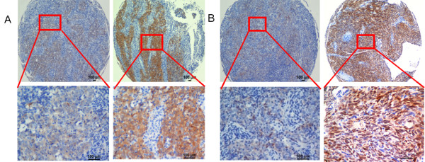
Methods: Immunohistochemistry (IHC) was employed to examine the expression patterns of Hsp70, human leukocyte antigen -A (HLA-A) in NPC tissue samples.
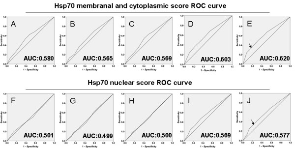
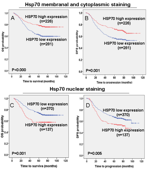
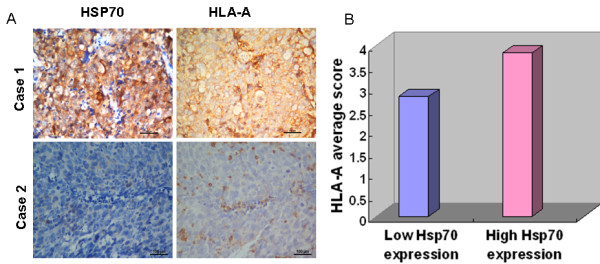
Results: The expression of Hsp70 exhibited different spatial patterns among nuclear, membrane and cytoplasm in 507 NPC tumor tissues. Kaplan-Meier survival analysis demonstrated that different Hsp70 expression patterns are correlated with different patient outcomes. High membranal and cytoplasmic levels of Hsp70 predicted good survival of patients. In contrast, high nuclear abundance of Hsp70 correlated with poor survival. Moreover, the membranal and cytoplasmic levels of Hsp70 were positively correlated with levels of the MHC I molecule HLA-A.
Conclusions: Different Hsp70 expression patterns had distinct predictive values. The different spatial abundance of Hsp70 may imply its important role in NPC development and provide insight for the development of novel therapeutic strategies involving immunotherapy for NPC.
Figures
Similar articles
-
[Study on the expression of HSP70 and HSP90beta in nasopharyngeal carcinoma and the clinical significance].Lin Chuang Er Bi Yan Hou Ke Za Zhi. 2005 Jul;19(14):640-2, 645. Lin Chuang Er Bi Yan Hou Ke Za Zhi. 2005. PMID: 16248461 Chinese.
-
Expression of human leukocyte antigen G is associated with prognosis in nasopharyngeal carcinoma.Int J Biol Sci. 2012;8(6):891-900. doi: 10.7150/ijbs.4383. Epub 2012 Jun 16. Int J Biol Sci. 2012. PMID: 22745579 Free PMC article.
-
[Correlation of heat shock protein 70 expression in nasopharyngeal carcinoma to immunoglobin A against viral capsid antigen of Epstein-Barr virus in sera and its clinical significance].Ai Zheng. 2008 Jun;27(6):650-3. Ai Zheng. 2008. PMID: 18570743 Chinese.
-
Loss of cytoplasmic KLF4 expression is correlated with the progression and poor prognosis of nasopharyngeal carcinoma.Histopathology. 2013 Sep;63(3):362-70. doi: 10.1111/his.12176. Epub 2013 Jun 12. Histopathology. 2013. PMID: 23758499
-
HSP70 and mucin 5B: novel protein targets of N,N'-dinitrosopiperazine-induced nasopharyngeal tumorigenesis.Cancer Sci. 2009 Feb;100(2):216-24. doi: 10.1111/j.1349-7006.2008.01028.x. Cancer Sci. 2009. PMID: 19068094 Free PMC article.
Cited by
-
Characterization of the dual functional effects of heat shock proteins (HSPs) in cancer hallmarks to aid development of HSP inhibitors.Genome Med. 2020 Nov 23;12(1):101. doi: 10.1186/s13073-020-00795-6. Genome Med. 2020. PMID: 33225964 Free PMC article.
-
Hsp70 in cancer: back to the future.Oncogene. 2015 Aug 6;34(32):4153-61. doi: 10.1038/onc.2014.349. Epub 2014 Oct 27. Oncogene. 2015. PMID: 25347739 Free PMC article. Review.
-
The Pro-Tumorigenic Role of Chemotherapy-Induced Extracellular HSP70 from Breast Cancer Cells via Intratumoral Macrophages.Cancers (Basel). 2023 Mar 22;15(6):1903. doi: 10.3390/cancers15061903. Cancers (Basel). 2023. PMID: 36980788 Free PMC article.
-
Molecular classification and molecular targeted therapy of cancer.Front Med. 2013 Jun;7(2):147-9. doi: 10.1007/s11684-013-0274-2. Front Med. 2013. PMID: 23686608 No abstract available.
-
Heat Shock Proteins 27, 70, and 110: Expression and Prognostic Significance in Colorectal Cancer.Cancers (Basel). 2021 Aug 31;13(17):4407. doi: 10.3390/cancers13174407. Cancers (Basel). 2021. PMID: 34503216 Free PMC article.
References
-
- Klibi J, Niki T, Riedel A, Pioche-Durieu C, Souquere S, Rubinstein E, Le Moulec S, Guigay J, Hirashima M, Guemira F, Adhikary D, Mautner J, Busson P. Blood diffusion and Th1-suppressive effects of galectin-9-containing exosomes released by Epstein-Barr virus-infected nasopharyngeal carcinoma cells. Blood. 2009;113(9):1957–1966. doi: 10.1182/blood-2008-02-142596. - DOI - PubMed
-
- Baujat B, Audry H, Bourhis J, Chan AT, Onat H, Chua DT, Kwong DL, Al-Sarraf M, Chi KH, Hareyama M, Pignon JP. MAC-NPC Collaborative Group. Chemotherapy in locally advanced nasopharyngeal carcinoma: an individual patient data meta-analysis of eight randomized trials and 1753 patients. Int J Radiat Oncol Biol Phys. 2006;64(1):47–56. doi: 10.1016/j.ijrobp.2005.06.037. - DOI - PubMed
-
- Al-Sarraf M, LeBlanc M, Giri PG, Fu KK, Cooper J, Vuong T, Forastiere AA, Adams G, Sakr WA, Schuller DE, Ensley JF. Chemoradiotherapy versus radiotherapy in patients with advanced nasopharyngeal cancer: phase III randomized Intergroup study 0099. J Clin Oncol. 1998;16(4):1310–1317. - PubMed
Publication types
MeSH terms
Substances
LinkOut - more resources
Full Text Sources
Research Materials
