. 2012 Aug 1;108(3):802-9.
doi: 10.1152/jn.00094.2012. Epub 2012 May 16.

Methamphetamine produces bidirectional, concentration-dependent effects on dopamine neuron excitability and dopamine-mediated synaptic currents

Affiliations

Methamphetamine produces bidirectional, concentration-dependent effects on dopamine neuron excitability and dopamine-mediated synaptic currents

Sarah Y Branch et al. J Neurophysiol. .

Abstract

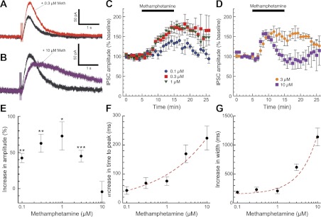
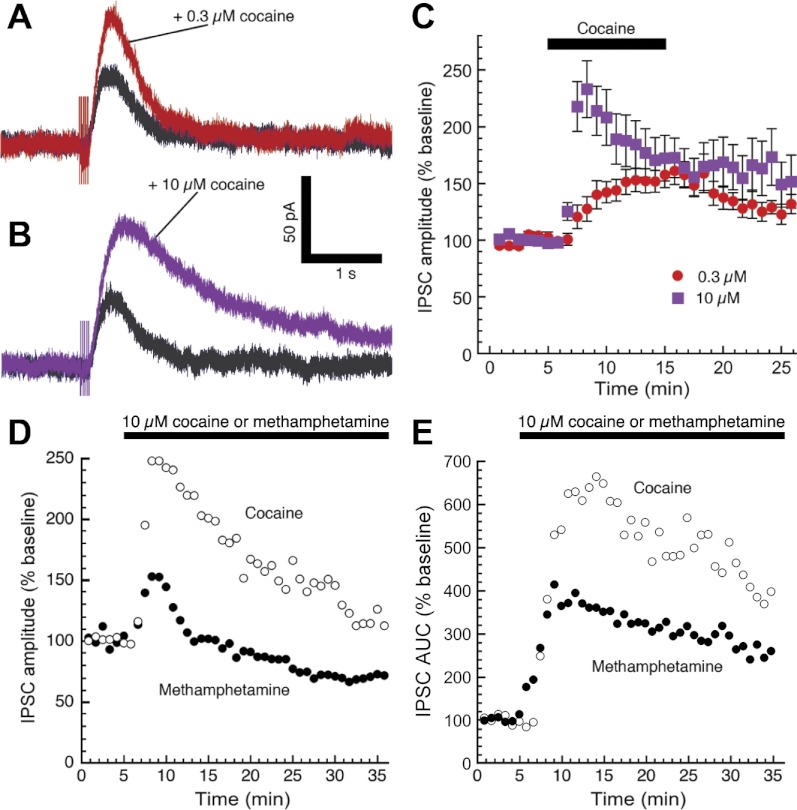
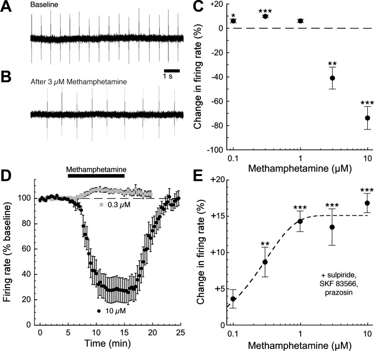
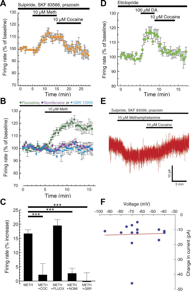
Amphetamine-like compounds are commonly used to enhance cognition and to treat attention deficit/hyperactivity disorder, but they also function as positive reinforcers and are self-administered at doses far exceeding clinical relevance. Many of these compounds (including methamphetamine) are substrates for dopamine reuptake transporters, elevating extracellular dopamine by inhibiting uptake and promoting reverse transport. This produces an increase in extracellular dopamine that inhibits dopamine neuron firing through autoreceptor activation and consequently blunts phasic dopamine neurotransmission, an important learning signal. However, these mechanisms do not explain the beneficial behavioral effects observed at clinically useful concentrations. In the present study, we have used patch-clamp electrophysiology in slices of mouse midbrain to show that, surprisingly, low concentrations of methamphetamine actually enhance dopamine neurotransmission and increase dopamine neuron firing through a dopamine transporter-mediated excitatory conductance. Both of these effects are reversed by higher concentrations of methamphetamine, which inhibit firing through dopamine D2 autoreceptor activation and decrease the peak amplitude of dopamine-mediated synaptic currents. These competing, concentration-dependent effects of methamphetamine suggest a mechanistic interplay by which lower concentrations of methamphetamine can overcome autoreceptor-mediated inhibition at the soma to increase phasic dopamine transmission.


Figures

Fig. 1.
Fig. 1.
Methamphetamine has bidirectional, concentration-dependent effects on dopamine inhibitory postsynaptic current (IPSC) amplitudes. We obtained whole cell patch-clamp recordings of dopamine-mediated IPSCs in substantia nigra and ventral tegmental area (VTA) dopamine neurons (Beckstead et al. 2004) in the presence of GABA, glutamate, and nicotinic acetylcholine receptor blockers. Bath perfusion of a low concentration of methamphetamine (0.3 μM, red trace) significantly enhanced the amplitude and slightly prolonged the duration of dopamine IPSCs (A and C; n = 6–13 cells from 3–9 mice). Perfusion of a high concentration of methamphetamine (10 μM, purple trace) briefly enhanced IPSC amplitudes but subsequently decreased IPSC amplitudes to baseline levels or slightly below (B and D; n = 6–12 cells from 3–6 mice). Thus methamphetamine exhibited an inverted U-shaped concentration-effect curve on dopamine IPSC amplitudes, determined as the average amplitude 10–12 min after the beginning of methamphetamine perfusion (E; paired t-tests for each concentration: *P < 0.05, **P < 0.01, ***P < 0.001). Methamphetamine effects on IPSC kinetics were examined by measuring time to peak (F) and width at 50% maximum amplitude (G). The slowing of IPSC kinetics was progressively enhanced by higher methamphetamine concentrations and was modeled well by a single exponential increase (red dashed lines).
Fig. 2.
Fig. 2.
Unlike methamphetamine, high concentrations of cocaine increase dopamine IPSC peak amplitudes. Perfusion of 0.3 μM cocaine [a dopamine transporter (DAT) inhibitor but not a DAT substrate] enhanced the amplitude and slightly prolonged the duration of dopamine IPSCs (A), similar to the effects of 0.3 μM methamphetamine shown in Fig. 1A. However, IPSC amplitudes were further elevated by high concentrations of cocaine (B and C; n = 9–13 cells from 6 mice), whereas the kinetics of the IPSC were substantially prolonged. In a separate experiment, either cocaine or methamphetamine (10 μM) was continuously perfused for 30 min. Summarized data indicate that during prolonged application of methamphetamine, the peak amplitude of dopamine IPSCs decreased (D) to a greater extent than the total charge transferred (area under the curve, E). Differences in the effects of the two psychostimulants were already evident during the first few minutes of perfusion. Error bars are omitted for clarity (n = 4–6 cells from 4–6 mice).
Fig. 3.
Fig. 3.
Bath perfusion of methamphetamine produces a slight reduction in maximal response to D2 receptor activation. A brief, maximal activation of D2 receptors was produced by a 2- to 3-s iontophoresis of exogenous dopamine (A; 1 M), repeated once every 5 min. Methamphetamine (10 μM) was bath perfused for 15 min. Summarized data (B; n = 8 cells from 6 mice) suggest that methamphetamine produced a significant but small (10–15%) reduction of maximal D2 receptor-mediated currents [1-way repeated-measures ANOVA (P = 0.042) followed by Dunnett's post hoc test: *P < 0.05, **P < 0.01].
Fig. 4.
Fig. 4.
Methamphetamine produces bidirectional, concentration-dependent effects on dopamine neuron firing rate. The rate of dopamine neuron pacemaker firing was monitored using the loose cell-attached patch technique (A). As expected, bath perfusion of high concentrations of methamphetamine (3–10 μM) reduced the firing rate of dopamine neurons, a consequence of increased extracellular concentrations of dopamine producing an activation of somatodendritic D2 autoreceptors (B). Surprisingly, however, lower concentrations of methamphetamine did not inhibit cell firing but actually produced a modest but significant increase in firing rate (C; n = 5–15 cells from 3–7 mice). D shows the time course of the excitatory and inhibitory effects of methamphetamine on firing rate (n = 12–15 cells from 6–7 mice). To eliminate the contribution of dopamine receptors to the effects of methamphetamine, we repeated the same experiment in the presence of the dopamine D2-type receptor antagonist sulpiride (200–500 nM), the D1/D5 receptor antagonist SKF 83566 (500 nM), and the α1-adrenergic receptor antagonist prazosin (100 nM). Under these conditions, bath perfusion of methamphetamine produced a concentration-dependent increase in pacemaker firing rate that was nearly maximal at 1 μM (E; n = 5–11 cells from 3–5 mice; 1-way ANOVA followed by Dunnett's post hoc test: *P < 0.05, **P < 0.01, ***P < 0.001).
Fig. 5.
Fig. 5.
DAT inhibitors block the dopamine neuron excitation produced by DAT substrates. Pacemaker firing of dopamine neurons was recorded in the presence of dopamine receptor antagonists (200–500 nM sulpiride, 500 nM SKF 83566, and 100 nM prazosin). Bath perfusion of methamphetamine (10 μM) increased firing rate, an effect that was quickly reversed by the nonselective DAT inhibitor cocaine (A; n = 5 cells from 2 mice). The methamphetamine-induced increase in firing was blocked by preincubation with either the norepinephrine/dopamine transporter inhibitor nomifensine (1–3 μM) or the selective DAT inhibitor GBR 12909 (1 μM), but not by the serotonin transporter inhibitor fluoxetine (800 nM; B). Summarized data are illustrated in C (1-way ANOVA followed by Dunnett's post hoc test: ***P < 0.001). In the presence of the D2 receptor antagonist eticlopride (100 nM), bath perfusion of the endogenous DAT substrate dopamine (100 μM) also increased dopamine neuron firing rate, an effect that was also rapidly reversed by cocaine (D; n = 6 cells from 3 mice). To investigate the voltage dependence of the DAT-mediated excitation, we obtained whole cell voltage-clamp recordings using the gramicidin perforated-patch technique in the presence of pharmacological blockers of dopamine receptors (sulpiride, SKF 83566, and prazosin). Under these conditions, bath perfusion of methamphetamine (10 μM) consistently produced a small inward current that was reversed by the DAT inhibitor cocaine (E). Methamphetamine produced inward currents at holding voltages ranging from −40 to −95 mV, suggesting that DAT-mediated excitation is not voltage dependent at subthreshold voltages (F). Each data point represents an individual perforated-patch experiment performed at the indicated holding voltage.

References

    1. Beckstead MJ, Gantz SC, Ford CP, Stenzel-Poore MP, Phillips PEM, Mark GP, Williams JT. CRF enhancement of GIRK channel-mediated transmission in dopamine neurons. Neuropsychopharmacology 34: 1926–1935, 2009 - PMC - PubMed
    1. Beckstead MJ, Grandy DK, Wickman K, Williams JT. Vesicular dopamine release elicits an inhibitory postsynaptic current in midbrain dopamine neurons. Neuron 42: 939–946, 2004 - PubMed
    1. Beckstead MJ, Phillips TJ. Mice selectively bred for high- or low-alcohol-induced locomotion exhibit differences in dopamine neuron function. J Pharmacol Exp Ther 329: 342–349, 2009 - PMC - PubMed
    1. Brien JF, Kitney JC, Peachey JE, Rogers BJ. Methamphetamine-induced behavioural effects and brain concentrations of methamphetamine and its metabolite amphetamine in mice. Res Commun Chem Pathol Pharmacol 22: 313–328, 1978 - PubMed
    1. Campbell AD, Kohl RR, McBride WJ. Serotonin-3 receptor and ethanol-stimulated somatodendritic dopamine release. Alcohol 13: 569–574, 1996 - PubMed

Publication types

MeSH terms