. 2012 Jun 1;11(11):2114-21.
doi: 10.4161/cc.20547. Epub 2012 Jun 1.

Sister chromatid cohesion establishment occurs in concert with lagging strand synthesis

Affiliations

Sister chromatid cohesion establishment occurs in concert with lagging strand synthesis

Soumya Rudra et al. Cell Cycle. .

Abstract

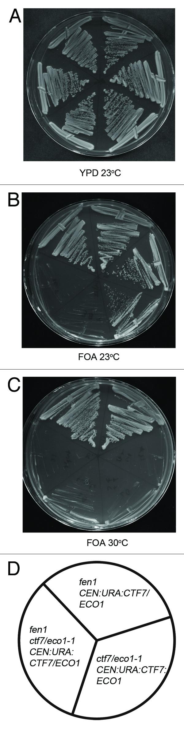
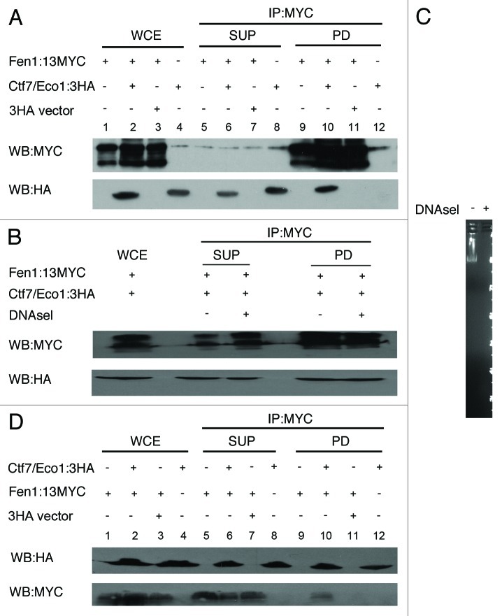
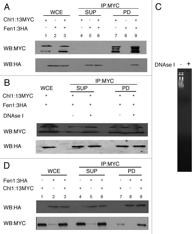
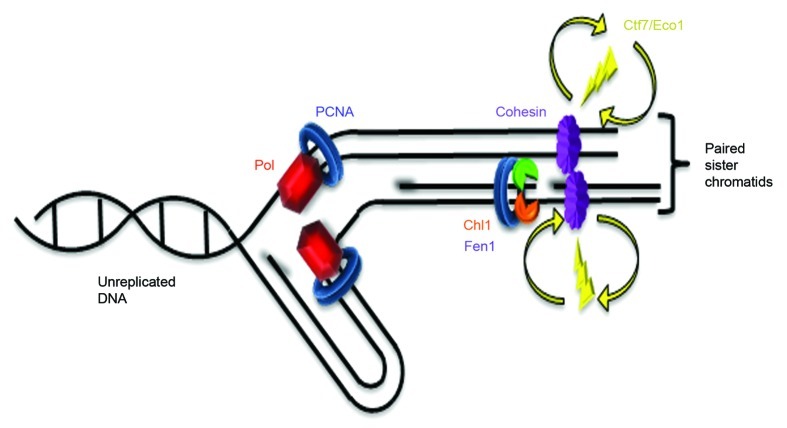
Cohesion establishment is central to sister chromatid tethering reactions and requires Ctf7/Eco1-dependent acetylation of the cohesin subunit Smc3. Ctf7/Eco1 is essential during S phase, and a number of replication proteins (RFC complexes, PCNA and the DNA helicase Chl1) all play individual roles in sister chromatid cohesion. While the mechanism of cohesion establishment is largely unknown, a popular model is that Ctf7/Eco1 acetylates cohesins encountered by and located in front of the fork. In turn, acetylation is posited both to allow fork passage past cohesin barriers and convert cohesins to a state competent to capture subsequent production of sister chromatids. Here, we report evidence that challenges this pre-replicative cohesion establishment model. Our genetic and biochemical studies link Ctf7/Eco1 to the Okazaki fragment flap endonuclease, Fen1. We further report genetic and biochemical interactions between Fen1 and the cohesion-associated DNA helicase, Chl1. These results raise a new model wherein cohesin deposition and establishment occur in concert with lagging strand-processing events and in the presence of both sister chromatids.


Figures

None
Figure 1.ctf7eco1–1 is synthetically lethal in combination with fen1. Yeast cells harboring ctf7eco1–1: ADE were crossed with fen1::KANr mutant cells and the resulting diploids were transformed with a CEN: URA3:CTF7ECO1 plasmid and sporulated. The resulting fen1 and ctf7eco1–1: ADE single mutants and fen1, ctf7eco1–1: ADE double mutants were plated on media with or without FOA (See also Table 1). Two independent isolates are shown for each strain. (A) Growth of fen1, ctf7eco1–1 single mutants and fen1::KAN ctf7eco1–1 CTF7: URA double mutants strains at 23°C on YPD. (B) Growth of fen1::KAN, ctf7eco1–1 single mutants and fen1 ctf7eco1–1: ADE CTF7: URA double mutants on FOA plates at 23°C. (C) Growth of fen1::KAN, ctf7eco1–1 single mutants and fen1 ctf7eco1–1: ADE CTF7: URA double mutants on FOA plates at 30°C (See also Fig. S1). (D) Schematic representation of fen1::KAN, ctf7eco1–1 single mutants and fen1 ctf7eco1–1: ADE CTF7: URA double mutant strains.
None
Figure 2. Fen1 and Ctf7/Eco1 physically associate in vivo. Cells expressing Fen1:13Myc and Ctf7/Eco1:3HA were mechanically lysed and clarified by centrifugation, the clarified whole cell extract was co-immunoprecipitated using anti-Myc beads (2A) and anti HA Beads (2D) and analyzed by immunoblotting for Fen1:13Myc and Ctf7/Eco1:3HA. Whole cell extracts (WCE, lanes 1–4), Supernatants (SUP, lanes 5–8) and pull down fractions (PD, 9–12) are shown. (A) Co-immunoprecipitation of Fen1:13Myc and Ctf7:3HA with anti-MYC beads. Cells expressing only 3HA tag (Lane 11) and cells expressing Ctf7:3HA but untagged Fen1 (Lane 12) were used to determine the specificity of the co-immunoprecipitation. (B) Clarified whole cell extracts of cells co-expressing Fen1:13Myc and Ctf7:/Eco1:3HA were treated with and without DNaseI before immunoprecipitation with anti-MYC beads. (C) 1 μg of λ DNA added in the clarified whole cell extract, with and without DNaseI treatment, run on a 1% agarose gel. (D) Reciprocal co-immunoprecipitation of cells expressing Fen1:13Myc and Ctf7:3HA using Anti-HA affinity beads. Cell expressing untagged Ctf7/Eco1 (Lane 9) and cells expressing 3HA tags alone (Lane 11) were used as a control to determine the specificity of the co-immunoprecipitation reaction.
None
Figure 3. Chl1:13Myc physically associate with Fen1:3HA in vivo. Cells expressing Chl1:13Myc and Fen1:3HA were mechanically lysed and clarified by centrifugation. The clarified whole cell extract was co-immunoprecipitated using anti-Myc beads (3A) and anti HA beads (3B) and analyzed by immunoblotting for Chl1:13Myc and Fen1:3HA. Whole cell extracts (WCE, lanes 1–3), Supernatants (SUP, lanes 4–6) and pull down fractions (PD, 7–9) are shown. (A) Co-immunoprecipitation of Chl1:13Myc and Fen1:3HA using anti-MYC beads. Cells expressing untagged Fen1 cell extracts were used to determine the specificity of the co-immunoprecipitation reaction (Lane 8). (B) Clarified whole cell extracts of cells co-expressing Chl1:13Myc and Fen1:3HA were treated with or without DNaseI treatment before co-immunoprecipitation with anti-MYC beads. (C) 1 μg of λ DNA added in the clarified whole cell extract with and without DNaseI treatment, run on a 1% agarose gel. (D) Reciprocal immunoprecipitation of cells co-expressing Chl1:13Myc and Fen1:3HA with Anti-HA beads. Cells expressing untagged Fen1 were used to determine the specificity of the co-immunoprecipitation reaction (Lane 7).
None
Figure 4. Cohesion establishment coupled to lagging strand processing. Replication fork (Pol = DNA polymerase coupled to PCNA) moves to the left: leading strand replication on the top and lagging strand replication on the bottom (RNA primers shadowed). Immediately behind the fork, PCNA associates with Fen1 (green) and Chl1 (orange). Ctf7/Eco1 (yellow) is not stably recruited to chromatin by any factor, but transiently interacts with chromatin to establish cohesion. Therefore, both cohesin deposition and subsequent cohesion establishment occur behind the replication fork. Cohesins (purple) depicted as unstructured to highlight the many models currently posited in the literature.,, MCM helicase, primase and RPA not shown (based on Burgers46).

Comment in

References

    1. Onn I, Heidinger-Pauli JM, Guacci V, Unal E, Koshland DE. Sister chromatid cohesion: a simple concept with a complex reality. Annu Rev Cell Dev Biol. 2008;24:105–29. doi: 10.1146/annurev.cellbio.24.110707.175350. - DOI - PubMed
    1. Skibbens RV. Mechanisms of sister chromatid pairing. Int Rev Cell Mol Biol. 2008;269:283–339. doi: 10.1016/S1937-6448(08)01005-8. - DOI - PubMed
    1. Unal E, Heidinger-Pauli JM, Koshland D. DNA double-strand breaks trigger genome-wide sister-chromatid cohesion through Eco1 (Ctf7) Science. 2007;317:245–8. doi: 10.1126/science.1140637. - DOI - PubMed
    1. Ström L, Karlsson C, Lindroos HB, Wedahl S, Katou Y, Shirahige K, et al. Postreplicative formation of cohesion is required for repair and induced by a single DNA break. Science. 2007;317:242–5. doi: 10.1126/science.1140649. - DOI - PubMed
    1. Dorsett D. Cohesin: genomic insights into controlling gene transcription and development. Curr Opin Genet Dev. 2011;21:199–206. doi: 10.1016/j.gde.2011.01.018. - DOI - PMC - PubMed

Publication types

MeSH terms