. 2012 May 16;32(20):6957-66.
doi: 10.1523/JNEUROSCI.5718-11.2012.

Subregional, dendritic compartment, and spine subtype specificity in cocaine regulation of dendritic spines in the nucleus accumbens

Affiliations

Subregional, dendritic compartment, and spine subtype specificity in cocaine regulation of dendritic spines in the nucleus accumbens

Dani Dumitriu et al. J Neurosci. .

Abstract

Numerous studies have found that chronic cocaine increases dendritic spine density of medium spiny neurons in the nucleus accumbens (NAc). Here, we used single-cell microinjections and advanced 3D imaging and analysis techniques to extend these findings in several important ways: by assessing cocaine regulation of dendritic spines in the core versus shell subregions of NAc in the mouse, over a broad time course (4 h, 24 h, or 28 d) of withdrawal from chronic cocaine, and with a particular focus on proximal versus distal dendrites. Our data demonstrate subregion-specific, and in some cases opposite, regulation of spines by cocaine on proximal but not distal dendrites. Notably, all observed density changes were attributable to selective regulation of thin spines. At 4 h after injection, the proximal spine density is unchanged in the core but significantly increased in the shell. At 24 h, the density of proximal dendritic spines is reduced in the core but increased in the shell. Such downregulation of thin spines in the core persists through 28 d of withdrawal, whereas the spine density in the shell returns to baseline levels. Consistent with previous results, dendritic tips exhibited upregulation of dendritic spines after 24 h of withdrawal, an effect localized to the shell. The divergence in regulation of proximal spine density in NAc core versus shell by cocaine correlates with recently reported electrophysiological data from a similar drug administration regimen and might represent a key mediator of changes in the reward circuit that drive aspects of addiction.


Figures

Figure 1.
Figure 1.
Experimental design. A, A 10× image of NAc core and shell microinjected neurons with corresponding diagram from the Paxinos mouse brain atlas (Paxinos and Franklin, 2001). Medial NAc neurons were microinjected +1.42 ± 0.25 mm anterior to bregma. Boxes represent the subcellular location of dendritic imaging, with red boxes representing proximal dendrites and blue box representing a dendritic tip. Scale bar, 100 μm. B, Projections of a high-resolution 3D confocal stack before and after deconvolution in the x–y and y–z direction. C, NeuronStudio 3D automatic identification, measurement, and classification of spines: yellow, thin; orange, mushroom; purple, stubby. D, Ten small-diameter and 10 large-diameter dendrites were analyzed semiautomatically with NeuronStudio and then manually by counting visible spines in 2D projected images. The percentage of 3D spines visible in 2D projected images is plotted, with circles indicating individual dendrites and lines indicating mean ± SEM. There is a significant difference between the two groups (p = 0.01), pointing to the difficulty in comparing 2D and 3D methods. *p < 0.05.
Figure 2.
Figure 2.
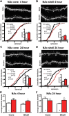
Proximal dendritic spine density and diameter are differentially regulated by cocaine in NAc core versus shell 4 and 24 h after the last injection. A–D, Top images show representative projected z-stacks from each experimental group. Cumulative frequencies are plotted using each analyzed dendrite from all animals in a group. Inset bar graphs show the average proximal spine density calculated per animal and then per group. A, B, At 4 h, cocaine significantly increases proximal spine density in the shell but not core. C, D, At 24 h, cocaine significantly decreases proximal spine density in the core and increases proximal spine density in the shell. E, F, Average proximal dendrite diameter is significantly increased in the shell at both 4 and 24 h after the last cocaine injection with a trend for an increase in the NAc core at 4 h. K–S test, *p < 0.05.
Figure 3.
Figure 3.
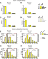
Cocaine selectively and oppositely regulates thin dendritic spine density and morphology in the proximal dendrites of NAc core and shell. A–D, Spine subtype densities based on an unbiased classifier in the core versus shell at 4 or 24 h after the last cocaine injection. A, B, At 4 h, proximal thin spine density is selectively and significantly upregulated by cocaine in shell but not core. C, D, At 24 h, cocaine significantly downregulates proximal thin spine density in the core, whereas the upregulation of thin spines in the shell is maintained. E–H, The spine head diameters of proximal thin and mushroom spines are plotted next to each other as cumulative distributions superimposed onto frequency plots in which the spines were binned by size first per animal and then by group using 0.1 μm bins (numbers below bar graph represent lower bound of bin). E, F, At 4 h, the cumulative frequencies of proximal thin spine head diameters in both core and shell are significantly left shifted. Binned data show a significant decrease in the frequency of thin spines with head diameters between 0.3 and 0.4 μm in both subregions and a significant increase in smaller thin spines in the shell. There is no change in the size of mushroom spines at this time point. G, H, At 24 h, the cumulative frequency of proximal thin spine head diameters is slightly but significantly right shifted in the core and unchanged in the shell. Binned data do not point to changes in frequency in any particular bin in the core. Interestingly, although mushroom spine size remains unaffected in the core, the cumulative frequency of the mushroom spine head diameter in the shell is significantly left shifted, which can be attributed to a selective increase in the smallest mushroom spines. K–S test, *p < 0.05, **p < 0.001.
Figure 4.
Figure 4.
Downregulation of proximal NAc core thin dendritic spine density persists after 28 d of withdrawal from chronic cocaine. A, B, Cumulative frequencies are plotted using each analyzed proximal dendrite from all animals in a group. Inset bar graphs show the average proximal spine density calculated per animal and then per treatment. Prolonged withdrawal is accompanied by persistent downregulation of proximal spines in the core, whereas shell spine density has returned to control levels. C, The downregulation of proximal spines in the core continues to be selective for a loss of thin spines. D, No changes in the density of any proximal spine subtype are observed in the shell. K–S test, *p < 0.05, **p < 0.001.
Figure 5.
Figure 5.
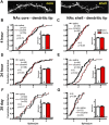
Distal dendritic spine density regulation by cocaine in NAc core versus shell 4 h, 24 h, and 28 d after the last injection. A, Images show representative projected z-stacks from dendritic tips in the core and shell. B–G, Cumulative frequencies are plotted using each analyzed dendrite from all animals in a group. Inset bar graphs show the average spine density calculated per animal and then per group. B, C, At 4 h, cocaine does not regulate distal spine density in either the core or the shell. D, E, At 24 h, cocaine significantly increases distal spine density in the shell with no effect in the core. F, G, After 28 d of withdrawal, cocaine does not regulate distal spine density in the core or shell. K–S test, *p < 0.05.
Figure 6.
Figure 6.
Summary diagram and table of the effects of chronic cocaine on proximal dendritic spine morphometrics. In the NAc core, chronic cocaine decreases proximal dendritic spine head diameter at 4 h and by 24 h causes a selective elimination of thin spines that is maintained for up to 1 month. Conversely, in the NAc shell, which at baseline has fewer proximal thin spines than the core, new thin spines are formed by 4 h and maintained for up to 24 h. There is also an increase in proximal dendritic diameter in the shell. However, unlike the core, the changes that occur in the shell are transient because they are not observed after prolonged withdrawal from cocaine. Red arrows indicate shrinkage or elimination, and green arrows indicate growth or sprouting. * Data adapted from Kourrich and Thomas (2009).

References

    1. Arellano JI, Benavides-Piccione R, Defelipe J, Yuste R. Ultrastructure of dendritic spines: correlation between synaptic and spine morphologies. Front Neurosci. 2007;1:131–143. - PMC - PubMed
    1. Azdad K, Chàvez M, Don Bischop P, Wetzelaer P, Marescau B, De Deyn PP, Gall D, Schiffmann SN. Homeostatic plasticity of striatal neurons intrinsic excitability following dopamine depletion. PLoS One. 2009;4:e6908. - PMC - PubMed
    1. Baker DA, McFarland K, Lake RW, Shen H, Tang XC, Toda S, Kalivas PW. Neuroadaptations in cystine-glutamate exchange underlie cocaine relapse. Nat Neurosci. 2003;6:743–749. - PubMed
    1. Bourne J, Harris KM. Do thin spines learn to be mushroom spines that remember? Curr Opin Neurobiol. 2007;17:381–386. - PubMed
    1. Branco T, Häusser M. The single dendritic branch as a fundamental functional unit in the nervous system. Curr Opin Neurobiol. 2010;20:494–502. - PubMed