. 2012 Aug;86(15):7806-17.
doi: 10.1128/JVI.00080-12. Epub 2012 May 16.

Modeling the intracellular dynamics of influenza virus replication to understand the control of viral RNA synthesis

Affiliations

Modeling the intracellular dynamics of influenza virus replication to understand the control of viral RNA synthesis

Frank S Heldt et al. J Virol. 2012 Aug.

Abstract

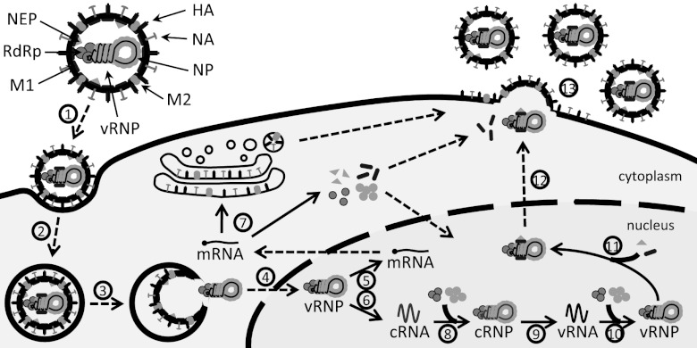
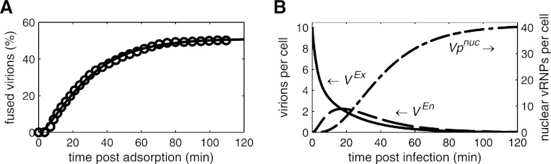
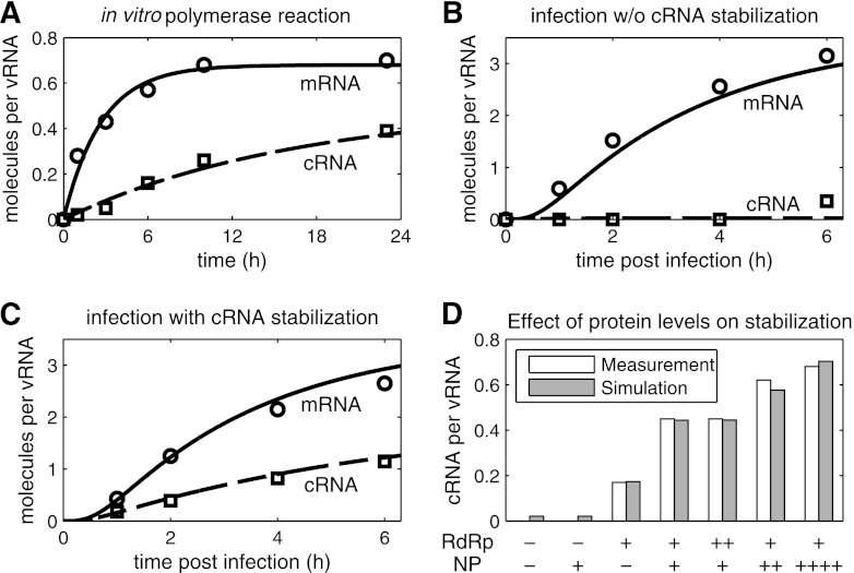
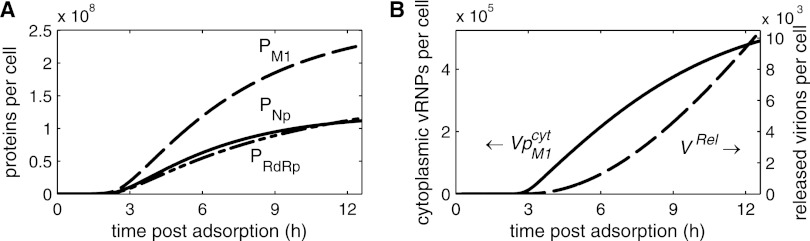
Influenza viruses transcribe and replicate their negative-sense RNA genome inside the nucleus of host cells via three viral RNA species. In the course of an infection, these RNAs show distinct dynamics, suggesting that differential regulation takes place. To investigate this regulation in a systematic way, we developed a mathematical model of influenza virus infection at the level of a single mammalian cell. It accounts for key steps of the viral life cycle, from virus entry to progeny virion release, while focusing in particular on the molecular mechanisms that control viral transcription and replication. We therefore explicitly consider the nuclear export of viral genome copies (vRNPs) and a recent hypothesis proposing that replicative intermediates (cRNA) are stabilized by the viral polymerase complex and the nucleoprotein (NP). Together, both mechanisms allow the model to capture a variety of published data sets at an unprecedented level of detail. Our findings provide theoretical support for an early regulation of replication by cRNA stabilization. However, they also suggest that the matrix protein 1 (M1) controls viral RNA levels in the late phase of infection as part of its role during the nuclear export of viral genome copies. Moreover, simulations show an accumulation of viral proteins and RNA toward the end of infection, indicating that transport processes or budding limits virion release. Thus, our mathematical model provides an ideal platform for a systematic and quantitative evaluation of influenza virus replication and its complex regulation.


Figures

Fig 1
Fig 1
Scheme of the influenza virus life cycle. For the sake of simplicity, only one vRNP in a virus particle is depicted, and nonstructural proteins are omitted. Solid arrows represent synthesis or protein binding. Dashed arrows indicate transport processes. Different steps are assigned by numbers (see the text for details): 1, attachment; 2, endocytosis; 3, fusion in late endosomes; 4, nuclear import; 5, transcription; 6, replication (cRNA synthesis); 7, protein translation; 8, cRNA encapsidation; 9, replication (vRNA synthesis); 10, vRNA encapsidation; 11, M1 and NEP binding; 12, nuclear export; and 13, virus assembly and budding.
Fig 2
Fig 2
Dynamics of virus entry. Lines represent simulation results. (A) Model fit to data (circles) for the fusion of R18-labeled influenza virus (strain NIB26) with endosomes in MDCK cells modified from those of Stegmann et al. (57). In brief, virus was added to MDCK cells at 0°C for 1 h to allow for virus adsorption, cells were washed, and warm buffer (37°C) was added. The percentage of fused out of total cell-associated virions is shown. (B) Simulated amounts of extracellular virions in the medium (VEx), virions in endosomes (VEn), and vRNPs in the nucleus (Vpnuc) for infection at an MOI of 10, neglecting viral protein synthesis and vRNP degradation.
Fig 3
Fig 3
Simulation of cRNA stabilization hypothesis. Experiments yielding NA gene-specific mRNA (○) and cRNA levels (□) were conducted by Vreede et al. using primer extension analysis (58, 61). We obtained relative RNA levels from these studies by densitometric analysis and normalized each data point to the constant vRNA signal. (A) Fit to data of an in vitro polymerase assay using virion-derived vRNPs (58). (B and C) Model fit to infection of 293T cells with influenza A/WSN/33 at an MOI of 5 (58). In brief, protein synthesis during infection was inhibited, and plasmids expressing NP, PA, and PB2 (B) or NP, PA, PB2, and PB1a (C) were transfected prior to infection. (D) Same as panel C, except that various amounts of plasmids expressing PA, PB2, PB1a (RdRp), NP, or empty vector (−) were transfected prior to infection. Bars represent the cRNA level at 2 hpi. PB1a, catalytically inactive mutant PB1-D445A/D446A which forms polymerase complexes that do not synthesize viral RNAs but stabilize cRNA.
Fig 4
Fig 4
Viral RNA synthesis during infection. (A and B) Model fit to vRNA (Δ), mRNA (○), and cRNA (□) levels of segment 5 (encoding NP) during an infection of MDCK cells with influenza A/WSN/33 at an MOI of 10 after 1 h of virus adsorption at 4°C. Data were determined by Kawakami et al. using strand-specific real-time RT-PCR (31). (C) Model prediction for the accumulation of M1 proteins (PM1) and vRNPs engaged in RNA synthesis (Vpnuc). (D) Comparison of model fit in panels A and B to data of Shapiro et al. for the synthesis rates of M1 proteins (◇) and mRNAs (×) of segment 7 (encoding M proteins) (53). In brief, BHK-21 cells were infected with influenza virus (WSN strain) at an MOI of 10 to 20, and virus was allowed to adsorb for 1 h at 4°C. Protein and mRNA levels were determined by pulse-chase experiments. In simulations, rM1Syn and rRM7Syn are the synthesis rates of M1 proteins (first term in equation 20) and of mRNAs of segment 7 (first term in equation 14, with i = 7), respectively. Data points and simulations are given as percentages of their maximums.
Fig 5
Fig 5
Dynamics of virus release. (A) Model prediction for the levels of M1 (PM1), NP (PNP), and viral polymerase complexes (PRdRp) based on the model fit shown in Fig. 4. (B) Level of cytoplasmic vRNPs (VpM1cyt) and cumulative amount of released progeny virions (VRel) for the simulation presented in Fig. 4.

References

    1. Akarsu H, et al. 2003. Crystal structure of the M1 protein-binding domain of the influenza A virus nuclear export protein (NEP/NS2). EMBO J. 22:4646–4655 - PMC - PubMed
    1. Amorim MJ, et al. 2011. A Rab11 and microtubule dependent mechanism for cytoplasmic transport of influenza A virus vRNA. J. Virol. 85:4143–4156 - PMC - PubMed
    1. Amorim MJ, Digard P. 2006. Influenza A virus and the cell nucleus. Vaccine 24:6651–6655 - PubMed
    1. Arava Y, et al. 2003. Genome-wide analysis of mRNA translation profiles in Saccharomyces cerevisiae. Proc. Natl. Acad. Sci. U. S. A. 100:3889–3894 - PMC - PubMed
    1. Babcock HP, Chen C, Zhuang X. 2004. Using single-particle tracking to study nuclear trafficking of viral genes. Biophys. J. 87:2749–2758 - PMC - PubMed