. 2012 Aug;86(16):8367-74.
doi: 10.1128/JVI.00423-12. Epub 2012 May 16.

Vpu mediates depletion of interferon regulatory factor 3 during HIV infection by a lysosome-dependent mechanism

Affiliations

Vpu mediates depletion of interferon regulatory factor 3 during HIV infection by a lysosome-dependent mechanism

Brian P Doehle et al. J Virol. 2012 Aug.

Abstract

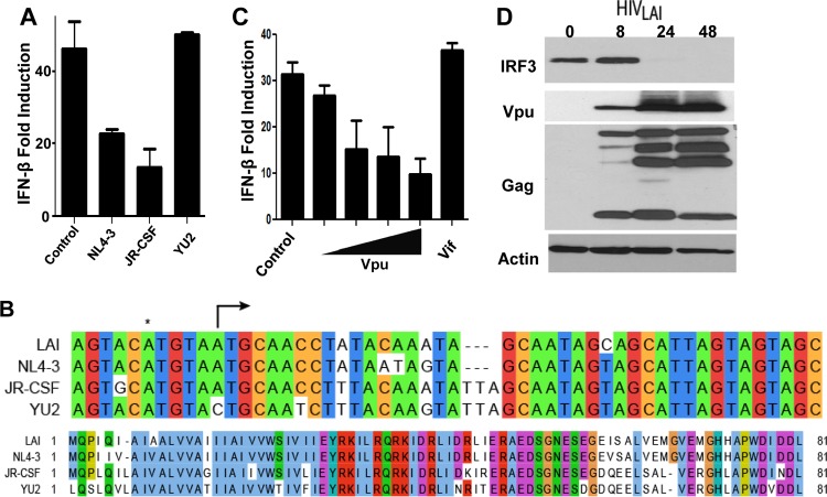
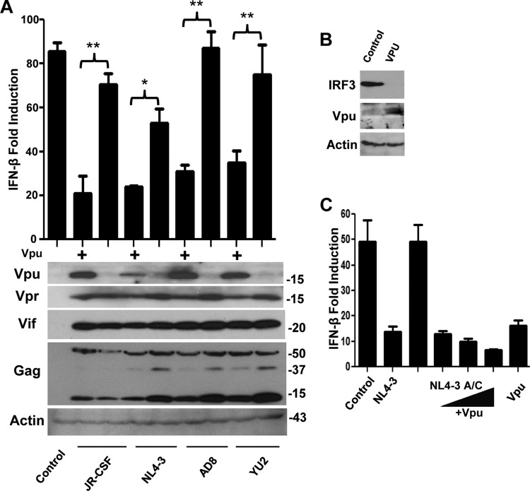
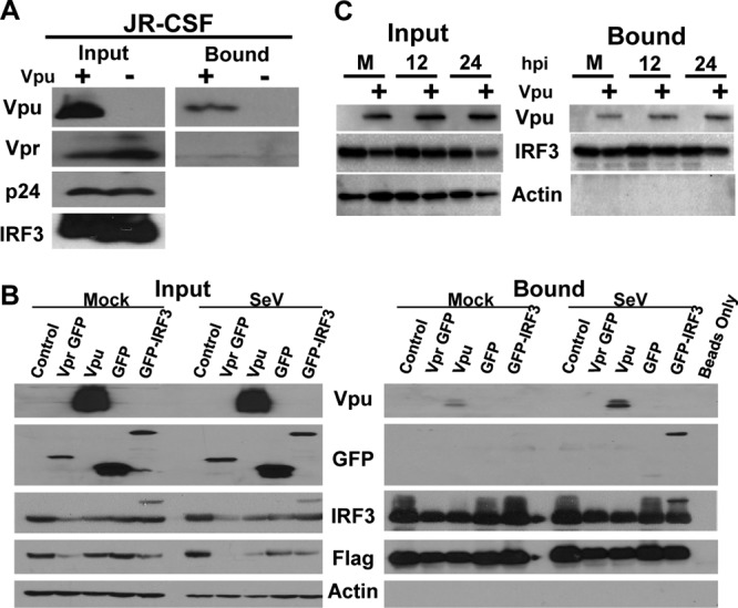
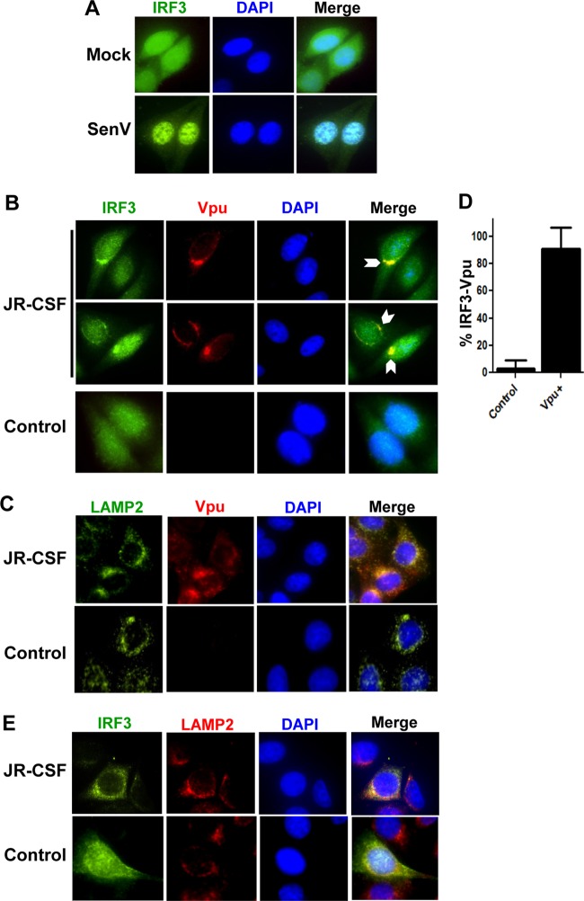
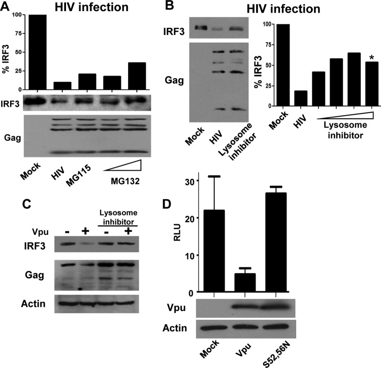
HIV has evolved sophisticated mechanisms to avoid restriction by intracellular innate immune defenses that otherwise serve to control acute viral infection and virus dissemination. Innate defenses are triggered when pattern recognition receptor (PRR) proteins of the host cell engage pathogen-associated molecule patterns (PAMPs) present in viral products. Interferon regulatory factor 3 (IRF3) plays a central role in PRR signaling of innate immunity to drive the expression of type I interferon (IFN) and interferon-stimulated genes (ISGs), including a variety of HIV restriction factors, that serve to limit viral replication directly and/or program adaptive immunity. Productive infection of T cells by HIV is dependent upon the targeted proteolysis of IRF3 that occurs through a virus-directed mechanism that results in suppression of innate immune defenses. However, the mechanisms by which HIV controls innate immune signaling and IRF3 function are not defined. Here, we examined the innate immune response induced by HIV strains identified through their differential control of PRR signaling. We identified viruses that, unlike typical circulating HIV strains, lack the ability to degrade IRF3. Our studies show that IRF3 regulation maps specifically to the HIV accessory protein Vpu. We define a molecular interaction between Vpu and IRF3 that redirects IRF3 to the endolysosome for proteolytic degradation, thus allowing HIV to avoid the innate antiviral immune response. Our studies reveal that Vpu is an important IRF3 regulator that supports acute HIV infection through innate immune suppression. These observations define the Vpu-IRF3 interface as a novel target for therapeutic strategies aimed at enhancing the immune response to HIV.


Figures

Fig 1
Fig 1
Role for Vpu in disruption of IRF3-dependent signaling. (A) Proviral constructs for HIV strains NL4-3, JR-CSF, and YU2 or control plasmid were transfected in 293 cells along with the IFN-β promoter luciferase construct and challenged with SeV to drive IRF3-dependent signaling. (B) (Top) Alignment of LAI, NL4-3, JR-CSF, and YU2 highlighting the start codon (arrow) for Vpu and the A-to-C transversion mutation found in the YU2 strain. *, upstream, nonproductive ATG. (Bottom) Amino acid alignment of Vpu from the same strains. (C) Vpu or Vif overexpression constructs tested as described for panel A. Vpu expression was titrated by expressing 50 ng to 375 ng of expression plasmid within equal DNA dosage transfections using control plasmid as filler; Vif expression from 375 ng of expression plasmid is shown. (D) Immunoblot analysis of either mock-infected HIV-1LAI-infected SupT1 cells or HIV-1LAI-infected SupT1 cells at 8, 24, or 48 h postinfection. Cell lysates were probed for IRF3, Vpu, HIV-1 Gag, or actin as a loading control. Luciferase reporter gene experiments were repeated 3 or more times, and representative immunoblot analyses are shown.
Fig 2
Fig 2
Vpu is necessary and sufficient for disruption of IRF3-dependent signaling and IRF3 depletion. (A) Wild-type and Vpu-deficient proviral mutants of JR-CSF, pNL4-3, AD8, and YU2 were transfected and tested for signaling in response to SeV as described for Fig. 1A; constructs were tested for expression of Vpu, Vpr, Vif, HIV-1 Gag, or actin by immunoblotting. *, P < 0.05; **, P < 0.01. (B) Cells were transfected as described for Fig. 1A and immunoblotted for IRF3, Vpu, and actin. (C) Cells were treated as described for panel A, with Vpu added in trans in increasing doses along with the Vpu-deficient pNL4-3 proviral construct. Luciferase reporter gene experiments were repeated 3 or more times, and representative immunoblot analyses are shown.
Fig 3
Fig 3
Vpu coimmunoprecipitates with IRF3, and the interaction is strengthened upon IRF3 activation. (A) 293 cells were transfected with Flag-tagged IRF3 as well as proviral constructs for JR-CSF and JR-CSF A/C (Vpu + and −, respectively). Cell lysates were exposed to anti-Flag beads for binding and then washed extensively. Input and bound fractions were immunoblotted for the presence of Vpu, Vpr, p24, and IRF3. (B) Vpu, Vpr, and control plasmids (vector control, GFP alone, GFP-IRF3) were transfected along with Flag-tagged IRF3 in 293 cells. At 24 h posttransfection, cells were either mock or SeV treated to activate IRF3. After 12 h, cells were harvested, IRF3 was immunoprecipitated, and samples were probed for Vpu, GFP, IRF3, Flag, and beta-actin. A portion of the total cell lysate was saved and probed as input. (C) Overexpressed Vpu was isolated with Flag-tagged IRF3 during activation of IRF3 with SeV for increasing times (mock [M], 12 h, or 24 h) posttreatment (hpi), as described for panel B.
Fig 4
Fig 4
IRF3 and Vpu colocalize with lysosomal markers during HIV protein expression. (A) Tzm-bl cells were mock or SeV infected overnight, stained with anti-IRF3 (green) and DAPI (blue), and visualized by immunofluorescence microscopy. (B) Tzm-bl cells were transfected with JR-CSF provirus for 24 h and stained with anti-Vpu (red), anti-IRF3 (green), and DAPI (blue). Cells were visualized with two fields presented for IRF3/Vpu staining. Arrows, areas of strong colocalization (C) Cells treated as described for panel B but stained with anti-Vpu (red), anti-LAMP2 (green), and DAPI (blue). (D) Quantification of the images in panel B with a total of 20 fields from 3 experiments of control or Vpu-positive cells is displayed. (E) Additional cells treated as described for panels B and C but stained with anti-LAMP2 (red), anti-IRF3 (green), and DAPI (blue). For all panels, representative cells are shown, with images of individual channels and a merged image of all three signals shown. Control cells were transfected with vector alone and treated as with their JR-CSF-matched staining panels.
Fig 5
Fig 5
Vpu promotes the endolysosomal degradation of IRF3. (A) SupT1 cells were infected with HIV-1LAI or mock treated. At 24 h after infection, HIV-1-infected samples were treated with the proteasome inhibitors MG115 and MG132 or mock treated (HIV-1) for an additional 8 h. Cells were harvested and probed for IRF3 and HIV-1 p24 levels. IRF3 levels are quantified and displayed as a percentage of the IRF3 of the mock-infected sample. (B) HIV-1LAI-infected SupT1 cells or mock-infected cells were treated with increasing doses of chloroquine. *, noticeable cell toxicity was apparent in culture. IRF3 levels were determined by immunoblotting and quantified as described for panel A. (C) 293 cells were transfected with HIVYU2 or HIVYU2 C/A provirus in the presence or absence of chloroquine. Lysates were immunoblotted for IRF3, p24 (HIV-1), and actin as a loading control. (D) IFN-β signaling determined in the presence of control, wt Vpu, or Vpu (S52, 56N) mutation plasmids, with cells treated as described in the legend to Fig. 1A. Immunoblot for Vpu and actin as controls for expression and loading. RLU, relative light units.

References

    1. Akira S, Uematsu S, Takeuchi O. 2006. Pathogen recognition and innate immunity. Cell 124:783–801 - PubMed
    1. Barro M, Patton JT. 2005. Rotavirus nonstructural protein 1 subverts innate immune response by inducing degradation of IFN regulatory factor 3. Proc. Natl. Acad. Sci. U. S. A. 102:4114–4119 - PMC - PubMed
    1. Bosinger SE, et al. 2004. Gene expression profiling of host response in models of acute HIV infection. J. Immunol. 173:6858–6863 - PubMed
    1. Dejucq N, Simmons G, Clapham PR. 2000. T-cell line adaptation of human immunodeficiency virus type 1 strain SF162: effects on envelope, Vpu and macrophage-tropism. J. Gen. Virol. 81:2899–2904 - PubMed
    1. Doehle BP, et al. 2012. Vpu-deficient HIV strains stimulate innate immune signaling responses in target cells. J. Virol. 8499–8506 - PMC - PubMed

Publication types

Substances

LinkOut - more resources