. 2012 Jul;23(13):2412-23.
doi: 10.1091/mbc.E11-12-0993. Epub 2012 May 16.

Endoglin regulates PI3-kinase/Akt trafficking and signaling to alter endothelial capillary stability during angiogenesis

Affiliations

Endoglin regulates PI3-kinase/Akt trafficking and signaling to alter endothelial capillary stability during angiogenesis

Nam Y Lee et al. Mol Biol Cell. 2012 Jul.

Abstract

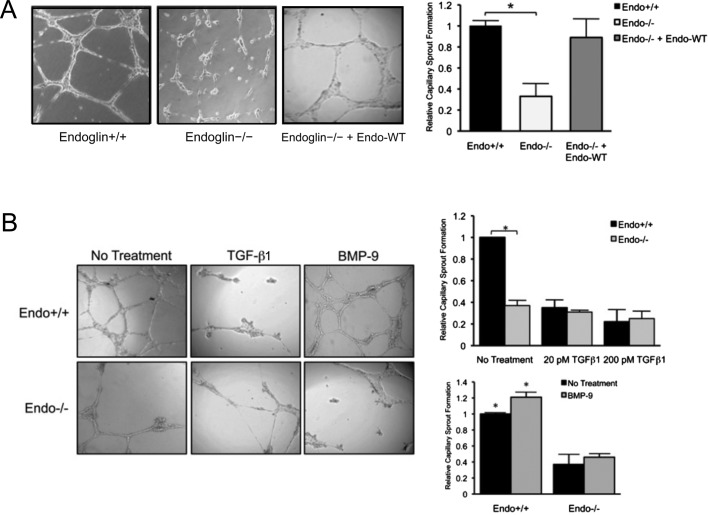
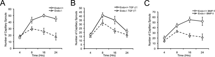
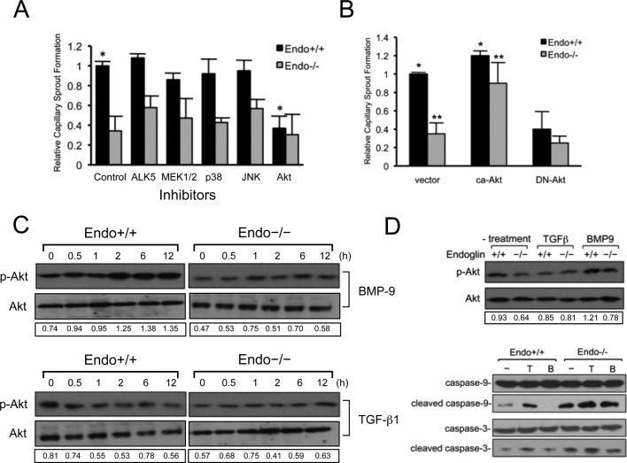
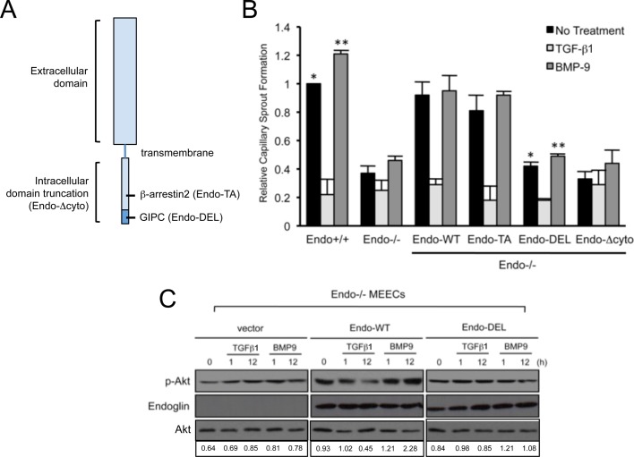
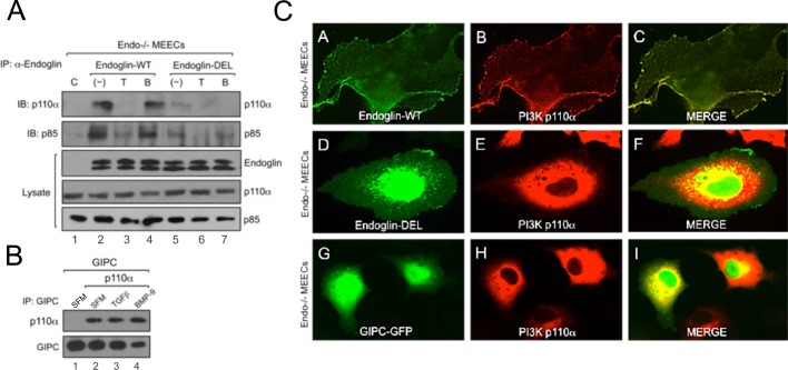
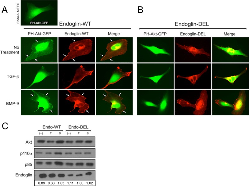
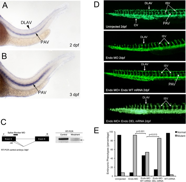
Endoglin (CD105) is an endothelial-specific transforming growth factor β (TGF-β) coreceptor essential for angiogenesis and vascular homeostasis. Although endoglin dysfunction contributes to numerous vascular conditions, the mechanism of endoglin action remains poorly understood. Here we report a novel mechanism in which endoglin and Gα-interacting protein C-terminus-interacting protein (GIPC)-mediated trafficking of phosphatidylinositol 3-kinase (PI3K) regulates endothelial signaling and function. We demonstrate that endoglin interacts with the PI3K subunits p110α and p85 via GIPC to recruit and activate PI3K and Akt at the cell membrane. Opposing ligand-induced effects are observed in which TGF-β1 attenuates, whereas bone morphogenetic protein-9 enhances, endoglin/GIPC-mediated membrane scaffolding of PI3K and Akt to alter endothelial capillary tube stability in vitro. Moreover, we employ the first transgenic zebrafish model for endoglin to demonstrate that GIPC is a critical component of endoglin function during developmental angiogenesis in vivo. These studies define a novel non-Smad function for endoglin and GIPC in regulating endothelial cell function during angiogenesis.


Figures

FIGURE 1:
FIGURE 1:
Endoglin promotes Matrigel-induced endothelial capillary sprouting and tube morphogenesis. (A) Comparison of capillary sprouting for Endo+/+ and Endo/ MEECs are shown, along with reconstituted expression of Endo-WT (human) in Endo/ MEECs. Graph represents the quantification, normalized to Endo+/+, of 10 independent experiments performed in triplicate. Error bars, SEM (see Materials and Methods). *p < 0.01 (B) Effects of ligands on capillary sprouting. Exogenous TGF-β1 (20 or 200 pM) and BMP-9 (16.5 nM) were added for 16 h. Quantification of capillary sprouting for Endo+/+ and Endo/ MEECs in the presence of each ligand is represented by graphs derived from six independent experiments performed in triplicate. *p< 0.05.
FIGURE 2:
FIGURE 2:
Endoglin modulates the stability of Matrigel-induced capillary sprouting and tube formation. (A) Comparison of capillary sprouting for Endo+/+ and Endo/ MEECs at 0–4, 8, 16, and 24 h. Data derived from the quantification of the total number of capillary sprouts of a representative experiment from three independent experiments performed in triplicate. Error bars, SEM of total scored sprouting branches (see Materials and Methods) (B) Exogenous TGF-β1 (50 pM) treatment for indicated times. (C) Exogenous BMP-9 (16.5 nM) treatment for indicated times.
FIGURE 3:
FIGURE 3:
Endothelial capillary stability requires endoglin-dependent signaling to the PI3K/Akt pathway. (A) The effects of inhibitors to ALK5 (10 μM SB431542), MEK1/2 (30 μM PD98059), p38 (10 μM SB203580), JNK (30 μM SP600125), and PI3K (5 μM LY294002) pathways in Matrigel-induced capillary sprouting. Graph represents the normalized compilation of three independent experiments performed in triplicate. Cells were treated with the inhibitors for 12–16 h and biochemically tested for their efficacy of inhibition. Error bars, SEM. *p < 0.05. (B) Akt signaling functions downstream of endoglin. Shown are Endo/ MEECs expressing vector, ca-Akt, or DN-Akt. Error bars, average SEM. *p < 0.05, **p < 0.03 (C) Biochemical analysis of Akt activation in response to TGF-β1 (50 pM) and BMP-9 (16.5 nM) using phospho-Akt antibody in Endo+/+ and Endo/ MEECs under monolayer culture conditions. Data are representative of at least three independent experiments. Densitometric analysis was based on band intensities of phospho-Akt relative to total Akt. (D) Direct biochemical analysis of Akt activation under no treatment, TGF-β1 (50 pM), or BMP-9 (16.5 nM) (top two panels and a representative sprout formation) and caspase cleavage (bottom three panels) upon capillary formation for 16 h. Data are representative of at least three independent experiments. Densitometric analysis was based on band intensities of phospho-Akt relative to total Akt.
FIGURE 4:
FIGURE 4:
Endoglin requires GIPC interaction for Akt activation and capillary sprout stabilization. (A) Schematic of endoglin structure. Shown are regions of truncation for the entire intracellular domain (Endo-Δcyto) and mutations on binding sites for β-arrestin2 (Endo-TA; amino acid residue substitution Thr-650 to Ala) and GIPC (Endo-DEL; amino acid truncation SMA656–658). (B) Endo/ MEEC assessed for capillary stability upon reconstituting expression of human endoglin, including the wild type (Endo-WT), a point mutant unable to bind β-arrestin2 (Endo-TA), a PDZ-motif truncation mutant unable to bind GIPC (Endo-DEL), or a mutant lacking the cytoplasmic domain (Endo-Δcyto). Graph represents three independent experiments performed in triplicate. Error bars, average SEM. *p < 0.01, **p < 0.01. (C) Biochemical analysis of Akt activation for the indicated times in human Endo-WT– and Endo-DEL– expressing Endo/ MEECs in response to TGF-β1 (50 pM) and BMP-9 (16.5 nM). Data are representative of at least three independent experiments. Densitometric analysis was based on band intensities of phospho-Akt relative to total Akt.
FIGURE 5:
FIGURE 5:
GIPC scaffolds PI3K to plasma membrane in an endoglin-dependent manner. (A) Endo/ MEECs expressing either human Endo-WT or Endo-DEL coimmunoprecipitated with endogenous p110α and p85 under no treatment, TGF-β1 (50 pM), or BMP-9 (16.5 nM) for 2 h. (B) Endo/ MEECs expressing FLAG-tagged GIPC coimmunoprecipitated with p110α in serum-free media (SFM; lanes 1 and 2), TGF-β1 (lane 3, 50 pM), or BMP-9 (lane 4, 16.5 nM). (C) Representative images of Endo/ MEECs expressing p110α with human Endo-WT (A–C), Endo-DEL (D–F), and GIPC-GFP (G–I).
FIGURE 6:
FIGURE 6:
Endoglin–GIPC interaction regulates Akt trafficking and activation. (A) Endo-WT expression with PH-Akt-GFP in Endo/ MEECs under no treatment or TGF-β1 (50 pM) or BMP-9 (16.5 nM) for 2 h. (B) Endo-DEL expression with PH-Akt-GFP in Endo/ MEECs under no treatment or TGF-β1 (50 pM) or BMP-9 (16.5 nM) for 2 h. (C) Subcellular fractionation followed by plasma membrane isolation of Endo/ MEECs expressing human Endo-WT or Endo-DEL under no treatment or TGF-β1 (50 pM) or BMP-9 (16.5 nM) for 4 h. Shown are transiently expressing human Endo-WT or Endo-DEL, along with endogenous levels of Akt, p110α, and p85 present in the isolated plasma membrane fraction. Densitometric analysis was based on band intensities of Akt relative to either Endo-WT or Endo-DEL in each lane.
FIGURE 7:
FIGURE 7:
Endoglin/GIPC interaction is required for angiogenesis in Fli1-EGFP zebrafish embryos. (A, B) Side views of endoglin expression in control embryos at 2 and 3 dpf, respectively. Black arrowheads (A) show robust and specific expression of endoglin at 2 dpf in the DLAV. Black arrows (A, B) show robust and strong expression of endoglin in the PAV, ventral vessel lateral to the notochord, adjacent to the myotome, at the level of the horizontal myoseptum (C) The MO targeting splice site is complementary to the fifth exon–intron boundary. RT-PCR was performed to confirm MO-targeting effects. Sequencing of the abnormal shorter transcript (black arrow) confirms that the natural splicing site is disrupted by the MO and a new cryptic splicing site is used in morphants, leading to the generation of an abnormal truncated transcript lacking the last 44 base pairs from exon 5. (D) Images of Fli1-EGFP–expressing control embryos, embryos injected with Endo-MO alone, or embryos rescued with human Endo mRNA at 48 hpf, as visualized by fluorescence microscopy. The Endo-MO–injected embryos display impaired sprouting of ISVs, an absence of the DLAV and the PTA, and a reduction of the caudal vein (CV) compared with controls. (E) Scoring of normal vs. mutant embryos based on the phenotype of Fli1-EGFP–expressing vasculature. Endo-WT mRNA rescues the MO phenotype, whereas Endo-DEL mRNA cannot. Each experimental condition is quantified based on the phenotype of at least 85 embryos.

References

    1. Bernabeu C, Conley BA, Vary CP. Novel biochemical pathways of endoglin in vascular cell physiology. J Cell Biochem. 2007;102:1375–1388. - PMC - PubMed
    1. Bourdeau A, Dumont DJ, Letarte M. A murine model of hereditary hemorrhagic telangiectasia. J Clin Invest. 1999;104:1343–1351. - PMC - PubMed
    1. Cheifetz S, Bellon T, Cales C, Vera SC, Bernabeu C, Massague J, Letarte M. Endoglin is a component of the transforming growth factor-beta receptor system in human endothelial cells. J Biol Chem. 1992;267:19027–19030. - PubMed
    1. Conley B, Koleva R, Simth J, Kacer D, Zhang D, Bernabeu C, Vary CP. Endoglin controls cell migration and composition of focal adhesions: function of the cytosolic domain. J Biol Chem. 2004;279:27440–27449. - PubMed
    1. David L, Feige JJ, Bailly S. Emerging role of bone morphogenetic proteins in angiogenesis. Cytokine Growth Factor Rev. 2009;20:203–212. - PubMed

Publication types

MeSH terms