. 2012 Jun 29;287(27):23119-27.
doi: 10.1074/jbc.M111.328294. Epub 2012 May 16.

A highly conserved cytoplasmic cysteine residue in the α4 nicotinic acetylcholine receptor is palmitoylated and regulates protein expression

Affiliations

A highly conserved cytoplasmic cysteine residue in the α4 nicotinic acetylcholine receptor is palmitoylated and regulates protein expression

Stephanie A Amici et al. J Biol Chem. .

Abstract

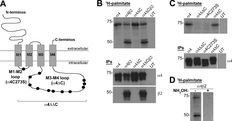
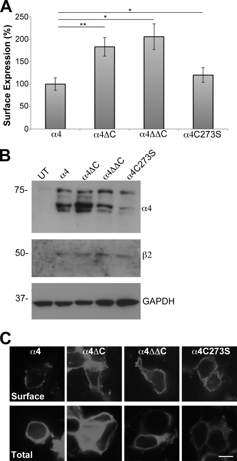
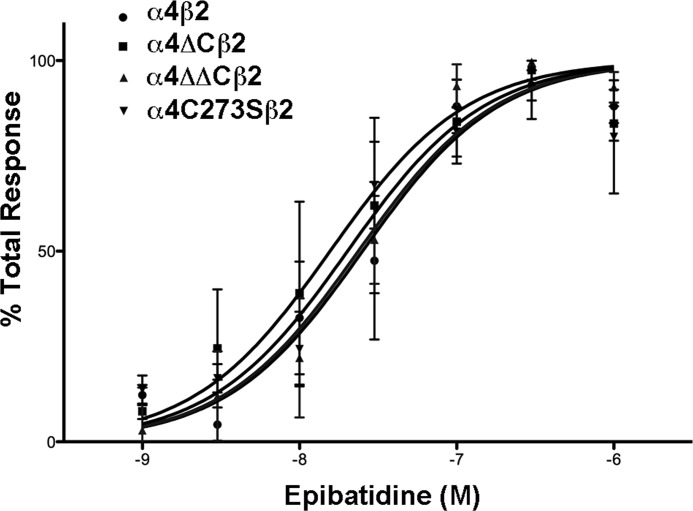
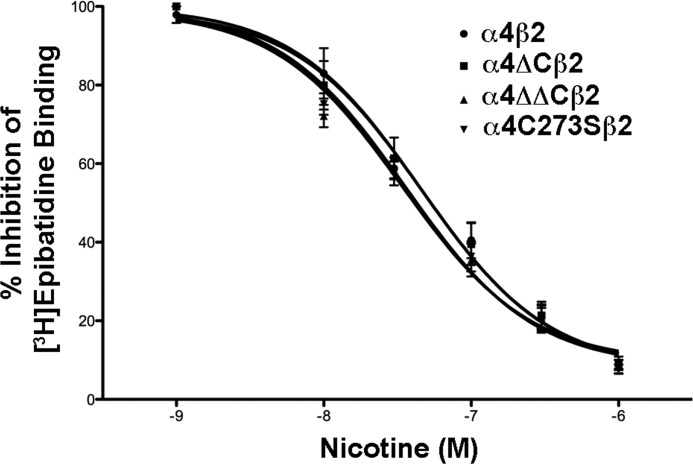
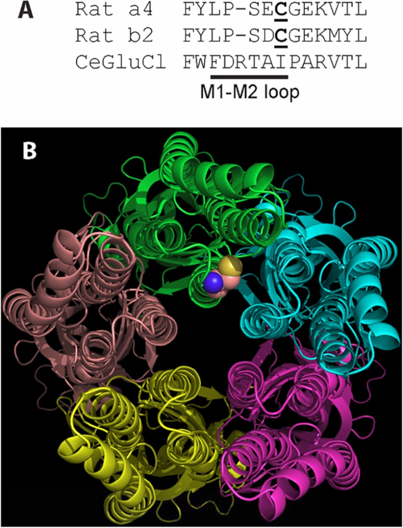
Nicotinic acetylcholine receptor (nAChR) cell surface expression levels are modulated during nicotine dependence and multiple disorders of the nervous system, but the mechanisms underlying nAChR trafficking remain unclear. To determine the role of cysteine residues, including their palmitoylation, on neuronal α4 nAChR subunit maturation and cell surface trafficking, the cysteines in the two intracellular regions of the receptor were replaced with serines using site-directed mutagenesis. Palmitoylation is a post-translational modification that regulates membrane receptor trafficking and function. Metabolic labeling with [(3)H]palmitate determined that the cysteine in the cytoplasmic loop between transmembrane domains 1 and 2 (M1-M2) is palmitoylated. When this cysteine is mutated to a serine, producing a depalmitoylated α4 nAChR, total protein expression decreases, but surface expression increases compared with wild-type α4 levels, as determined by Western blotting and enzyme-linked immunoassays, respectively. The cysteines in the M3-M4 cytoplasmic loop do not appear to be palmitoylated, but replacing all of the cysteines in the loop with serines increases total and cell surface expression. When all of the intracellular cysteines in both loops are mutated to serines, there is no change in total expression, but there is an increase in surface expression. Calcium accumulation assays and high affinity binding for [(3)H]epibatidine determined that all mutants retain functional activity. Thus, our results identify a novel palmitoylation site on cysteine 273 in the M1-M2 loop of the α4 nAChR and determine that cysteines in both intracellular loops are regulatory factors in total and cell surface protein expression of the α4β2 nAChR.


Figures

FIGURE 1.
FIGURE 1.
The α4 nAChR subunit is palmitoylated at Cys273. A, schematic of the α4 nAChR subunit, identifying the cysteines in the cytoplasmic loops that were mutated to serine residues: M1-M2 loop (C273S); and M3-M4 loop (C370S, C401S, C412S, C425S, C446S, C452S, C488S, C496S, C533S, C535S, C537S). B–D, tsA 201 cells were transiently transfected with N-terminally FLAG-tagged α4 (lane 1), α4 and β2 (lane 2), α4ΔC (lane 3), or α4ΔC and β2 (lane 4) nAChR subunits (B and D) or with N-terminally FLAG-tagged α4, α4ΔΔC, α4C273S, or α4ΔC nAChR subunits (C). UT denotes untransfected cells processed in parallel as a control (B and C). The cells were metabolically labeled with 1 millicurie/ml [3H] palmitate, solubilized, and the FLAG-tagged subunits were captured by IP with FLAG M2 beads. The IP eluates were separated by SDS-PAGE, dried under vacuum, exposed to film at −80 °C, and developed after 21 days (B and C, top panels). 5 μl of IP eluate (10% of total IP) was run on a SDS-PAGE and analyzed by Western analysis using anti-FLAG antibody and anti-β2 antibody (B and C, bottom panels). D, lysates from α4β2 expressing cells were treated with 1 m Tris-HCl (control) or 1 m hydroxylamine (NH2OH) to confirm thioester linkage to intracellular cysteines. Molecular mass is in kDa along the left side of the blot.
FIGURE 2.
FIGURE 2.
Removal of cysteines from the α4 nAChR cytoplasmic loops increases the surface expression of the receptor. A, tsA 201 cells were transfected with untagged β2 and N-terminally FLAG-tagged α4, α4ΔC, α4ΔΔC, or α4C273S nAChR subunits. Cell surface expression of α4β2 was measured using an enzyme-linked immunoassay using an mAb against the extracellular domain of the β2 nAChR subunit (mAb 295). The absorbance data were normalized, and the WT α4β2 nAChR was set to 100%, and data were expressed as means ± S.E. The α4ΔCβ2, α4ΔΔCβ2 and α4C273Sβ2 all had significantly higher surface expression than α4β2. *, p < 0.05; **, p < 0.001 (n = 6). B, transiently transfected tsA 201 cells were lysed in 750 μl of 2% Triton X-100 buffer. Soluble fractions (20 μl) were analyzed by SDS-PAGE and Western blotting. Blots were cut horizontally so that the same blot could be probed with either mouse monoclonal anti-FLAG antibody to detect the α4 nAChR, goat polyclonal anti-β2 antibody to detect the β2 nAChR or rabbit polyclonal anti-GAPDH antibody. Lysate from untransfected (UT) cells was run as a negative control. Molecular mass is in kDa along the left side of the blot. C, single-plane confocal images of transiently transfected tsA 201 cells were captured with a spinning disc confocal microscope. Top panel, to detect α4β2 nAChR expression on the cell surface, fixed, non-permeabilized cells were immunolabeled with mAb 295 followed by Alexa Fluor 594-conjugated goat anti-rat secondary antibody. Bottom panel, cells in a parallel set of wells were fixed, permeabilized, and immunolabeled with mAb 295 and Alexa Fluor 594-conjugated goat anti-rat secondary antibody to detect total α4β2 nAChR expression. All of the images were taken for the same exposure time with the same camera settings and processed identically. Scale bar, 10 μm.
FIGURE 3.
FIGURE 3.
2-Bromopalmitate significantly decreases the surface expression of α4β2 nAChRs. A, tsA 201 cells were transfected with untagged β2 and N-terminally FLAG-tagged α4, α4ΔC, α4ΔΔC, or α4C273S nAChR subunits and incubated with 120 μm 2-BP or DMSO (vehicle control) overnight at 30 °C. Cell surface expression of α4β2 was measured using an enzyme-linked immunoassay using an mAb against the extracellular domain of the β2 nAChR subunit (mAb 295). The absorbance data were converted to expression ratios by dividing the 2-BP-treated cells by the DMSO-treated cells for each construct (expressed as percentages). 2-BP significantly decreased the surface expression of all of the constructs. The values are from three separate experiments and expressed as the means ± S.E. B, tsA 201 cells were transiently transfected with untagged β2 nAChR and with N-terminally FLAG-tagged α4, α4ΔC, α4ΔΔC, or α4C273S. The constructs were incubated with DMSO (vehicle control) (left blots, −) or with 120 μm 2-BP (right blots, +). The cells were lysed in 750 μl of 2% Triton X-100 buffer. Soluble fractions (20 μl) were analyzed by SDS-PAGE and Western blotting. Blots were cut horizontally, so that the same blot could be probed with either mouse monoclonal anti-FLAG antibody (to detect the α4 nAChR) or rabbit polyclonal anti-GAPDH antibody. Molecular mass is in kDa along the left side of the blot. UT, untransfected.
FIGURE 4.
FIGURE 4.
There is no significant change in the EC50 for α4 nAChR cysteine mutants. Cultured tsA 201 cells expressing the various α4 constructs plus β2 were loaded with calcium5 NW dye for 60 min, stimulated with either 1, 3, 10, 30, 100, 300, or 1000 nm epibatidine, and intracellular calcium was measured via fluorescence. Results are expressed as percentage of epibatidine peak fluorescence levels after subtracting basal fluorescence. Values represent means ± S.E., n = 4 for α4 and α4ΔC; n = 3 for α4ΔΔC and α4C273S. The data were fitted to a normalized dose-response curve using the equation Y = 100/(1 + 10((log EC50X)·HS), where Y is the percentage of the maximal effect at a given concentration (X), and HS is the Hill slope (Hill slope = 1).
FIGURE 5.
FIGURE 5.
Cysteine mutations in the α4 nAChR subunit do not affect α4β2 nAChR ligand binding affinity. Cultured tsA 201 cells expressing the various α4 constructs plus β2 were fixed with paraformaldehyde, permeabilized, and incubated with 400 pm epibatidine in the presence or absence of 10, 30, 100, 300, or 1000 nm nicotine overnight at 4 °C. After several washes, the amount of bound epibatidine was quantified by scintillation counting. The data shown was normalized to the amount bound in the absence of nicotine and represent the mean ± S.E., n = 6 for α4; n = 5 for α4ΔC; n = 3 for α4ΔΔC and α4C273S. Curves were fitted to a one-site competitive binding using a Kd = 0.015 nm, using the equation Y = 100/(1 + 10((log IC50X)·HS), where Y is the percentage of the maximal effect at a given concentration (X), and HS is the Hill Slope (Hill slope = 1).
FIGURE 6.
FIGURE 6.
Palmitoylation of the α4 nAChR subunit does not affect the targeting of the receptor to presynaptic terminals. Single plane images of neurons that were transfected at 10 DIV, coplated with tsA 201 cells at 12 DIV and fixed, permeabilized, and immunostained at 14 DIV. A, neurons expressing α4β2 nAChRs were coplated with tsA 201 cells expressing neuroligin-1-HA (nlg1). B, neurons expressing α4β2 nAChRs and neurexin-1β VSV (nrx1β) were coplated with tsA 201 cells expressing neuroligin-1-HA. C, neurons expressing α4C273Sβ2 nAChRs and neurexin-1β were coplated with tsA 201 cells expressing neuroligin-1-HA. α4β2 nAChR and neurexin-1β-expressing cocultures exhibit enhanced targeting of α4β2 nAChRs to synapses (arrows). Antibody combinations were as follows: anti-β2 nAChR (mAb 295, β2), anti-VSV-G (nrx1β), and anti-HA (nlg1) antibodies. Scale bar, 10 μm.
FIGURE 7.
FIGURE 7.
The position of the palmitoylated cysteine likely allows ready access by the hydrophobic palmitoyl moiety to the hydrophilic cytoplasmic space and ion permeation path but does not allow ready access to the lipid bilayer. A, the primary sequence comparison for α4, β2, and a Cys-loop receptor from C. elegans (19) shows the residues of and adjacent to the M1-M2 loop. The palmitoylated cysteine of α4 and homologous cysteine of β2 are underlined. The spatial position of α4Cys273 in B is based on this primary sequence homology and the x-ray crystal structure of the receptor from C. elegans (Protein Data Bank code 3RHW). B, as seen from the cytoplasmic side of the receptor from C. elegans, the likely position of α4Cys273 is adjacent to the transmembrane domains, the cytoplasmic space, and the ion permeation path at the center of the figure. Only the main chains of the subunits are displayed except for the homologous position of α4Cys273, which is displayed as a space-filling model of Cys at the distal end of the M1-M2 loop of the green chain. M1 is at the 9 o'clock position of the green chain, M2 is at 6 o'clock, and M3 is at 3 o'clock.

References

    1. Millar N. S. (2003) Assembly and subunit diversity of nicotinic acetylcholine receptors. Biochem. Soc. Trans. 31, 869–874 - PubMed
    1. Ren X. Q., Cheng S. B., Treuil M. W., Mukherjee J., Rao J., Braunewell K. H., Lindstrom J. M., Anand R. (2005) Structural determinants of α4β2 nicotinic acetylcholine receptor trafficking. J. Neurosci. 25, 6676–6686 - PMC - PubMed
    1. Xu J., Zhu Y., Heinemann S. F. (2006) Identification of sequence motifs that target neuronal nicotinic receptors to dendrites and axons. J. Neurosci. 26, 9780–9793 - PMC - PubMed
    1. Charollais J., Van Der Goot F. G. (2009) Palmitoylation of membrane proteins (Review). Mol. Membr. Biol. 26, 55–66 - PubMed
    1. el-Husseini Ael-D., Bredt D. S. (2002) Protein palmitoylation: A regulator of neuronal development and function. Nat. Rev. Neurosci. 3, 791–802 - PubMed

Publication types

MeSH terms

LinkOut - more resources