. 2012 Jun 15;188(12):6071-83.
doi: 10.4049/jimmunol.1102066. Epub 2012 May 16.

Stromal endothelial cells establish a bidirectional crosstalk with chronic lymphocytic leukemia cells through the TNF-related factors BAFF, APRIL, and CD40L

Affiliations

Stromal endothelial cells establish a bidirectional crosstalk with chronic lymphocytic leukemia cells through the TNF-related factors BAFF, APRIL, and CD40L

Montserrat Cols et al. J Immunol. .

Abstract

Chronic lymphocytic leukemia (CLL) is a clonal B cell disorder of unknown origin. Accessory signals from the microenvironment are critical for the survival, expansion, and progression of malignant B cells. We found that the CLL stroma included microvascular endothelial cells (MVECs) expressing BAFF and APRIL, two TNF family members related to the T cell-associated B cell-stimulating molecule CD40L. Constitutive release of soluble BAFF and APRIL increased upon engagement of CD40 on MVECs by CD40L aberrantly expressed on CLL cells. In addition to enhancing MVEC expression of CD40, leukemic CD40L induced cleavases that elicited intracellular processing of pro-BAFF and pro-APRIL proteins in MVECs. The resulting soluble BAFF and APRIL proteins delivered survival, activation, Ig gene remodeling, and differentiation signals by stimulating CLL cells through TACI, BAFF-R, and BCMA receptors. BAFF and APRIL further amplified CLL cell survival by upregulating the expression of leukemic CD40L. Inhibition of TACI, BCMA, and BAFF-R expression on CLL cells; abrogation of CD40 expression in MVECs; or suppression of BAFF and APRIL cleavases in MVECs reduced the survival and diversification of malignant B cells. These data indicate that BAFF, APRIL, and CD40L form a CLL-enhancing bidirectional signaling network linking neoplastic B cells with the microvascular stroma.


Figures

FIGURE 1
FIGURE 1
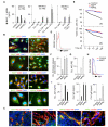
MVECs infiltrate CLL tissues and promote malignant B cell survival and proliferation. A. Immunofluorescence analysis of CLL lymph node (LN; one of 5 cases), spleen (SP; one of 3 cases) and bone marrow (BM; one of 3 cases) tissue samples stained for CD11c, CD21, CD31, CD68, CD206, elastase, factor VIII, vW factor or CD206. DAPI (blue) counterstains nuclear DNA. Original magnification, x10. B. CLL LN and SP tissue samples (one of 5 and 3 cases, respectively) stained for IgD (green), factor VIII or CD206 (red), and DAPI (blue). Original magnification, x40. C. CLL and normal LN tissue samples (5 cases in each group) stained for CD54, CD105, CD144 (red), and DAPI (blue). Original magnification, x40. D. Flow cytometric analysis of CD31, CD54, CD102, CD105, CD144 and CD206 expression by UVECs, SMVECs and LMVECs. Grey solid profile, isotype-matched control. E. Time course analysis of viable trypan blue-negative CLL cells (one of 5 cases) exposed to standard medium (SM, orange line), endothelial medium (EM, black line), conditioned medium from endothelial cell cultures (CM, blue line), or SMVECs (red line). F. MTT proliferation assay with CLL cells from mutated (MU, left panel) or unmutated (UM, right panel) samples (5 cases in each group) co-cultured with endothelial medium alone (control), UVECs or SMVECs. Results are expressed as fold induction release of MTT-derived formazan by proliferating CLL cells. Data are from one of three or five experiments yielding similar results (A-E) or summarize three different experiments (F). Error bars, s.e.m.; * P < 0.05 (ANOVA followed by one-tailed unpaired Student’s t-test).
FIGURE 2
FIGURE 2
MVECs promote division, activation and differentiation of CLL cells. A. Immunofluorescence analysis of CLL lymph node (LN) tissue samples (one of 5 cases) stained for Ki-67 (green or red), factor VIII (red) and IgD (blue or green). DAPI (blue) counterstains nuclei. Original magnification, x10 (left panel) or x63 (right panels). Arrowheads indicate proliferating CLL cells. B. 3HTdR incorporation of B cells from CLL patients or healthy individuals (5 cases in each group) cultured for 4 days with endothelial medium alone (control) or SMVECs. C. Southern blot analysis of germline Iγ1-Cγ1, Iγ2-Cγ2 and Iγ3-Cγ3 transcripts as well as Iγ1/2-Cμ, Iγ3-Cμ and Iα-Cμ switch circle transcripts RT-PCR amplified from CLL cells (one of 5 cases with little or no constitutive CSR) cultured for 4 days with endothelial medium alone (control), UVECs or SMVECs. β-actin is a loading control. bp, base pairs. D. QRT-PCR analysis of AID mRNA from CLL cells (5 cases) cultured for 4 days as in C. RE, relative expression as compared to freshly isolated CLL cells. E. CLL LN tissue (one of 5 cases) stained for AID (green), factor VIII (red) and Pax5 (blue). Arrowheads point to an intercellular bridge that connects factor VIII-positive MVECs with an AID-positive CLL cell, AID being a hallmark of ongoing B cell activation. F. ELISA of IgM, IgG and IgA secreted by CLL cells (5 cases) cultured for 7 days as in C. G. Flow cytometric analysis of IgG and IgA on CLL cells (one of 5 cases) cultured with endothelial medium alone (red open profile) or UVECs (blue open profile) for 7 days. Grey solid profile, isotype-matched control. H. Flow cytometric analysis of CD19 and CD38 on CLL cells (one of 5 cases) cultured for 7 days as in C. Data are from one of five experiments yielding similar results (A, C, E, G, H) or summarize three different experiments (B, D, F). Error bars, s.e.m.; * P < 0.05 (ANOVA followed by one-tailed unpaired Student’s t-test).
FIGURE 3
FIGURE 3
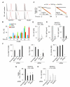
MVECs enhance BAFF ad APRIL release by interacting with CLL cells. A. ELISA of BAFF and APRIL secreted by UVECs, SMVECs or LMVECs cultured for 7 days in the absence (left panel) or presence (right panel) of CLL cells (3 cases) or CLL cell-derived conditioned medium (CM) (3 cases). Cultures were performed in plates with or without a transwell system (TW, no TW, respectively). B. Immunofluorescence analysis of BAFF or APRIL (green), CD31, vWF, CD63 or TGN-46 (red), and DAPI (blue) in MVECs. Original magnification, x40. C. Immunofluorescence analysis of CLL lymph node (LN; 5 cases) and spleen (SP; 3 cases) tissue samples stained for BAFF or APRIL (green), factor VIII or CD206 (red) and DAPI (blue). Original magnification, x40. D. Flow cytometric analysis of BAFF on SMVECs. Gray open profile, isotype-matched control; red open profile; BAFF. E. Time course analysis of viable trypan blue-excluding CLL cells from unmutated (UN) or mutated (MU) samples (one of 5 cases in each group) cultured with endothelial medium in the presence or absence of BAFF or APRIL. F. Formazan release assay with CLL cells (5 cases) cultured for 4 days as in D. G. QRT-PCR analysis of AID mRNA from CLL cells (5 cases) cultured for 4 days as in D. RE, relative expression compared to freshly isolated CLL cells. H. Flow cytometric analysis of surface IgA on CLL cells (one of 5 cases) cultured for 4 days with endothelial medium alone (red open profile) or BAFF (blue open profile). Gray solid profile, isotype-matched control. I. ELISA of IgM, IgG and IgA secreted by CLL cells cultured for 7 days as in E. Data are from one of three or five experiments yielding similar results (B-E and H) or summarize three different experiments (A, F, G and I). Error bars; s.e.m.; * P < 0.05 (ANOVA followed by one-tailed unpaired Student’s t-test).
FIGURE 4
FIGURE 4
MVECs stimulate CLL cells by engaging TACI, BCMA and BAFF-R through BAFF and APRIL. A. Flow cytometric analysis of BAFF-R, TACI and BCMA (red lined open profile) proteins on CLL cells from UN (upper row) or MU (lower row) samples (one of 4 cases in each group; see Table 1). Gray solid profile, isotype-matched control. B. Time course QRT-PCR analysis of BAFF-R, TACI and BCMA mRNAs from CLL cells (3 cases) exposed to CD40L. RE, relative expression compared to freshly isolated CLL cells. C. Viable trypan blue-negative CLL cells (one of 3 cases) cultured in endothelial medium with or without UVECs or SMVECs and in the presence or absence of control Fc5 or TACI-Ig for 0 or 144 hours. D. QRT-PCR analysis of AID mRNA from CLL cells (3 cases) cultured for 4 days in endothelial medium with SMVECs and control Fc5 or TACI-Ig. RE, relative expression compared to freshly isolated CLL cells. E. ELISA of IgM and IgA secreted by CLL cells (3 cases) cultured for 7 days as in D. F. QRT-PCR of BAFF-R, TACI and BCMA mRNAs in CLL cells (3 cases) nucleofected with scrambled, BAFF-R-, TACI- or BCMA-targeting siRNAs. RE, relative expression compared to non-nucleofected CLL cells. G. Viable trypan blue-negative CLL cells (3 cases) nucleofected with scrambled, BAFF-R-, TACI- or BCMA-targeting siRNAs and exposed to SMVECs for 4 days. H. QRT-PCR analysis of AID mRNA from CLL cells (3 cases) treated and cultured as in G. RE, relative expression compared to non-nucleofected CLL cells. Data are from one of eight (A) or three (C) experiments yielding similar results or summarize three different experiments (B and D-H). Error bars, s.e.m.; * P < 0.05 (ANOVA followed by one-tailed unpaired Student’s t-test).
FIGURE 5
FIGURE 5
MVECs express CD40 and up-regulate CD40L on CLL cells. A. Upper panels: immunofluorescence analysis of CLL lymph node (LN; 5 cases) or normal tonsillar (TO; 5 cases) tissues stained for CD40 (green), factor VIII (red), and IgD (blue). Bottom panels: flow cytometric analysis of CD40 (red open profile) on UVECs, SMVECs and LMVECs. Gray solid profile, isotype-matched control. Original magnification, x10 (upper panels) and x40 (bottom panels). B. Upper panels: CLL bone marrow (BM; 3 cases) stained for IgD or Pax5 (green), CD40L (red or blue), CD3 (yellow), and Ki-67 (blue). Inset in large left panel shows merged IgD and CD40L. Original magnification, x10 (left panels) or x63 (right panels). Bottom panels: CD40L (red open profile) on CLL cells (3 cases). Gray solid profile, isotype-matched control. Numbers indicate specific CLL cases. C. Left panels: normal tonsillar (TO; 5 cases) tissue stained for IgD (green), AID (red) and CD40L (blue). Original magnification, x10 (upper panel) or x20 (lower panel). Right panels: CD40L (red open profile) on naïve, marginal zone or memory B cells from a healthy spleen (3 cases). Gray solid profile, isotype-matched control. D. QRT-PCR of CD40L mRNA in naïve, marginal zone or memory B cells from healthy spleens (3 cases), freshly isolated CLL cells (6 cases, each analyzed in triplicates), or CLL cells exposed to SMVECs (2 cases, each analyzed in triplicates) for 4 days. CLL cells were pre-incubated with control vehicle (DMSO) or an NF-κB inhibitor (IKK inhibitor III) for 3 hours, washed, and then co-cultured with SMVECs. Results are normalized to freshly isolated naïve B cells. Numbers indicate CLL cases. E. QRT-PCR of CD40 mRNA in UVECs, SMVECs or LMVECs incubated with endothelial medium alone (control) or CD40L. F. Flow cytometric analysis of CD40 on SMVECs cells incubated with control Ig or CD40-Ig in the presence of CD40Lhigh CLL cells (blue open profile; one of three cases) or absence of CD40Lhigh CLL cells (red open profile) for 48 hours. Gray solid profile, isotype-matched control. Data are from one of six experiments yielding similar results (A-C, E, F) or summarize three different experiments (D). Error bars, s.e.m.; * P < 0.05 (ANOVA followed by one-tailed unpaired Student’s t-test).
FIGURE 6
FIGURE 6
MVECs enhance cleavase-dependent processing of BAFF and APRIL in response to CD40L. A. Left panel: time course QRT-PCR analysis of BAFF and APRIL mRNAs from SMVECs incubated with CD40L. Right panel: QRT-PCR analysis of BAFF mRNA from SMVECs exposed to CLL cells with low or high CD40L expression (3 cases in each group). RE, relative expression compared to unstimulated SMVECs. B. ELISA of BAFF and APRIL from UVECs or SMVECs incubated for 0, 3 or 6 hours with CD40L. C. Furin cleavage sites (blue arrows) and putative TACE cleavage sites (green arrows) of pro-BAFF and pro-APRIL proteins. The TACE cleavage site (red arrow) of the pro-TNF protein is shown for comparison. Numbers indicate amino acid residues in the membrane-proximal stalk region of BAFF, APRIL and TNF. D. Immunoblot of full-length pro-BAFF and pro-APRIL proteins from total lysates of UVECs and SMVECs incubated with medium alone (control) or CD40L for 6 hours. β-actin is a loading control. kDa, kilodaltons. E. ELISA of BAFF and APRIL in the supernatants from SMVECs incubated with or without CD40L and in the presence or absence of vehicle (DMSO), furin inhibitor (choromethylketone, CMK) or TACE inhibitor (TAPI-2). F. Immunofluorescence analysis of CLL lymph node (LN; 3 cases) stained for TACE or furin (green), Factor VIII (red), and IgD (blue). Original magnification, x10. G. QRT-PCR of TACE and furin in UVECs and SMVECs incubated for 1 or 5 hours with CD40L. Results are normalized to β-actin mRNA. RE, relative expression compared to unstimulated cells. H. Immunoblot of TACE and furin from SMVECs incubated with CD40L for various time points. Actin is a loading control. In mid panel, 96, 90 and 60 kDa bands correspond to precursor, mature and cleaved furin proteins. I. Viable trypan blue-excluding CLL cells (one of 3 cases) cultured with SMVECs, UVECs or MVECs in the presence or absence of control Ig or CD40-Ig for 0, 24 or 48 hours. J. Viable CLL cells (one of 3 cases) cultured with SMVECs, nucleofected with scrambled (scr) or CD40 siRNA for 0, 24 or 48 hours. Data are from one of three experiments yielding similar results (C, D, F and H-J) or summarize three different measurements (A, B, E and G). Error bars, s.e.m.; * P < 0.05 (ANOVA followed by one-tailed unpaired Student’s t-test).
FIGURE 7
FIGURE 7
MVECs up-regulate CD40L expression on CLL cells through BAFF and APRIL. A. Flow cytometric analysis of CD40L, IgG, IgA, BAFF-R, TACI and BCMA on CLL cells (one of 6 cases) incubated with endothelial medium alone (red open profile, also shown in center and right panels), SMVECs plus control Fc5 (blue open profile), or SMVECs plus TACI-Ig (blue open profile) for 7 days. Gray solid profile, isotype-matched control. B. Flow cytometric analysis of CD40L on CLL cells (one of 6 cases) incubated with endothelial medium alone (red open profile; also shown in center and right panels) or endothelial medium supplemented with BAFF or APRIL (blue open profile) for 7 days. Gray solid profile, isotype-matched control. C. Viable trypan blue-excluding CLL cells (one of 6 cases) cultured with control Ig or CD40-Ig for 0, 24 or 48 hours. Data in A-C are from one of six experiments yielding similar results. D. Model summarizing crosstalk between MVECs and CLL cells via CD40L, BAFF and APRIL.

Similar articles

Cited by

References

    1. Chiorazzi N, Rai KR, Ferrarini M. Chronic lymphocytic leukemia. N Engl J Med. 2005;352:804–815. - PubMed
    1. Damle RN, Wasil T, Fais F, Ghiotto F, Valetto A, Allen SL, Buchbinder A, Budman D, Dittmar K, Kolitz J, Lichtman SM, Schulman P, Vinciguerra VP, Rai KR, Ferrarini M, Chiorazzi N. Ig V gene mutation status and CD38 expression as novel prognostic indicators in chronic lymphocytic leukemia. Blood. 1999;94:1840–1847. - PubMed
    1. Hamblin TJ, Davis Z, Gardiner A, Oscier DG, Stevenson FK. Unmutated Ig V(H) genes are associated with a more aggressive form of chronic lymphocytic leukemia. Blood. 1999;94:1848–1854. - PubMed
    1. Wiestner A, Rosenwald A, Barry TS, Wright G, Davis RE, Henrickson SE, Zhao H, Ibbotson RE, Orchard JA, Davis Z, Stetler-Stevenson M, Raffeld M, Arthur DC, Marti GE, Wilson WH, Hamblin TJ, Oscier DG, Staudt LM. ZAP-70 expression identifies a chronic lymphocytic leukemia subtype with unmutated immunoglobulin genes, inferior clinical outcome, and distinct gene expression profile. Blood. 2003;101:4944–4951. - PubMed
    1. Furman RR, Asgary Z, Mascarenhas JO, Liou HC, Schattner EJ. Modulation of NF-kappa B activity and apoptosis in chronic lymphocytic leukemia B cells. J. Immunol. 2000;164:2200–2206. - PubMed

Publication types

MeSH terms

Substances